Original Article

Inhibitory effect of an ethanol extract mixture of Aralia elata leaf, Chaenomeles sinensis fruit and Glycyrrhizae radix on Aβ (25–35)-induced memory impairment

Yeon Hee Seong 1 , * https://orcid.org/0000-0001-9088-8987
Author Information & Copyright
1College of Veterinary Medicine, Chungbuk National University, Cheongju 28644, Korea
*Corresponding author: Yeon Hee Seong, College of Veterinary Medicine, Chungbuk National University, Cheongju 28644, Korea, Tel: +82-43-261-2968, E-mail: vepharm@cbnu.ac.kr

© Research Institute of Veterinary Medicine, Chungbuk National University. All rights reserved.

Received: May 20, 2021; Revised: Jun 07, 2021; Accepted: Jun 08, 2021

Published Online: Jun 30, 2021

Abstract

The young shoots of Aralia elata, Chaenomeles sinensis fruit and Glycyrrhizae radix are edible and traditionally used as anti-inflammatory and antioxidant agents. The present study was performed to investigate the protective effect of an ethanol extract mixture of these three medicinal plants (ACG) against amyloid β protein (Aβ) (25–35)-induced memory impairment in an ICR mouse model. Memory impairment was induced by intracerebroventricular microinjection of 15 nmol Aβ (25–35) and assessed using the passive avoidance test and the Morris water maze test. The step-through latency in the passive avoidance test was decreased and the latency to reach the hidden platform in the Morris water maze test was increased in mice treated with Aβ (25–35), indicating memory impairment. This memory impairment induced by Aβ (25–35) was significantly prevented by chronic treatment with ACG (10, 25, and 50 mg/kg, p.o., 8 days). In memory impaired mice brain, cholinesterase activity and concentration of thiobarbituric acid reactive substance, a lipid peroxidation marker, were increased and glutathione level was decreased. These biochemical changes in Aβ (25–35)-treated mice were reversed by chronic administration of ACG. The present results suggest that antioxidant and anti-cholinesterase activities of ACG might be responsible for the inhibition of Aβ (25–35)-induced memory impairment and that ACG preparation may have a therapeutic role in preventing the progression of Alzheimer’s disease.

Keywords: Aralia elata; Chaenomeles sinensis fruit; Glycyrrhizae radix; antidementia; amyloid β protein

Introduction

Alzheimer’s disease (AD) is a multifactorial neurodegenerative disorder with progressive and devastating memory impairment. The AD patient brain is characterized by amyloid β protein (Aβ) deposits, neurofibrillary tangles and loss of basal forebrain cholinergic neurons [1]. Both in vivo and in vitro studies have reported the toxic effects of Aβ or Aβ peptide fragments in brain suggesting an important role of Aβ in the pathogenesis of AD [2]. The mechanisms underlying Aβ neurotoxicity are complex but may involve oxidative stresses associated with reactive oxygen species (ROS) generation and decreased levels of glutathione (GSH) and superoxide dismutase (endogenous antioxidants). Aβ has been reported to produce hydrogen peroxide and lipid peroxide in neurons [3], superoxide in microglia [4] and proinflammatory cytokines [5]. This Aβ-induced oxidative stress is indexed by protein oxidation, lipid peroxidation, DNA oxidation, and neuronal cell death [68]. Therefore, antioxidants such as α-tocopherol and anti-inflammatory agents such as indomethacin reportedly slow the progression of AD [9, 10].

Aralia elata Seem (Araliaceae) is widely distributed in oriental countries such as Korea, Japan and China. The young shoots of Aralia elata are a popular edible plant in the spring. Several bioactive constituents such as saponins, alkaloids, glycosides, palmitic acid, and linoleic acid from Aralia elata have been reported to have anti-inflammatory and anti-oxidative effects and inhibit neurodegeneration [1113]. Chaenomeles sinensis (Thouin) Koehne fruit (Rosaceae) is a valuable source of health food and Chinese medicine. Chaenomeles sinensis fruit is rich in dietary fibre, organic acids and bioactive pentacyclic triterpene acids such as oleanolic acid and ursolic acid and large amounts of bioactive phenolic acids and vitamin C [14, 15]. These compounds are related to anti-inflammatory and antioxidant effect of Chaenomeles sinensis fruit [1618]. Phytochemical researches have revealed that the major constituents of Glycyrrhizae radix (Leguminosae), the root of Glycyrrhiza uralensis, are flavonoids and triterpenoid saponins, which possess diverse pharmacological properties such as anti-ulcer, anti-inflammatory, antispasmodic, antioxidation, anti-cancer, anti-depression and memory enhancement [1924].

In previous reports, Chaenomeles sinensis fruit was demonstrated to show neuroprotective effects against Aβ (25–35)-induced neurotoxicity in in vivo and in vitro [25]. Glycyrrhizae radix prevented Aβ (25–35)-induced neurotoxicity in cultured neurons [26]. Glycyrrhizae radix have been proved for its memory-enhancing effect and its anticholinesterase activity [23, 27]. Studies have reported neuronal protective effects of Aralia elata in some animal models [13, 28]. Therefore, it was hypothesized that mixture of these three plants, possessing anti-oxidant and anti-inflammatory activities, might show more effective and synergistic neuroprotective effect with the reduction of possible individual toxicity due to large quantity. The present study was conducted to confirm the inhibitory effect of an ethanol extract mixture of Aralia elata, Chaenomeles sinensis fruit, and Glycyrrhizae radix, which was named as ACG, on memory impairment induced by a single intracerebroventricular (i.c.v.) injection of Aβ (25–35) in mouse model of AD-type amnesia [29].

Materials and Methods

Plant materials and extraction and reagents

The dried and sliced Chaenomeles sinensis fruit and Glycyrrhizae radix were purchased from Daegu Oriental Pharm at Daegu, Korea. The young shoots of Aralia elata were collected at Keryong Mountain in Daejeon, Korea. These plants were identified by Dr. Bangyeon Hwang, Chungbuk National University. Each 200 g of the three plants was mixed, extracted with 95% ethanol (3 L × 24 h × 3) at room temperature and filtered through filter paper (Advantec MFS, CA, USA). The filtrate was concentrated under reduced pressure using a rotary evaporator (Heidolph Instruments GmbH & Co,. Schwabach, Germany) to yield an ethanol extract (ACG, 55 g), which was then stored at room temperature until required. Aβ (25–35) was purchased from Bachem (Bubendorf, Switzerland). Acetylthiocholine iodide and 5,5’-dithiobis-2-nitrobenzoic acid (DTNB) were purchased from Sigma Chemical (St. Louis, MO, USA). Thiobarbituric acid (TBA) was purchased from Tokyo Kasei Kogyo (Tokyo, Japan).

Experimental animals

Male ICR mice (5-week-old) were supplied by Daehan BioLink (Chungbuk, Korea) and housed in an environmentally controlled room at 22 ± 2°C and 55 ± 5% RH under a 12-h light/dark cycle with ad libitum access to food and water. Mice were allowed to adapt to the experimental environment for a week.

Induction of memory impairment in mice and administration of ACG

Memory impairment was induced by i.c.v. injection of 15 nmol of the aggregated form of Aβ (25–35), as previously described [2]. ACG was administered orally 30 min before Aβ (25–35) injection on experimental day 1 (ED1) and then further administered daily for 7 days (to ED8). ACG was suspended in distilled water. Sham-controls were injected i.c.v. with physiological saline instead of 15 nmol of Aβ (25–35). Mice were randomly divided into five groups ; sham-operated (the sham control group), the 15 nmol Aβ (25–35) group, the 15 nmol Aβ (25–35) + 10 mg/kg ACG group, the 15 nmol Aβ (25–35) + 25 mg/kg ACG group, and the 15 nmol Aβ (25–35) + 50 mg/kg ACG groups. Distilled water as vehicle was administered orally to the sham control and Aβ (25–35) control groups using the same schedule as ACG administration.

Memory assessment

Passive avoidance test was performed using a two-compartment shuttle chamber device (Avoidance System version 1.1, B. S Technolab, Seoul, Korea), one chamber was illuminated and the other was unlit and equipped with a grid floor and shock generator [2]. Thirty minutes after administering ACG on ED8, the mice were trained to perform the step-through passive avoidance task with the electric shock system activated (0.5 mA, 3 s; the acquisition trial). Retention trials were performed 24 h after the acquisition trial and the step-though latencies were recorded (maximum test time was set at 300 s).

A Morris water maze test was performed to measure spatial learning and memory of the mice according to the Morris method [30] with slight modification. The experimental apparatus consisted of a circular water tank (90-cm diameter, height of 40 cm) filled with water (25 ± 2°C) to a depth of 30 cm. An escape platform with an 8-cm diameter was submerged 1 cm below the surface of the water and placed in the middle of the same quadrant throughout the training phase. Mice were pretested in same condition prior to ACG and/or Aβ (25–35) injection to identify the similar ability to find hidden platform on ED1. Mice were allowed 4 training trials per day after ACG administration for 5 consecutive days (from ED4 to ED8). For each trial, the mice were placed into the water from one of the four starting points on the edge of the tank. The time required to escape onto the hidden platform was recorded. If the mouse located the platform, it was allowed to remain there for 10 s. If the mouse failed to find the platform within 120 s, it would be gently guided to the platform where it was allowed to stay for 10 s, and the escape latency was recorded as 120 s.

Measurement of cholinesterase activity and oxidative stress in mouse brain

After the passive avoidance test retention trial on ED9, mice were deeply anesthetized with diethyl ether, brains were quickly removed, and brain homogenates were prepared using five volumes of 0.1 M phosphate buffer (pH 7.4) in ice bath. Cholinesterase activity and levels of reduced GSH in mouse brain were determined spectrophotometrically using Ellman reagent [31]. Acetylthiocholine iodide was used as a substrate to measure cholinesterase activity. Extent of lipid peroxidation was assayed by measuring TBA reactive substance (TBARS) levels at 532 nm as described by Yoshioka et al. using 1,1,3,3-tetramethoxypropane as a standard [32]. Protein concentrations were determined using Lowry’s method [33].

Statistical analysis

Results are presented as means ± S.E.M. The significances of differences were assessed by one-way analysis of variance (ANOVA) followed by Tukey’s tests. p-values of <0.05 were considered significant.

Results

Inhibitory effect of ACG on Aβ (25–35)-induced memory impairment in mice

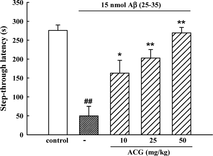
In the initial acquisition trial of the passive avoidance task, the step-through latency did not differ among the 5 groups. However, in the retention trial, step-through latency in the Aβ (25–35)-control group was significantly shorter than that in the sham control group (49.7 ± 25.5 s versus 275.8 ± 14.5 s) indicating that Aβ (25–35) had induced memory impairment. Mice administered ACG daily 8 days showed significant memory improvements (162.7 ± 34.0, 202.7 ± 22.5 and 269.2 ± 14.4 s for 10, 25, and 50 mg/kg, respectively (Fig. 1).

jbtr-22-2-78-g1
Fig. 1. Inhibitory effect of ACG on Aβ (25–35)-induced memory impairment in mice. Learning and memory performance were assessed using the passive avoidance test (n = 10 – 12). Values are expressed as means ± S.E.M. ##p<0.01 vs. sham control; *p<0.05 and **p<0.01 vs. 15 nmol Aβ (25–35) control.
Download Original Figure

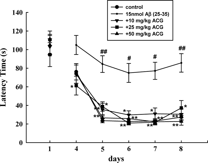
During the first trials using the Morris water maze test, latency time required to reach the hidden platform did not significantly differ among all the groups. The sham mice rapidly learned the location of the hidden platform, reaching it within 30 s on ED5 compared to the first trials (110.3 ± 6.1 s). Mice that received Aβ (25–35) showed a significantly delayed escape latency time from ED5 to ED8 compared to that of the sham-control group, indicating that Aβ (25–35) caused remarkable cognitive deficit. In contrast, chronic administration of ACG (10, 25, and 50 mg/kg) significantly decreased the Aβ (25–35)-induced prolonged latency time (ED5, 6, 7, and 8; Fig. 2).

jbtr-22-2-78-g2
Fig. 2. Inhibitory effect of ACG on Aβ (25–35)-induced spatial memory impairment in mice. The spatial learning and memory performance were assessed using a Morris water maze test (n = 7–9). Values are expressed as means ± S.E.M. #p<0.05 and ##p<0.01 vs. sham control; *p<0.05 and **p<0.01 vs. 15 nmol Aβ (25–35) control.
Download Original Figure
Inhibitory effect of ACG on Aβ (25–35)-induced increase of cholinesterase activity

Cholinesterase activity in the brains of mice in the Aβ (25–35)-treated group was significantly higher than that in the sham control group (9.3 ± 0.5 μmol/h/mg protein versus 5.1 ± 0.3 μmol/h/mg protein). The increased cholinesterase activity was significantly inhibited by 25 and 50 mg/kg ACG, showing 7.5 ± 0.2 and 7.2 ± 0.3 μmol/h/mg protein, respectively (Table 1).

Table 1. Effect of ACG on brain cholinesterase activity in Aβ (25–35)-treated mouse brain
Group Dose (mg/kg) Cholinesterase activity (μmol/h/mg protein)
Sham - 5.1 ± 0.3
Aβ (25–35) 15 nmol/animal 9.3 ± 0.5##
+ ACG 10 8.8 ± 0.2
+ ACG 25 7.5 ± 0.2**
+ ACG 50 7.2 ± 0.3**

Cholinesterase activities in mouse brain homogenates were measured after the passive avoidance test retention trial. Values are expressed as the means ± S.E.M. of acetylthiocholine concentrations hydrolyzed by cholinesterase preparation of brain homogenate for 1 h (n = 6 mice/group).

## p<0.01 vs. sham control,

** p<0.01 vs. 15 nmol Aβ (25–35) control.

Download Excel Table
Inhibitory effect of ACG on Aβ (25–35)-induced GSH depletion and lipid peroxidation

GSH contents of the brains from Aβ (25–35)-treated mice was significantly lower compared to that of the sham group. ACG (25 and 50 mg/kg) inhibited the decrease of GSH levels in Aβ (25–35)-treated brains. TBARS concentration in the brain of Aβ (25–35)-treated mice significantly increased compared to that of the sham group. ACG (25 and 50 mg/kg) suppressed the elevation of TBARS concentration induced by Aβ (25–35) (Table 2).

Table 2. Inhibitory effects of ACG on Aβ (25–35)-induced reductions in GSH levels and increases in TBARS levels in mouse brain
Group Dose (mg/kg) GSH (nmol/mg protein) TBARS (nmol/mg protein)
Sham - 9.6 ± 0.4 0.17 ± 0.02
Aβ (25–35) 15 nmol 7.9 ± 0.2## 0.23 ± 0.02#
+ ACG 10 8.9 ± 0.6 0.19 ± 0.01
+ ACG 25 9.1 ± 0.3* 0.17 ± 0.01**
+ ACG 50 10.5 ± 0.6** 0.16 ± 0.01**

GSH and TBARS levels were measured after the passive avoidance test retention trial. Values are expressed as means ± S.E.M. (n = 6 mice/group).

# p<0.05

## p<0.01 vs. sham control;

* p<0.05

** p<0.01 vs. 15 nmol Aβ (25–35) control.

Download Excel Table

Discussion

Aβ peptides (1–40 and 1–42 amino acid), cleaved from amyloid protein precursor, are the major constituents of senile plaques occurring in AD and playing a critical role as the principal toxic species to neuronal cell death and breakage of neurite [34]. The 11 amino acid sequence Aβ (25–35) exhibits neurotoxic properties of full-length sequence Aβ (1–42) [35]. It is widely acknowledged that mice i.c.v. injected with Aβ (25–35) can mimic some cognitive deficits in AD [36, 37], showing memory impairment in different behavioral paradigms such as Morris water maze and passive avoidance tests [38, 39]. In the present study, i.c.v. injection of Aβ (25–35) to mice produced memory impairment in the passive avoidance test and Morris water maze test. Chronic treatment with ACG effectively protected the mice against Aβ (25–35)-induced memory deficit.

Cholinergic transmission is crucial to learning and memory, and its alteration is considered main causes of cognitive disorder in AD. I.c.v. injection of Aβ (25–35) in rats resulted in learning and memory deficits that were accompanied by reduction of choline-acetyltransferase activity [40]. Accumulation of Aβ in AD brain is associated with elevation of cholinesterase and results in learning and memory deficits [41]. Thus, some cholinesterase inhibitors, tacrine (Cognex®) and donepezil (Aricept®), have been used for the treatment of AD [4143]. In the present study, the cholinesterase activity in brain tissue was increased in Aβ (25–35)-treated mice, and ACG (25 and 50 mg/kg) significantly inhibited the increase of cholinesterase. This result suggests that ACG could ameliorate memory impairment by inhibiting cholinesterase activity.

Studies have suggested that oxidative stress is responsible for the onset of the cognitive dysfunction during the progression of AD [3, 4, 7, 44]. Extracellular Aβ accumulation may directly or indirectly alter N-methyl-D-aspartic acid (NMDA) type glutamate receptor-mediated cytosolic increases in Ca2+, which is rapidly taken up by mitochondria and endoplasmic reticulum. Ca2+ overload of mitochondria causes ROS generation [45], and excessive production of ROS and impairment of the ROS removal system in the AD brain result in protein, nucleic acid, and neuronal membrane lipid damage, which in turn, cause neuronal cell death [46]. Therefore, a number of antioxidants have been reported to protect cells from Aβ (25–35)-induced toxicity [9, 47, 48]. GSH, a major intracellular antioxidant, plays a key role in the removal of ROS and protection of cells from oxidative stress. GSH level, thus, is the first indicator for oxidative stress in the brain [49]. TBARS level, commonly used as lipid peroxidation marker, has been elevated in the AD brain tissue [44]. In the present study, brain GSH contents in the Aβ (25–35)-treated group were significantly reduced, and ACG administration significantly prevented these decreases. Additionally, Aβ (25–35)-induced increase in TBARS concentration was significantly decreased by administration of ACG.

The three constituent plants of ACG and their active principles have been reported for neuroprotective activities. It is well known that Aralia elata contains antioxidants, such as congmuyanosides, echinocystic acid and oleanolic acid [12, 50]. Ethyl acetate fraction from Aralia elata (100 mg/kg) improved learning, memory ability, and spatial cognition through inhibition of cholinesterase activity and antioxidant effect [51]. Chaenomeles sinensis fruit is rich in antioxidant components such as flavonoids, phenolics, total triterpenes, and vitamin C [52]. In a previous report, we demonstrated that Chaenomeles sinensis fruit has protective effect on Aβ (25–35)-induced neurotoxicity in in vitro and in vivo [25]. Chaenomeles sinensis fruit inhibited Aβ (25–35)-induced increase of intracellular Ca2+ concentration ([Ca2+]i), ROS generation, and, in result, neuronal death in cultured neurons. Furthermore, Chaenomeles sinensis fruit (50 mg/kg) suppressed Aβ (25–35)-induced increase of brain cholinesterase activity and memory impairment. Glycyrrhizae radix contains many licorice flavonoids including liquiritin, isoliquiritin, liquiritigenin and isoliquiritigenin [53]. Glycyrrhizae radix and isoliquiritigenin prevented Aβ (25–35)-induced neuronal apoptotic death by interfering with the increases of [Ca2+]i and ROS in cultured cortical neurons [26]. Memory enhancing activity of Glycyrrhizae radix in mice was shown at dosage of higher than 150 mg/kg [23, 54]. Maximal effect of ACG was shown at the lower dosage (50 mg/kg) than the sum of maximal effect dosage of each plant. Therefore, it is suggested that the preparation of ACG might reveal synergistic effect of three medicinal plants in protection of Aβ-induced memory impairment through the inhibition of ROS generation and cholinergic dysfunction. Abundant antioxidants contained in this preparation might be responsible for the prevention of Aβ (25–35)-induced neurotoxicity. In conclusion, it is evident that ACG could provide a marked protection against Aβ (25–35)-induced memory deficit. This result may explain the inhibitory action of ACG on the progression of AD.

Conflict of Interest

The authors declare no potential conflict of interest.

Ethics Approval

All procedures involving experimental animals complied with the animal care guidelines issued by the U.S. National Institute of Health and those issued by the Animal Ethics Committee of Chungbuk National University (CBNUA-177-1001-01).

References

1.

Ballard C, Gauthier S, Corbett A, Brayne C, Aarsland D, Jones E. Alzheimer’s disease. Lancet 2011;377:1019-1031.

2.

Cho SO, Ban JY, Kim JY, Jeong HY, Lee IS, Song KS, Bae K, Seong YH. Aralia cordata protects against amyloid β protein (25–35)–induced neurotoxicity in cultured neurons and has antidementia activities in mice. J Pharmacol Sci 2009;111:22-32.

3.

Behl C, Davis JB, Lesley R, Schubert D. Hydrogen peroxide mediates amyloid β protein toxicity. Cell 1994;77:817-827.

4.

McDonald DR, Brunden KR, Landreth GE. Amyloid fibrils activate tyrosine kinase-dependent signaling and superoxide production in microglia. J Neurosci 1997;17:2284-2294.

5.

Gitter BD, Cox LM, Rydel RE, May PC. Amyloid beta peptide potentiates cytokine secretion by interleukin-1 beta-activated human astrocytoma cells. Proc Natl Acad Sci USA 1995;92:10738-10741.

6.

Butterfield DA, Boyd-Kimball D. Amyloid β-peptide (1-42) contributes to the oxidative stress and neurodegeneration found in Alzheimer disease brain. Brain Pathol 2004;14:426-432.

7.

Singh M, Nam DT, Arseneault M, Ramassamy C. Role of by-products of lipid oxidation in Alzheimer’s disease brain: a focus on acrolein. J Alzheimers Dis 2010;21:741-756.

8.

Markesbery WR. Oxidative stress hypothesis in Alzheimer’s disease. Free Radic Biol Med 1997;23:134-147.

9.

Sano M, Ernesto C, Thomas RG, Klauber MR, Schafer K, Grundman M, Woodbury P, Growdon J, Cotman CW, Pfeiffer E, Schneider LS, Thal LJ. A controlled trial of selegiline, alpha-tocopherol, or both as treatment for Alzheimer’s disease. The Alzheimer’s disease cooperative study. N Engl J Med 1997;336:1216-1222.

10.

Gasparini L, Ongini E, Wenk G. Non-steroidal anti-inflammatory drugs (NSAIDs) in Alzheimer’s disease: old and new mechanisms of action. J Neurochem 2004;91:521-536.

11.

Shikov AN, Pozharitskaya ON, Makarov VG. Aralia elata var. mandshurica (Rupr. & Maxim.) J.Wen: an overview of pharmacological studies. Phytomedicine 2016;23:1409-1421.

12.

Zhang M, Liu G, Tang S, Song S, Yamashita K, Manabe M, Kodama H. Effect of five triterpenoid compounds from the buds of Aralia elata on stimulus-induced superoxide generation, tyrosyl phosphorylation and translocation of cytosolic compounds to the cell membrane in human neutrophils. Planta Med 2006;72:1216-1222.

13.

Kim SJ, Kim MJ, Choi MY, Kim YS, Yoo JM, Hong EK, Ju S, Choi WS. Aralia elata inhibits neurodegeneration by downregulating O-GlcNAcylation of NF-κB in diabetic mice. Int J Ophthalmol 2017;10:1203-1211.

14.

Hamauzu Y, Yasui H, Inno T, Kume C, Omanyuda M. Phenolic profile, antioxidant property, and anti-influenza viral activity of Chinese quince (Pseudocydonia sinensis Schneid.), quince (Cydonia oblonga Mill.), and apple (Malus domestica Mill.) fruits. J Agric Food Chem 2005;53:928-934.

15.

Grygorieva O, Klymenko S, Vergun O, Mňahončáková E, Brindza J, Terentjeva M, Ivanišová E. Evaluation of the antioxidant activity and phenolic content of Chinese quince (Pseudocydonia sinensis Schneid.) fruit. Acta Sci Pol Technol Aliment 2020;19:25-36.

16.

Du H, Wu J, Li H, Zhong PX, Xu YJ, Li CH, Ji KX, Wang LS. Polyphenols and triterpenes from Chaenomeles fruits: chemical analysis and antioxidant activities assessment. Food Chem 2013;141:4260-4268.

17.

Han YK, Kim YS, Natarajan SB, Kim WS, Hwang JW, Jeon NJ, Jeong JH, Moon SH, Jeon BT, Park PJ. Antioxidant and anti-inflammatory effects of Chaenomeles sinensis leaf extracts on LPS-stimulated RAW 264.7 cells. Molecules 2016;21:422.

18.

Zhang R, Zhan S, Li S, Zhu Z, He J, Lorenzo JM, Barba FJ. Anti-hyperuricemic and nephroprotective effects of extracts from Chaenomeles sinensis (Thouin) Koehne in hyperuricemic mice. Food Funct 2018;9:5778-5790.

19.

Wu H, Zhang CE, Yu XH, Ma GC, Wang JB, Ma ZJ, Zhao KJ. Anti-inflammatory and immunomodulatory effects of Tripterygium wilfordii processed with licorice on mice model of ulcerative colitis. Zhongguo Zhong Yao Za Zhi 2019;44:3435-3440.

20.

Sato Y, He JX, Nagai H, Tani T, Akao T. Isoliquiritigenin, one of the antispasmodic principles of Glycyrrhiza ularensis roots, acts in the lower part of intestine. Biol Pharm Bull 2007;30:145-149.

21.

Tang SY, Whiteman M, Peng ZF, Jenner A, Yong EL, Halliwell B. Characterization of antioxidant and antiglycation properties and isolation of active ingredients from traditional chinese medicines. Free Radic Biol Med 2004;36:1575-1587.

22.

Wang W, Hu X, Zhao Z, Liu P, Hu Y, Zhou J, Zhou D, Wang Z, Guo D, Guo H. Antidepressant-like effects of liquiritin and isoliquiritin from Glycyrrhiza uralensis in the forced swimming test and tail suspension test in mice. Prog Neuropsychopharmacol Biol Psychiatry 2008;32:1179-1184.

23.

Dhingra D, Parle M, Kulkarni SK. Memory enhancing activity of Glycyrrhiza glabra in mice. J Ethnopharmacol 2004;91:361-365.

24.

Ahn J, Um M, Choi W, Kim S, Ha T. Protective effects of Glycyrrhiza uralensis Fisch. on the cognitive deficits caused by β-amyloid peptide 25–35 in young mice. Biogerontology 2006;7:239-247.

25.

Jung MH, Song KS, Seong YH, Inhibitory effect of Chaenomeles sinensis fruit on amyloid β protein (25–35)-induced neurotoxicity in cultured neurons and memory impairment in mice. Korean J Med Crop Sci 2012;20:8-15.

26.

Lee HK, Yang EJ, Kim JY, Song K, Seong YH. Inhibitory effects of Glycyrrhizae radix and its active component, isoliquiritigenin, on Aβ (25–35)-induced neurotoxicity in cultured rat cortical neurons. Arch Pharm Res 2012;35:897-904.

27.

Liu GX, Zhao ZF, Xie J, Sang J, Liang YF, Qian MC, Li CQ. Virtual screen of effective AChE inhibitory constituents from Glycyrrhizae radix et Rhizoma based on pharmacophore and molecular docking. Zhongguo Zhong Yao Za Zhi 2020;45:2431-2438.

28.

Kim SJ, Yoo WS, Kim H, Kwon JE, Hong EK, Choi M, Han Y, Chung I, Seo S, Park J, Yoo JM, Choi WS. Aralia elata prevents neuronal death by downregulating tonicity response element binding protein in diabetic retinopathy. Ophthalmic Res 2015;54:85-95.

29.

Stepanichev MY, Moiseeva YV, Lazareva NA, Onufriev MV, Gulyaeva NV. Single intracerebroventricular administration of amyloid-beta (25–35) peptide induces impairment in short-term rather than long-term memory in rats. Brain Res Bull 2003;61:197-205.

30.

Morris R. Developments of a water-maze procedure for studying spatial learning in the rat. J Neurosci Methods 1984;11:47-60.

31.

Ellman GL, Burkhalter A, LaDou J. A fluorometric method for the determination of hippuric acid. J Lab Clin Med 1961;57:813-818.

32.

Yoshioka T, Kawada K, Shimada T, Mori M. Lipid peroxidation in maternal and cord blood and protective mechanism against activated-oxygen toxicity in the blood. Am J Obstet Gynecol 1979;135:372-376.

33.

Lowry OH, Rosebrough NJ, Farr AL, Randall RJ. Protein measurement with the Folin phenol reagent. J Biol Chem 1951;193:265-275.

34.

Selkoe DJ. Alzheimer’s disease: genes, proteins, and therapy. Physiol Rev 2001;81:741-766.

35.

Pavía J, Alberch J, Alvárez I, Toledano A, de Ceballos ML. Repeated intracerebroventricular administration of β-amyloid(25–35) to rats decreases muscarinic receptors in cerebral cortex. Neurosci Lett 2000;278:69-72.

36.

Kwon SH, Lee HK, Kim JA, Hong SI, Kim SY, Jo TH, Park YI, Lee CK, Kim YB, Lee SY, Jang CG. Neuroprotective effects of Eucommia ulmoides Oliv. bark on amyloid beta(25–35)-induced learning and memory impairments in mice. Neurosci Lett 2011;487:123-127.

37.

Li J, Liu Y, Li W, Wang Z, Guo P, Li L, Li N. Metabolic profiling of the effects of ginsenoside Re in an Alzheimer’s disease mouse model. Behav Brain Res 2018;337:160-172.

38.

Ruan CJ, Si JY, Zhang L, Chen DH, Du GH, Sun L. Protective effect of stilbenes containing extract-fraction from Cajanus cajan L. on Aβ(25–35)-induced cognitive deficits in mice. Neurosci Lett 2009;467:159-163.

39.

Kim JY, Lee HK, Jang JY, Yoo JK, Seong YH. Ilex latifolia prevents amyloid β protein (25–35)-induced memory impairment by inhibiting apoptosis and tau phosphorylation in mice. J Med Food 2015;18:1317-1326.

40.

Nitta A, Itoh A, Hasegawa T, Nabeshima T. β-Amyloid protein-induced Alzheimer’s disease animal model. Neurosci Lett 1994;170:63-66.

41.

Maurice T, Lockhart BP, Privat A. Amnesia induced in mice by centrally administered β-amyloid peptides involves cholinergic dysfunction. Brain Res 1996;706:181-193.

42.

Bonner LT, Peskind ER. Pharmacologic treatments of dementia. Med Clin North Am 2002;86:657-674.

43.

Tan ECK, Hilmer SN, Garcia-Ptacek S, Simon Bell J. Current approaches to the pharmacological treatment of Alzheimer’s disease. Aust J Gen Pract 2018;47:586-592.

44.

Butterfield DA, Drake J, Pocernich C, Castegna A. Evidence of oxidative damage in Alzheimer’s disease brain: central role for amyloid β-peptide. Trends Mol Med 2001;7:548-554.

45.

Ferreiro E, Baldeiras I, Ferreira IL, Costa RO, Rego AC, Pereira CF, Oliveira CR. Mitochondrial- and endoplasmic reticulum-associated oxidative stress in Alzheimer’s disease: from pathogenesis to biomarkers. Int J Cell Biol 2012;2012:735206.

46.

Miranda S, Opazo C, Larrondo LF, Muñoz FJ, Ruiz F, Leighton F, Inestrosa NC. The role of oxidative stress in the toxicity induced by amyloid β-peptide in Alzheimer’s disease. Prog Neurobiol 2000;62:633-648.

47.

Melo JB, Agostinho P, Oliveira CR. Involvement of oxidative stress in the enhancement of acetylcholinesterase activity induced by amyloid beta-peptide. Neurosci Res 2003;45:117-127.

48.

Jang JH, Surh YJ. Protective effect of resveratrol on β-amyloid-induced oxidative PC12 cell death. Free Radic Biol Med 2003;34:1100-1110.

49.

Woltjer RL, Nghiem W, Maezawa I, Milatovic D, Vaisar T, Montine KS, Montine TJ. Role of glutathione in intracellular amyloid-α precursor protein/carboxy-terminal fragment aggregation and associated cytotoxicity. J Neurochem 2005;93:1047-1056.

50.

Suh SJ, Jin UH, Kim KW, Son JK, Lee SH, Son KH, Chang HW, Lee YC, Kim CH. Triterpenoid saponin, oleanolic acid 3-O-β-d-glucopyranosyl(1→3)-α-l-rhamnopyranosyl(1→2)-α-l-arabinopyranoside (OA) from Aralia elata inhibits LPS-induced nitric oxide production by down-regulated NF-κB in raw 264.7 cells. Arch Biochem Biophys 2007;467:227-233.

51.

Kwon BS, Kim JM, Park SK, Kang JY, Kang JE, Lee CJ, Park SH, Park SB, Yoo SK, Lee U, Kim DO, Heo HJ. Chronic alcohol exposure induced neuroapoptosis: diminishing effect of ethyl acetate fraction from Aralia elata. Oxid Med Cell Longev 2019;2019:7849876.

52.

Miao J, Zhao C, Li X, Chen X, Mao X, Huang H, Wang T, Gao W. Chemical composition and bioactivities of two common Chaenomeles fruits in China: Chaenomeles speciosa and Chaenomeles sinensis. J Food Sci 2016;81:H2049-H2058.

53.

Asl MN, Hosseinzadeh H. Review of pharmacological effects of Glycyrrhiza sp. and its bioactive compounds. Phytother Res 2008;22:709-724.

54.

Chakravarthi KK, Avadhani R. Beneficial effect of aqueous root extract of Glycyrrhiza glabra on learning and memory using different behavioral models: an experimental study. J Nat Sci Biol Med 2013;4:420-425.