ARTICLE

Analysis of porcine macrophage immune response to antigenic molecules and short chain fatty acids

Na-Eun Han1, Eun-Joo Lee1, Kwan-Sik Park1, In-Sook Jeon1, Hak-Kyo Lee2, Ki-Duk Song2, Joong-Kook Choi1,*
Author Information & Copyright
1Division of Biochemistry, College of Medicine, Chungbuk National University, Cheongju 362-763, Korea
2Department of Animal Biotechnology, Chonbuk National University, Iksan 570-752, Korea
*Corresponding Author : Joong-Kook Choi,Division of Biochemistry, College of Medicine, Chungbuk National University, Cheongju 362-763, Korea Tel: +82-43-261-2844, Fax: +82-43-273-9710,jkchoi@chungbuk.ac.kr

© Research Institute of Veterinary Medicine, Chungbuk National University All rights reserved. This is an Open-Access article distributed under the terms of the Creative Commons Attribution Non-Commercial License (http://creativecommons.org/licenses/by-nc/3.0/) which permits unrestricted non-commercial use, distribution, and reproduction in any medium, provided the original work is properly cited.

Received: May 25, 2015; Revised: Jun 26, 2015; Accepted: Jun 29, 2015

Abstract

Macrophages play an important role in both the innate and adaptive immune responses. These include phagocytosis, killing of microorganisms, antigen presentation, and induction of immune cytokines and antimicrobial genes. Macrophage activity is reported to be controlled by diverse exogenous antigenic or endogenous metabolic molecules, and the underlying mechanisms are well documented in human and mouse macrophage cells. Bacterial lipopolysaccharide (LPS) is known to be one of the most potent stimuli activating macrophages through the toll like receptor 4 (TLR4) signaling pathway. There are other antigenic molecules, such as muramyl dipeptide (MDP) and outer membrane protein A (OmpA), that are also known to activate immune cells. On the other hand, short chain fatty acids (SCFAs) such as acetate and butyrate are produced by gut microbiota and control host energy metabolism and signal transduction through GPR receptors. However, there are few studies demonstrating the effects of these molecules in macrophages from domestic animals, including domestic pigs. In this study, we attempted to characterize gene expression regulation in porcine macrophages (PoM2, Pig Monocytes clone 2) following treatment with LPS, MDP, OmpA, and two short chain fatty acids using porcine genome microarray and RT-PCR techniques. A number of novel porcine genes, including anti-microbial peptides and others, appeared to be regulated at the transcriptional level. Our study reports novel biomarkers such as SLC37A2, TMEN184C, and LEAP2 that are involved in the porcine immune response to bacterial antigen LPS and two short chain fatty acids.

Keywords: LPS; macrophage; immune response; DNA microarray; short chain fatty acid

Introduction

The immune system in humans is classified into two krsubsystems, innate versus adaptive immune and humoral versus cell-mediated immunity. The immune system constantly interacts with pathogenic and non-pathogenic bacteria, including Gram-negative/positive bacteria [1]. Although commensal strains such as lactic acid bacteria and bifidobacteria are tolerated in the gut, pathogenic strains are restrained by the immune system. However, neither commensals nor pathogenic bacteria are tolerated by the immune system outside of the gastrointestinal track. Commensal bacteria are reported to inhibit growth of pathogenic bacteria through acidic metabolites and subsequent creation of an acidic gut environment [2].

Bacterial infection across the intestinal surface barrier into the underlying gut tissue stimulates activation of nearby immune cells by bacterial antigens such as peptidoglycan, outer membrane protein A (OmpA), flagella, and bacterial DNA [3-5]. Lipopolysaccharide (LPS)/OmpA and muramyl dipeptide (MDP) are structural components of pathogenic bacteria that act as potent antigenic activators of the immune system as well as downstream signal toll like receptor (TLR) pathways [6-9]. On the other hand, short chain fatty acids (SCFAs) are known to be the most abundant microbial metabolites in the intestine, leading to activation of MAPK signaling and rapid production of chemokines and cytokines [10]. These pathways mediate protective immunity and tissue inflammation in mice [11]. Acetic or butyric acids are known to induce unique intracellular pH changes [12] or anti-inflammatory activity [13].

Macrophages, a type of white blood cells found in all tissues, engulf and digest cellular debris, foreign substances, microbes, and cancer cells in a process called phagocytosis [14]. Macrophages play a critical role in non-specific defense (innate immunity) and help initiate specific defense mechanisms (adaptive immunity) by recruiting other immune cells such as lymphocytes. In addition, they play an important anti-inflammatory role and can reduce immune reactions through release of cytokines. However, most of these studies were performed in human lymphocytes, including macrophages [15].

There is a need to identify novel biomarkers of the immune response in domestic animals. In the present study, we attempted to characterize the gene expression profile of porcine macrophages treated with LPS/OmpA/MDP or acetic/butyric acid by DNA microarray and RT-PCR assay.

Materials and Methods

Cell Culture and Reagents

Monocytes/macrophages from pig blood (PoM2) (Pig Monocytes clone 2) were established at the University of Maribor Slovenia (Gradišnik et al., 2006). They were grown in advanced Dulbecco’s Modified Eagle’s Medium (DMEM) (Sigma-Aldrich, Grand Island, USA), supplemented with 5% fetal calf serum (Lonza, Basel, Switzerland), L-glutamine (2 mM, Sigma), penicillin (100 U/mL, Sigma), and streptomycin (1 mg/mL, Fluka, Buchs, Switzerland).

LPS and MDP were purchased from Sigma Chemical Co. (Mo., USA). OmpA antigen was kindly provided by Professor Kang, Ho Young at Pusan National University.

DNA Microarray

AffymetrixGeneChip® Porcine Genome Array was used to analyze the gene expression profile of the porcine macrophage cell line-Pom2. After stimulation with bacterial antigen such as LPS, OmpA, or MDP for 6 hrs, total RNAs were extracted with an RNA Blood Mini Kit (QIAGEN, USA) for analysis of differentially expressed genes by an AffymetrixGeneChip® Scanner 3000 7G, Affymetrix Command Console1.1 and MAS5 Normalization (Macrogen, Seoul, Korea).

Reversre Transcriptase (RT) PCR

POM2 cells were seeded in a 35-mm dish at a density of 2 × 10⁵ cells/well overnight, followed by treatment with PBS, MDP (1 μg/mL), LPS (1 μg/mL), or OmpA (5 μg/mL) for 6 hrs. Total RNAs were extracted with an RNA Blood Mini Kit (QIAGEN, USA) before cDNA synthesis with AccuPower rocket script RT/PCR premix (Bioneer) and 10 ng of total RNA and 10 μM of 5’ and 3’ primers (each) in a total volume of 20 μL. PCR reaction was as follows: cDNA synthesis for 42ºC/40 min and pre-denaturation for 95ºC/5 min, followed by 35 cycles of denaturation for 95ºC/30 sec, annealing for 54ºC/30 sec, and extension for 72ºC/30 sec. PCR reaction (10 μL) was run on 1.5% agarose gels for electrophoresis and stained with ethidium bromide for image analysis.

Results

Analysis of Differentially Expressed Porcine Genes

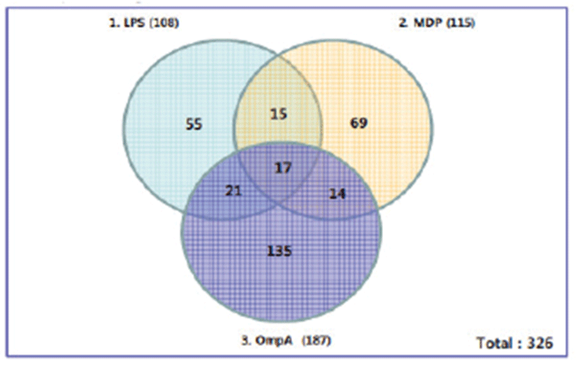
Gene expression assay using immune cells is a common way to investigate the diverse roles of macrophages in bacterial infections or antigenic challenges. Thus, we chose DNA microarray to chart the expression levels of porcine genes in response to bacterial antigens such as LPS, MDP, and OmpA. Pom2 macrophage cells were treated for 6 hrs with each antigen, after which total RNAs were extracted for GeneChip® Porcine Genome Array analysis. Of about 20,000 genes in the array, a total of novel 326 genes were found to be differentially expressed by at least 2-fold upon antigen stimulation compared to the control. Among these, LPS differentially regulated the expression of total 108 genes; 86 genes were up-regulated and 22 genes were down-regulated (Fig. 1). The list in Table 1 shows 16 novel genes representing diverse biological processes that were well annotated and up-regulated by at least 2-fold upon LPS stimulation. Pappalysin-2 is a metalloproteinase involved in cartilage development and angiogenesis without any known role in the immune response. SLC37A2 (Sugar phosphate exchanger) is a novel gene involved in glucose 6 phosphate transport but without any apparent role in the immune response. IL-23 along with IL-17 may participate in an acute response to infection in peripheral tissues, whereas CD1d is known to mediate presentation of primarily lipid and glycolipid antigens of self or microbial origin to T cells. Expression levels of the 16 genes above were also up-regulated by 2-folds upon MDP and OmpA stimulation

jbtr-16-2-47-g1
Fig. 1. Analysis of differentially expressed porcine genes in response to bacterial antigens. Pig Monocytes clone 2 was treated with three bacterial antigens, including LPS, MDP, and OmpA, under the conditions indicated in the Materials and Methods. Subsequently, PoM2 cells were lysed and total RNAs extracted for gene expression analysis by AffymetrixGeneChip® Porcine Genome Array. The number of genes showing at least 2-fold differentially regulated expression in response to the three antigens is presented in each circle of the diagram.
Download Original Figure
Table 1. List of representative porcine genes expressed differentially by at least 2-fold in response to LPS stimulation of PoM2 cells
UniGene ID Gene Title Folds Change
Ssc.10776 pappalysinPappalysin-2 like 3.3
Ssc.5887 SLC37A2 (Sugar phosphate exchanger 2-like) 2.6
Ssc.48532 TMEM184C (Transmembrane protein 184C) 2.7
Ssc.15990 retinoid Retinoid X receptor, alpha 2.1
Ssc.52038 CD1d molecule 2.6
AW414558 protein Protein capicua homolog 2.2
Ssc.18952 Ig alpha heavy chain 2.2
CK455989 MAP1B (microtubule-associated protein 1B) 2.3
Ssc.22131 PCA (Putative cation transporting ATPase) 2.9
Ssc.12344 RhoJ-like 2.3
Ssc.39853 DNA-directed RNA polymerase subunit beta 2.1
Ssc.54823 perlecan Perlecan (heparan sulfate proteoglycan 2) 2.0
Ssc.5924 Mitogen-activated protein kinase 3 2.0
Ssc.7825 interleukin Interleukin 23 3.8
Ssc.8222 L-threonine O-3-phosphate decarboxylase 3.0
Ssc.9629 dual Dual specific tyr-phos-regulated kinase 2-like 2.5
Download Excel Table
Gene Expression Analysis of Porcine Genes identified in DNA microarray by RT-PCR

Based on the list of up-regulated porcine genes, we confirmed the DNA microarray data by RT-PCR. Using the gene-specific primers shown in Table 2, 10 novel porcine genes along with one control GAPDH gene were chosen for RT-PCR analysis (Fig. 2). Pom2 macrophage cells were stimulated with each of the three antigens above before total RNA extraction. Out of the 10 candidate genes for bacterial antigen responsive biomarkers, SLC37A2, PAPPA2, TMEM 184C, and Syntaxin-binding protein 5 (Stx5) were shown to be up-regulated by RT-PCR. Although the expression level of porcine Stx5 was modestly up-regulated in the DNA microarray analysis (1.2-fold), expression of its human homolog increased by more than 4-fold in human macrophage THP1 cells compared to the negative control (results from another DNA microarray experiment and data not shown). Further, its expression level was up-regulated by an average of 5-fold in response to each antigen, as shown in Fig. 2.

jbtr-16-2-47-g2
Fig. 2. RT-PCR analysis of porcine genes identified in DNA microarray. The differentially up-regulated genes identified by DNA microarray analysis were investigated by RT-PCR using gene-specific primers as indicated in Table 1. As a control, porcine GAPDH gene was included. PoM2 cells were stimulated for 6 hrs as indicated in the Materials and Methods, followed by RNA extraction by an RNA Blood Mini Kit for RT-PCR. Out of 10 porcine genes, SLC37A2, PAPPA2, TMEM184C, and Syntaxin-binding protein 5 were found to be up-regulated, whereas expression of the other five genes remained unchanged.
Download Original Figure
Table 2. List of PCR primers of genes identified from porcine DNA microarray analysis
NCBI No. Gene Gene symbol Sequences of primers (5’-3’) Location Size of amplified products (bp)
Ssc.5887 Sugar phosphate exchanger 2-like SLC37A2 CAGAAGACAGCCAGGACCTC 1031 249
CAGCCTCCTTGGCAGTAAAG 1279
Ssc.10776 Pappalysin-2 like PAPPA2 CCTTGGGCTTATCTTCCACA 133 205
TCCCCTGAAACTCAACTGCT 337
Ssc.15990 Retinoid X receptor, alpha RXR-alpha CTGCAAGGGCTTCTTCAAAC 199 206
TGGACTCCACCTCGTTCTCT 404
Ssc.18952 Immunoglobulin alpha heavy chain constant region IGHA ATGCTTCAGCTGCTCCATCT 263 203
GGCCTCTGTGTCTTCCTGTC 465
Ssc.22131 Putative cation transporting ATPase, P-type PCA1 TCACCAAGTGAAGGTTCGTG 423 171
CCAAAGGCACTGATCACAGA 593
Ssc.48532 Transmembrane protein 184C, inhibit cell growth TMEM184C CCCATGGGTCACTATCAAGG 1727 221
GGCTTGTCCCAAGCATAAAA 1947
Ssc.52038 CD1d molecule CD1d CACGTCTCTGGCTTCTACCC 902 204
GATGATGTCCTGGCCTCCTA 1105
AW414558 Protein capicua homolog TCACAGTACCCCCTCAGGAC 1100 245
GAACGTAACACGCTCCAGGT 1344
CK455989 Microtubule-associated protein 1B MAP1B TTCCAGAAAATGGGAGTTGG 1347 217
TTTTCTCTGCAGGGTTTGCT 1563
Ssc. 4770 Syntaxin binding protein 5 TTCCATTTGTCCTTGGCTCT 149 232
GAAATGCAAACCAAGGGAAT 380
ssc. GAPDH Glyceraldehyde-3-phosphate dehydrogenase GAPDH GTCGGTTGTGGATCTGACCT 714 210
AGCTTGACGAAGTGGTCGTT 923
Download Excel Table

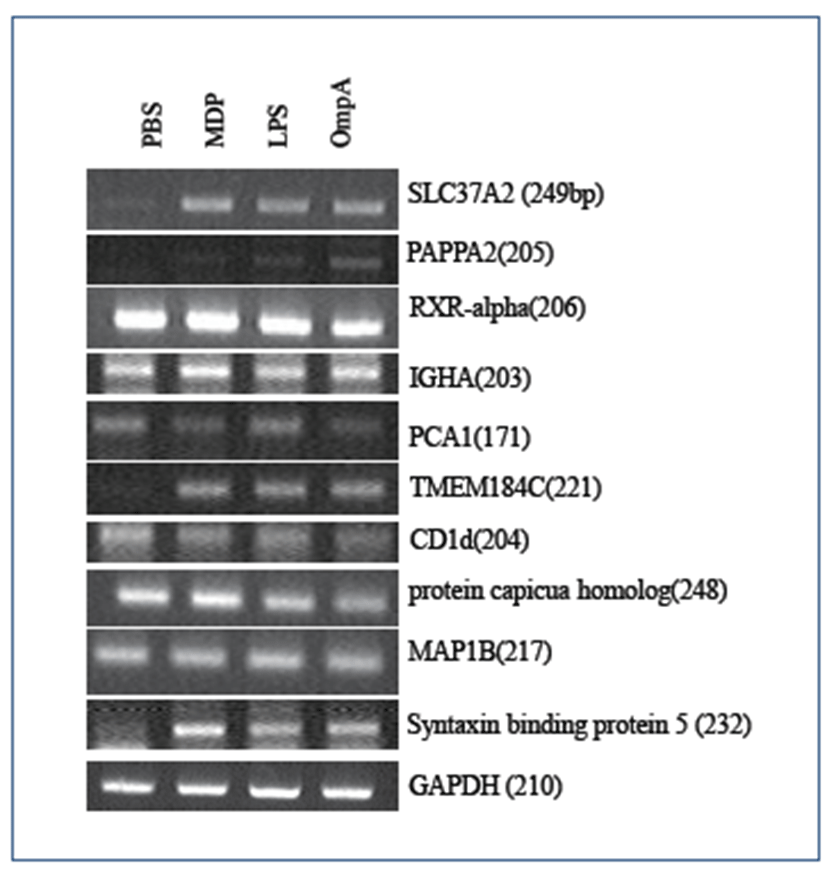
Regardless of the class of each antigen (lipopolysaccharide vs proteinaceous), the three antigens had similar effects on the expression of the four genes (Fig. 2). However, it is unclear why the RXR-alpha, IGHA, PCA1, CD1d, protein capicua homolog, and MAP1B genes did not show any differential expression patterns compared to the control.

Expression Profile of Porcine Antimicrobial Genes

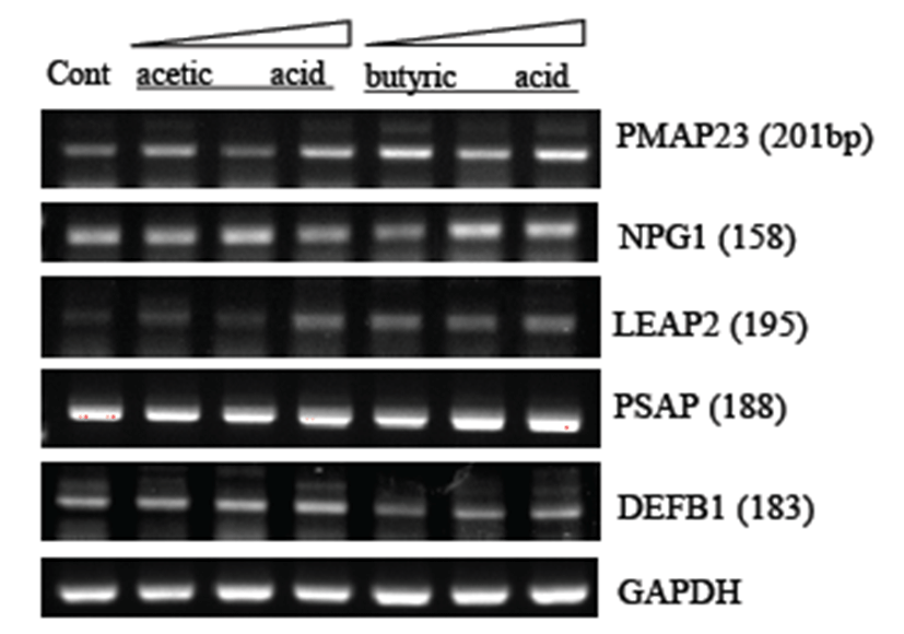
SCFAs are abundant in the intestines of vertebrates and are proposed to play a role in the immune response in addition to their direct anti-microbial activity. Therefore, we investigated the effects of two major SCFAs, acetic and butyric acid, on expression of major porcine antimicrobial peptide (AMP) genes. Concentrations of acids were determined based on the minimal amount of each acid that lowered the pH of the culture media (DMEM with 10% FBS) and induced a color change by the pH indicator phenol red. The pH levels of the culture media containing 0.5, 2, and 5 mM of each acid were 8.41, 8.22, and 7.86 for acetic acid as well as 8.12, 8.04, and 7.75 for butyric acid, respectively, whereas the pH of control DMEM with 10% FBS was 8.37.

The porcine genome has been sequenced, but its annotation is incomplete. Professor Kim, Herbal at Seoul National Univ. helped us find human homologs of the antimicrobial genes listed in Table 3. Pom2 cells were treated with the acids at final concentrations of 0.5, 2, and 5mM for 15 hrs before total RNA extraction and RT-PCR analysis using gene-specific primers (Table 3). Out of seven porcine AMP genes, including PMAP-23, NPG1, LEAP2, NPG3, BPIFA1, PSAP, and DEFB1, expression of three genes (PMAP23, NPG1, and LEAP2) was up-regulated by butyric and acetic acids. Butyric acid appeared to have better activity than acetic acid in this experiment.

Table 3. Porcine antimicrobial peptide genes identified through comparison of porcine genes with human homologs
NCBI No. Gene Gene symbol Sequences of primers (5’-3’) Location Size of amplified products (bp)
ENSSSCG00000021877 Antibacterial protein PMAP-23 TCACGGTGAAGGAGACTGTG 246 201
GTTTCTGTGGCCGACGTACT 446
ENSSSCG00000023733 Protegrin 1 NPG1 TAGGTTCTGCGTCTGTGTCG 420 138
TTCACCGTCTACCAGGGAAC 557
ENSSSCG00000014290 Liver expressed antimicrobial peptide 2 LEAP2 AACTGAGTTCCGCCAAGAGA 105 195
TGTCGCAGGTAAGTGCTGTC 299
TTCACCGTCTACCAGGGAAC 557
ENSSSCG00000010281 Prosaposin PSAP TCTGTGAGGTGTGCAAGAGG 1241 188
CATCACCTCCACCAGGATCT 1428
ENSSSCG00000020997 Defensin, beta 1 DEFB1 GGAAGAGGCTCAGTCAGTGG 210 231
GGCACAGGTAACAGGACCAT 440
ssc. GAPDH Glyceraldehyde-3-phosphate dehydrogenase GAPDH GTCGGTTGTGGATCTGACCT 714 210
AGCTTGACGAAGTGGTCGTT 923
Download Excel Table

Discussion

Identification of biomarkers is regarded as an important step towards disease characterization and management of disease in animals. Post-genomic technologies have promoted the development of strategies aimed at identifying specific and sensitive biomarkers from the thousands of molecules present in tissues or biological fluids [16].

jbtr-16-2-47-g3
Fig. 3. Expression analysis of porcine antimicrobial genes in response to acetic and butyric acids. Pom2 cells (1 × 10⁶/mL) were treated with 0.5, 2, or 5 mM of acetic or butyric acid for 15 hrs and then washed once with PBS before cell harvesting and one-step RT-PCR reaction according to the manufacturer’s protocol (Bioneer premix kit) using gene-specific primers (as indicated in Table 2). Control (Cont.) cells were treated with PBS and subjected to the same procedure as above. The number in the bracket after the gene name indicates the expected PCR product size. Porcine GAPDH was included as a control.
Download Original Figure

In this study, we searched for porcine biomarkers showing induced expression in response to three bacterial antigens (LPS, MDP, and OmpA) as well as bacterial metabolites of short chain fatty acids (acetic and butyric acid).

Porcine DNA microarray analysis of Pom2 cells stimulated by these three antigens revealed a total of 326 porcine genes showing at least 2-fold increased expression (Fig. 1). Expression of 16 genes increased by 2-fold regardless of the type of antigen; LPS and MDP are components of peptidoglycan, whereas OmpA is the major protein in the outer membrane region. We also examined half of the 16 representative genes by RT-PCR. Out of 10 porcine genes, four genes (SLC37A2, PAPPA2, TMEM184C, and Stx5) showed differential expression (Table 2 and Fig. 2). Although we used the same RNA samples for DNA microarray and RT-PCR experiments, it is unclear why the other six genes did not show differential expression. There might have been differences in the probing region of each transcript by either the probes of the DNA microarray or RT-PCR primers. Splicing variations might also have caused these differences. Recently, the expression of 22 inducible genes, which include VCAM1, HMOX1, and Serglycin, was reported to be induced in response to Salmonella or LPS in porcine alveolar macrophages. Interestingly, 13 genes, including IL1-a/-b, CD14, OPN, and VCAM, were found to be putative NF-kappaB targets [17]. However, there is no report on the role of NFkB in the transcriptional regulation of the four genes identified in this study (SLC37A2, PAPPA2, TMEM184C, and Stx5).

SCFAs have been reported to be major intestinal microbial metabolites and include acetic/butyric acids. These appear to be absorbed into the body either through specific receptors or passively. Most SCFA studies have focused on their direct inhibitory effects on pathogenic microbes [18], but recent studies have investigated their effects on the host metabolism, including gluconeogenesis, insulin secretion, and fatty acid metabolism [19-21].

Until now, there is no report that describes their effect on innate immunity. Therefore, we studied the effects of SCFAs on expression of a set of antimicrobial peptide (AMP) genes in Pom2 porcine macrophage cells (Table 3). Out of five porcine homologs of human AMP genes investigated by RT-PCR, three genes (PMAP23, NPG1, and LEAP2) were found to be up-regulated by both acetic and butyric acids (Fig. 3). It remains to be characterized which signaling pathway is involved in the modulation of AMP expression and the innate immune response against any invading microbe. It will be interesting to compare the innate immune response between human and porcine macrophages for either antigens or bacterial pathogens. In conclusion, we report here a set of novel porcine biomarkers involved in the innate immune response of porcine macrophages. This finding may lead us to better understand the porcine immune response and develop diagnostic biomarkers for the prevention or diagnosis of pathogenic bacteria such as Salmonella typhimurium.

Acknowledgements

Acknowledgements

This research was supported by the research grant of Chungbuk National University in 2012 (to JK Choi) and by grants from the Next-Generation BioGreen 21 Program (No. PJ01104401 and PJ01110901), Rural Development Administration, Republic of Korea (to HK Lee and KD Song respectively).

REFERENCES

1.

Jacobs JP, Braun J. Immune and genetic gardening of the intestinal microbiome. FEBS Lett. 2014; 588:4102-4111.

2.

Fayol-Messaoudi D, Berger CN, Coconnier-Polter MH, Liévin-Le Moal V, Servin AL. pH-, Lactic Acid-, and Non-Lactic Acid-Dependent Activities of Probiotic Lactobacilli against Salmonella enterica Serovar Ty-phimurium. Appl Environ Microbiol. 2005; 71:6008-6013.

3.

Weintraub A. Immunology of bacterial polysaccharide antigens. Carbohydr Res. 2003; 338:2539-2547.

4.

Duan Q, Zhou M, Zhu X, Bao W, Wu S, Ruan X, Zhang W, Yang Y, Zhu J, Zhu G. The flagella of F18ab Escherichia coli is a virulence factor that contributes to infection in a IPEC-J2 cell model in vitro. Vet Microbiol. 2012; 160:132-140.

5.

Srikanth CV, Cherayil BJ. Intestinal innate immunity and the pathogenesis of Salmonella enteritis. Immunol Res. 2007; 37:61-78.

6.

Bhowmick R, Pore D, Chakrabarti MK. Outer membrane protein A (OmpA) of Shigella flexneri 2a induces TLR2-mediated activation of B cells involvement of protein tyrosine kinase, ERK and NF-kappaB. PLoS One. 2014; 9:e109107.

7.

Bode JG, Ehlting C, Haussinger D. The macrophage response towards LPS and its control through the p38(MAPK)-STAT3 axis. Cell Signal. 2012; 24:1185-1194.

8.

Lee SI, Kang SK, Jung HJ, Chun YH, Kwon YD, Kim EC. Muramyl dipeptide activates human beta defensin 2 and pro-inflammatory mediators through Toll-like receptors and NLRP3 inflammasomes in human dental pulp cells. Clin Oral Invest. 2015; 19:1419-1428.

9.

Lu YC, Yeh WC, Ohashi PS. LPS/TLR4 signal transduction pathway. Cytokine. 2008; 42:145-151.

10.

Natarajan N, Pluznick JL. From microbe to man the role of microbial short chain fatty acid metabolites in host cell biology. Am J Physiol Cell Physiol. 2014; 307:979-985.

11.

Kim MH, Kang SG, Park JH, Yanagisawa M, Kim CH. Short-chain fatty acids activate GPR41 and GPR43 on intestinal epithelial cells to promote inflammatory responses in mice. Gastroenterology. 2013; 145:396-406.

12.

Ishii S, Kishi M, Yamagami K, Okada S, Abe K, Misaka T. The use of mammalian cultured cells loaded with a fluorescent dye shows specific membrane penetration of undissociated acetic acid. Biosci Biotechnol Bio chem. 2012; 76:523-529.

13.

Mishiro T, Kusunoki R, Otani A, Ansary MM, Tongu M, Harashima N, Yamada T, Sato S, Amano Y, Itoh K, Ishihara S, Kinoshita Y. Butyric acid attenuates intestinal inflammation in murine DSS-induced colitis model via milk fat globule-EGF factor 8. Lab Invest. 2013; 93:834-843.

14.

Artis D, Spits H. The biology of innate lymphoid cells. Nature. 2015; 517:293-301.

15.

Bain CC, Mowat AM. The monocyte-macrophage axis in the intestine. Cell Immunol. 2014; 291:41-48.

16.

Moore RE, Kirwan J, Doherty MK, Whitfield PD. Biomarker discovery in animal health and disease the application of post-genomic technologies. Biomark In sights. 2007; 2:185-196.

17.

Kyrova K, Stepanova H, Rychlik I, Polansky O, Leva L, Sekelova Z, Faldyna M, Volf J. The response of por-cine monocyte derived macrophages and dendritic cells to Salmonella Typhimurium and lipopolysaccharide. BMC Vet Res. 2014; 10:244-252.

18.

den Besten G, van Eunen K, Groen AK, Venema K, Reijngoud DJ, Bakker BM. The role of short-chain fatty acids in the interplay between diet, gut microbiota, and host energy metabolism. J Lipid Res. 2013; 54:2325-2340.

19.

Hijova E, Chmelarova A. Short chain fatty acids and colonic health. Bratisl Lek Listy. 2007; 108:354-358.

20.

Sakakibara S, Yamauchi T, Oshima Y, Tsukamoto Y, Kadowaki T. Acetic acid activates hepatic AMPK and reduces hyperglycemia in diabetic KK-A(y) mice. Bio chem Biophys Res Commun. 2006; 344:597-604.

21.

Ge H, Li X, Weiszmann J, Wang P, Baribault H, Chen JL, Tian H, Li Y. Activation of G protein-coupled receptor 43 in adipocytes leads to inhibition of lipolysis and suppression of plasma free fatty acids. Endocrinology. 2008; 149:4519-4526.