


Introduction
Canine uterine diseases such as cystic endometrial hyperplasia (CEH), mucometra, hydrometra, and pyometra are common in countries where neutering healthy dogs are not generally practiced [1]. Mucometra, hydrometra, and pyometra are defined by the type of fluid present in the uterus and the degree of mucin hydration [2]. Hematometra, in which the uterine content is hemorrhagic, is rarely reported [3]. By definition, pyometra is the accumulation of pus within the uterine lumen, typically occurring during or immediately following a period of high progesterone levels [4].
Clinically, a bitch with pyometra may present with lack of appetite, depression, polydipsia, lethargy, abdominal distension, and possible vaginal discharge [5]. Symptoms of pyometra are classified by the patency of the uterine cervix. Closed-cervix pyometra is a medical emergency that requires rapid intervention to prevent sepsis and potential death [5].
The diagnosis of canine uterine disease is best made with ultrasonography and radiology [6]. Pyometra presents as an enlarged uterus with convoluted tubular horns filled with anechoic to echogenic fluid, in which case movement, characterized by slow and swirling patterns, is often noted; mucometra and hydrometra, however, present with thin uterine walls and echogenic or anechoic fluid, respectively [7]. In hematometra, complete blood count and serum chemistry results can vary. Mild normocytic, normochromic, and nonregenerative anemia often develops secondary to chronic disease and generally resolves following treatment [3]. In hematometra, physical examination findings and clinical signs are inconclusive, but the bitch can suffer from anemia with frank vulvar hemorrhage of an open-cervix type. A diagnosis of hematometra is not made until a laparotomy is performed and gross findings are assessed.
Although pyometra can occur at every age, cases of pyometra or hematometra complex in a young dog have not been previously reported. This case describes CEHpyometra/ hematometra in a young Poongsan dog.
Case Report
An 8-month-old, intact female Poongsan dog presented with severe, continuous hemorrhagic vulvar discharge and anemia. Hemorrhagic vulvar discharge was continued for 5 days before presentation. The initial diagnostic evaluation included abdominal ultrasonography, radiography, and physical and laboratory examinations. An activated partial thromboplastin time test was performed to rule out a coagulation disorder. Because of the severe anemia and dehydration, blood cross-matching was performed in anticipation of a blood transfusion.
The electrolyte levels were low: Na and Cl ion levels were lower than the normal reference range (Table 1). However, alkaline phosphatase (ALP) levels were much higher than the normal reference range. At presentation, a complete blood cell count was performed, revealing a white blood cell (WBC) count of 60.19 × 10³/μL (reference range, 5.05-16.76 × 10³/μL), a packed cell volume (PCV) of 11.8% (reference range, 37.3%-61.7%), a hemoglobin level of 4.6 g/dL (reference range, 13.1-20.5 g/ dL), and a platelet count of 48 × 103/μL (reference range, 148-484 × 103/μL) (Table 2). These laboratory results corresponded to a recent hemorrhagic episode. On the basis of the laboratory results and patient history, we performed a whole blood transfusion on day 0 (day of presentation). Additionally, metronidazole (15 mg/kg, IV, BID, TRIZELE InjⓇ; JW Pharmaceutical Co., Ltd.) and cefotaxime (50 mg/kg, IV, TID, CEFOTAXIMENARIUM Ⓡ; Wooridul Pharmaceutical Co., Ltd.) were administered for vaginal and systemic infections, respectively. Because of continuous vaginal hemorrhage, cimetidine (10 mg/kg, IV, BID, H-2 InjⓇ; JW Pharmaceutical Co., Ltd.), vitamin K (2.5 mg/kg, SC, TID, VITAMIN K1 InjⓇ; Dae Han Pharm Co., Ltd.), vitamin B (0.3 mL/body, IV, SID, BEECOMHEXA InjⓇ; Yuhan Co., Ltd.), taurine (3 mL/body, IV, BID, TARUINE-F InjⓇ; Samyang Anipharm Co., Ltd.), and desmopressin (1 μg/kg, SC, Once, MINIRIN InjⓇ; Ferring Pharmaceuticals Korea Inc.) were administered for anemia. On day 2, laboratory results showed abnormalities: the WBC (60.19 × 10³/μL) was higher than the normal reference range, the PCV was 16.3%, the hemoglobin level was 5.4 g/dL, and the platelet count was 102 × 103/μL. The PCV, hemoglobin level, and platelet count were all lower than the normal reference range. Because of the sustained decline in PCV, the bitch was given a whole blood transfusion over 6 h.
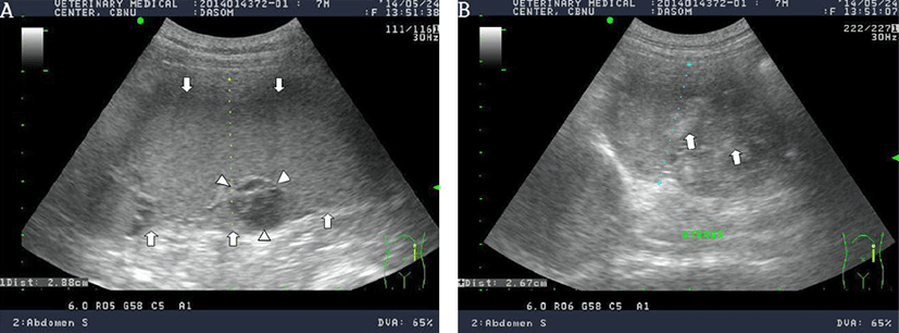
On abdominal ultrasonography, an enlarged uterus was observed. The left uterine horn was enlarged and filled with echogenic fluid. Its diameter was approximately 3 cm. An anechoic cyst and echogenic mass were seen in the uterine wall and uterine cavity, respectively (Fig. 1).
Based on patient history, physical examination, abdominal ultrasonography, radiography, and laboratory examination, the dog was diagnosed with pyometra, mucometra, hydrometra, and hematometra. For surgical treatment, an ovariohysterectomy was performed. Tramadol (3 mg/kg, IV, TID, MARITROL InjⓇ; Jeil Pharm. Co. Ltd.), cefotaxime (50 mg/kg, IV, TID), metronidazol (10 mg/kg, IV, BID), and cimetidine (10 mg/kg IV, BID) were administered preoperatively. The bitch was anesthetized by inhalation of isoflurane. Propofol (PROVIVEⓇ Inj; 10 mg/mL, Myungmoon Pharm) was administered intravenously until the lack of jaw tone, gagging reflex, and coughing reflex was observed. After these reflexes disappeared, an endotracheal tube was inserted. Anesthesia was maintained with isoflurane (TERRELLTM, Piramal Critical Care, Bethlehem).
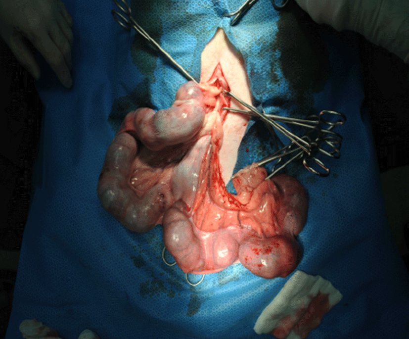
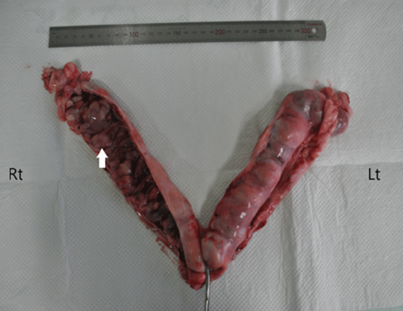
At laparotomy, the uterus was enlarged and filled with fluid; evidence of hemorrhage was present (Fig. 2). Postoperatively, the uterine horn was incised, revealing an accumulation of blood clots. The endometrium contained blood and blood clots and had a cystic appearance. Additionally, thick and yellowish pus had accumulated in the uterine cavity. The diameter of the uterine horn was approximately 3 cm and was filled with cyst, pus, and blood clots (Fig. 3). The incised uterine horn contained a cyst. Gross findings revealed a definitive diagnosis of cystic endometrial hyperplasia-pyometra complex with severe hemorrhage, termed hematometra.
Postoperatively, we prescribed tramadol (3 mg/kg, IV, TID), cefotaxime (50 mg/kg, IV, QID), cimetidine (10 mg/kg, IV, BID), metronidazol (15 mg/kg, IV, BID), vitamin B (0.3 mL/body, IV, BID), vitamin K (2 mg/kg, SC, TID), and taurine (3 mL/body, IV, BID). The bitch was maintained on IV fluids (normal saline 0.9%, 35 mL/h and albumin 30 mL/h). In addition, antibiotics (metronidazol, 15 mg/kg, IV, BID, and cefotaxime, 50 mg/kg, IV, QID) were administered for 2 days. The patient showed normal condition at the last follow-up, 7 days postoperatively.
Discussion
Jindo and Poongsan dogs are the most popular breeds in Korea. Poongsan dogs are commonly used for hunting and are a representative native breed in North Korea [8]. Canine CEH-pyometra complex is a commonly diagnosed disease of the reproductive system but can be life-threatening [9-11]. Hematometra is rare among cases of CEH-pyometra complex. Pyometra is typically considered a disease of intact, middle-aged bitches, but it can occur in younger dogs [19]. In this case, Poongsan dog developed CEH-pyometra complex at 8-month-old, which is younger than previously reported [3, 12-14].
Hematometra is regarded as an emergency because early diagnosis and rapid therapeutic intervention are necessary to prevent a fatal outcome [1]. Differential diagnosis of mucometra, hydrometra, hematometra, and pyometra can be made based on cytological examination, complete blood count, serum chemistry analysis, urinalysis, and ultrasonography [15]. In the present case, cytological examination, complete blood count, serum chemistry analysis, and ultrasonography were performed for the differential diagnosis of mucometra, hydrometra, hematometra, and pyometra.
Hematometra and metrorrhagia are uncommon clinical presentations [3]. Reported etiologies include postpartum subinvolution of placental sites [16, 17], anticoagulant rodenticide toxicity [18] and other acquired coagulation deficiencies, uterine trauma, neoplasia [19, 20], placental necrosis, congenital coagulation deficiency [15], idiopathic prepubertal metrorrhagia [21], postpartum endometritis [8], and uterine serosal inclusion cysts [22]. Therefore, a differential diagnosis according to these etiologies should be performed. In the present case, these diseases were ruled out by thorough laboratory and physical examinations.
Symptom onset of pyometra in dogs is gradual and insidious [23]. Generally, clinical signs include vomiting, abdominal distension, dehydration, anorexia, and polyuria/ polydipsia [23-25]. In the present case, clinical signs included vomiting, anorexia, depression, dehydration, and continuous hemorrhage.
When hematometra is suspected, ovariohysterectomy should be performed as soon as possible due to lifethreatening complications associated with bacteremia and endotoxemia [3]. Bitches who are seriously ill should be medically stabilized with appropriate intravenous fluid therapies and broad-spectrum antibiotics prior to surgery [5]. A whole blood transfusion should be performed for bitches with severe anemia. In the present case, the dog received a whole blood transfusion to correct the severe anemia prior to surgery, and intravenous fluids were administered to address the dehydration. Subsequently, an ovariohysterectomy was performed. After surgery, gross findings revealed that one part of the uterine lumen was filled with blood clots and that another part of the uterine lumen was filled with pus. Further, the endometrium contained many cysts. The bitch was finally diagnosed with concurrent pyometra and hematometra. This case describes evidence of hematometra with severe vulvar hemorrhage in a young Poongsan dog.