
 PDF Download
 PubReader
 Export Citation
 Email the Author
 Share Facebook
 Share Twitter
 Cited by 0 Article
 Metrics (View:35)







INTRODUCTION
3-Hydroxy-2-naphthoic acid (CAS No. 90-70-6) is a yellow crystalline powder with a melting point of 222°C –223°C, molecular weight of 188.18 g/mol, and molecular formula of C11H8O3 [1]. It is mainly used as an intermediate in the production of dyes, pigments, insecticides, and pharmaceuticals [2, 3]. Its global production capacity was approximately 30,000 tons per year [3]; however, it has declined in recent years. Despite this reduction, concerns regarding its toxicity remain.
Toxicological information on 3-hydroxy-2-naphthoic acid in humans and experimental animals is limited. In humans, exposure can irritate the skin, eyes, respiratory tract, and gastrointestinal system [4]. According to the Organization for Economic Co-operation and Development (OECD) and the Screening Information Dataset [3], several toxicity studies on 3-hydroxy-2-naphthoic acid have been conducted. The acute oral LD50 of 3-hydroxy-2-naphthoic acid in rats ranges from 823 to 1,040 mg/kg body weight. A 10% 3-hydroxy-2-naphthoic acid solution was found to be lethal to guinea pigs 24 h after dermal exposure. In rabbits, this compound caused mild skin irritation and severe ocular damage including corneal opacity. Repeated oral administration induced liver toxicity and adrenal effects in female rats, with a no-observed-effect level (NOEL) of 12 mg/kg/day in female and 60 mg/kg/day in male rats. Inhalation of the compound for 10 days resulted in renal toxicity and necrosis. A one-generation fertility and developmental toxicity study conducted based on the OECD Test Guideline (TG) 415 showed that 3-hydroxy-2-naphthoic acid did not affect parental reproductive performance. However, at a dose of 200 mg/kg/day, the offspring exhibited reduced body weight, growth retardation, and malformations. The NOEL for reproductive toxicity was 200 mg/kg/day, whereas that for offspring toxicity was 50 mg/kg/day. The NOEL for systemic toxicity was 12.5 mg/kg/day in male and 50 mg/kg/day in female rats, with forestomach lesions observed at higher doses.
OECD TG 415 and OECD TG 421 differ fundamentally in their study objectives, duration, and scope of evaluation. OECD TG 415 was designed as a one-generation reproductive toxicity study that follows parental animals through mating, gestation, and lactation and evaluates the growth and development of their offspring over a substantial portion of the reproductive cycle. In contrast, OECD TG 421 is a short-term reproductive and developmental toxicity screening test intended to identify preliminary hazard signals, including effects on mating, fertility, gestation, parturition, and early postnatal development. Because TG 421 includes fewer animals, a shorter exposure and observation period, and more limited offspring evaluations than TG 415, it cannot provide a definitive assessment of the full reproductive cycle. However, it offers a more rapid and resource-efficient approach for initial hazard identification and for determining whether further comprehensive reproductive toxicity testing is warranted [5, 6].
Although the reproductive and developmental toxicities of 3-hydroxy-2-naphthoic acid were previously investigated in a one-generation study conducted according to OECD TG 415 [3], no study conducted according to OECD TG 421 has been reported. Therefore, the present study evaluated the potential reproductive and developmental toxicity of 3-hydroxy-2-naphthoic acid in Sprague–Dawley rats in compliance with Good Laboratory Practice and OECD TG 421 to provide screening-level information for initial hazard and risk assessment.
MATERIALS AND METHODS
3-Hydroxy-2-naphthoic acid was purchased from Sigma-Aldrich (St. Louis, MO, USA). Test compounds (batch no. BCBZ3916) was 99.5% pure and stored at room temperature. It was suspended in dimethyl sulfoxide (DMSO; Sigma-Aldrich), and dosing formulations were prepared by analyzing the resulting solution’s homogeneity and stability.
Six-week-old-specific-pathogen-free Sprague–Dawley rats of both sexes were obtained from ORIENTBIO (Seongnam, Korea) and acclimated for 5 weeks prior to dosing. The animal room was maintained under controlled environmental conditions: temperature 20°C–24°C, relative humidity 35%–55%, a 12-h light/dark cycle, and fresh air ventilation at 10–15 air changes per hour. Rats were housed individually in stainless steel wire-mesh cages (width 350 mm × length 260 mm × height 180 mm) with unrestricted access to a commercial rodent diet (Altromin 1314 IRR, Altromin Spezialfutter, Lage, Germany) and drinking water provided in polycarbonate bottles.
Doses were individually calculated based on the most recent weight data of each animal, with a dose volume of 5 mL/kg/day. Rats received daily oral gavage of 3-hydroxy-2-naphthoic acid at the doses of 0, 12.5, 50, or 200 mg/kg/day. The control rats were administered DMSO. Each dose was administered to 24 animals (12 males and 12 females). Dose levels were selected based on the findings of the previous one-generation study [3]. The test substance was orally administered once daily. Males were treated for 42 days, from 2 weeks before mating until 14 days after mating. Females were treated from 2 weeks before mating until postpartum day 13. However, females in which mating was not confirmed during the mating period were administered the test substance daily until 25 days after the end of the mating period, and females in which mating was confirmed but no signs of delivery were observed were administered the test substance daily until gestation day 25. All procedures were performed in accordance with the OECD TG 421 and GLP guidelines. All the procedures were approved by the Institutional Animal Care and Use Committee (IACUC; protocol no. ABIACUCRA21093).
All the animals were monitored daily for clinical signs throughout the study period. Body weights of the male rats were recorded on the first day of dosing, weekly during the dosing period, and on the day of necropsy. Body weights of the female rats were recorded on the first day of dosing, weekly throughout the dosing period (excluding the mating period), on gestational day (GD) 0, 7, 14, and 20, on lactational day (LD) 0, 4, and 13, and on the day of necropsy. Also, food consumption was measured in a similar manner.
Male and female rats were mated in a 1:1 ratio until successful copulation occurred or a maximum of 2 weeks. Successful mating in females was confirmed twice daily based on the presence of a vaginal plug or sperm in a vaginal smear. GD 0 was defined as the day when mating was confirmed.
The ovaries and uterus of each female rats were excised and examined for implantation site status. Each litter was examined for its birth rate on postnatal day (PND) 0; anogenital distance on PND 4; nipple/areolae retention on PND 12; survival rate and sex ratio on PND 0 and 14; and gross abnormalities and body weight on PND 0, 4, and 13. Pups were euthanized using CO2 and externally examined on PND 4. The male rats were necropsied 2 weeks after the mating period, whereas the female rats were necropsied on PND 14 by exsanguination under isoflurane anesthesia. Necropsy was performed as soon as possible when all pups of a dam died during the lactation period, whereas it was performed 26 days after the end of the mating period for the female rats that showed no signs of copulation during the mating period and on GD 26 for the female rats that exhibited evidence of copulation but showed no signs of parturition.
Blood samples were collected from pups on PND 13 and from adult males (excluding moribund or dead animals) to assess the levels of serum thyroid hormones (T4 and TSH).
Weights were measured for the thyroid glands, prostate, seminal vesicles with coagulating glands, testes, and ovaries/uterus with the cervix. Organs with gross abnormalities were also analyzed. All organs were preserved in 10% neutral-buffered formalin, except for the testes and epididymides, which were fixed in the Bouin’s solution before being transferred to 10% neutral-buffered formalin. For pups, one male and one female per litter were weighed on PND 13, and only the thyroid gland was preserved in 10% neutral-buffered formalin.
All collected organs were processed routinely, embedded in paraffin, sectioned at 3–4 μm, stained with hematoxylin and eosin, and examined microscopically under low- to high-power magnification.
All statistical analyses were performed using the SPSS software version 19.0 (IBM, Chicago, IL, USA). Differences among groups were evaluated using one-way analysis of variance. The homogeneity of variances was assessed using the Levene’s test. Post-hoc comparisons were conducted using the Scheffé’s multiple range test for homogeneous variances, whereas the Dunnett’s T3 test, with each group compared to the control, was applied for heterogeneous variances.
RESULTS
In the group that received 3-hydroxy-2-naphthoic acid at the dose of 200 mg/kg/day, One female rat was found dead in a prone position on GD 19, after exhibiting salivation, which started at dosing day (DD) 11 and was followed by decreased locomotor activity, emaciation, and the absence of stool. Intermittent salivation occurred in a dose-dependent pattern in male rats at 50 mg/kg/day and in both sexes at 200 mg/kg/day (Table 1).
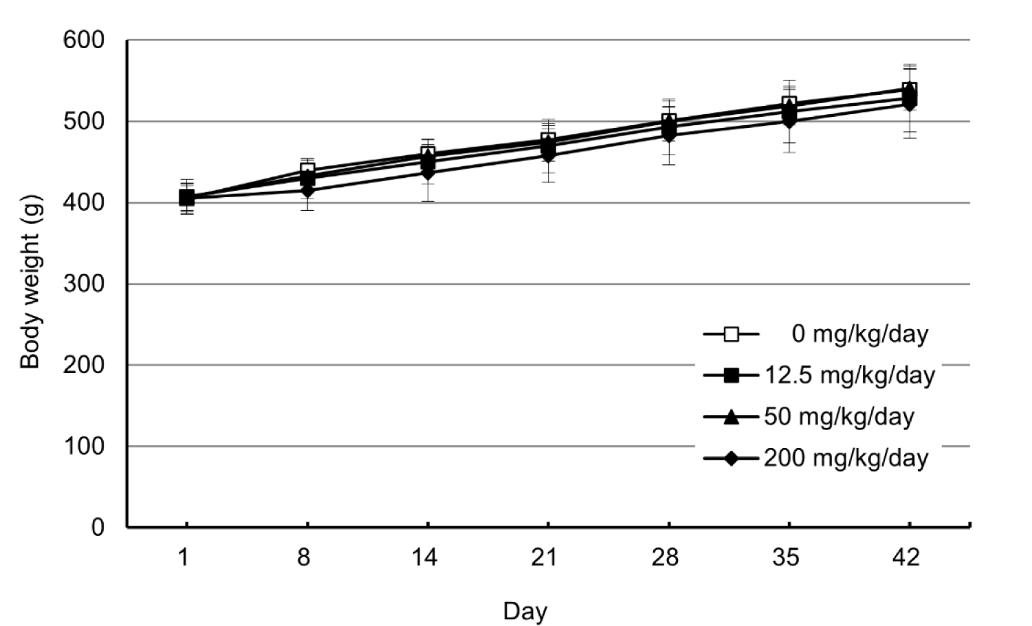
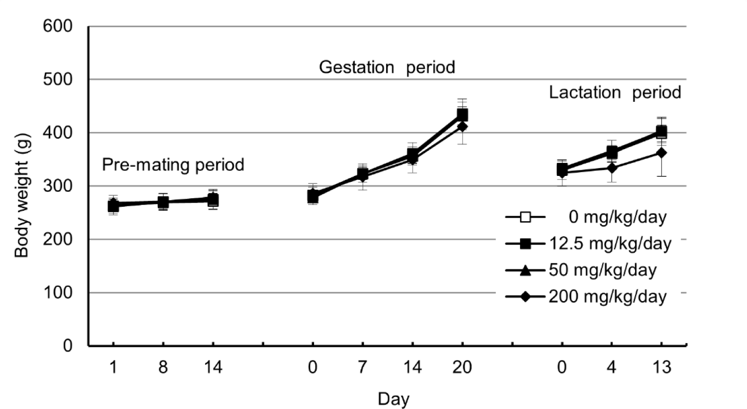
The male rats in the group that received 3-hydroxy-2-naphthoic acid at the dose of 200 mg/kg/day showed a trend of decreasing body weight from DD 8–42, whereas the female rats in the same group exhibited decreased body weight during GD 7–20 and LD 0–13, although the differences were not statistically significant (Figs. 1 and 2).
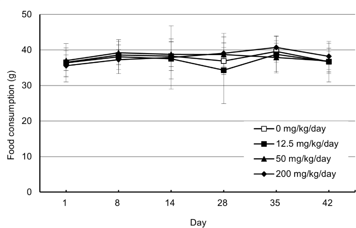
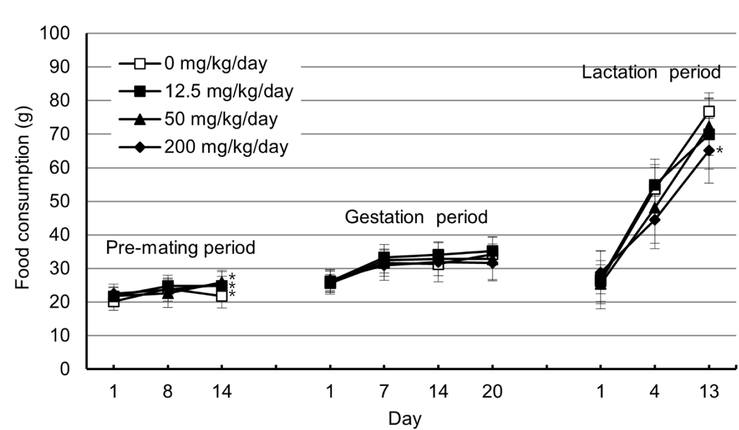
In the male rats, no significant differences in food consumption were observed in the groups that received 3-hydroxy-2-naphthoic acid at the doses of 12.5, 50, or 200 mg/kg/day throughout the dosing period compared with the control group. In the female rats, food consumption significantly increased on day 14 during the pre-mating period in all treatment groups. However, a significant decrease in food consumption was observed on LD 13 in the group that received 3-hydroxy-2-naphthoic acid at the dose of 200 mg/kg/day compared with the control group (Figs. 3 and 4).
The mean estrous cycle lengths of the female rats in the groups that received 3-hydroxy-2-naphthoic acid at the doses of 12.5, 50, and 200 mg/kg/day did not differ from those of the female rats in the control group during the dosing period (Table 2), and all females were in diestrus at necropsy. Mating and fertility indices (Table 3). Implantation, delivery parameters, birth and survival rates, litter size, sex ratios, anogenital distance on PND 4, and nipple retention on PND 12 were not significantly different among the groups, except for one dam in the group that received 3-hydroxy-2-naphthoic acid at the dose of 200 mg/kg/day that lost its entire litter on the day of delivery (Tables 4, 5, 6, and 7). No external abnormalities were observed in pups of any groups (Data was not shown). However, the pups’ body weights in the group that received 3-hydroxy-2-naphthoic acid at the dose of 200 mg/kg/day were lower than those in the control group in both sexes on PND 0, 4, and 13 (Figs. 5 and 6).
The viability index on PND 0 (%) = (Number of pups born alive on PND 0 / Total Number of pups born) × 100.
| Group | Dose (mg/kg/day) | No of nipples | |
|---|---|---|---|
| G1 | 0 | Mean | 0.0 |
| S.D. | 0.0 | ||
| N | 10 | ||
| G2 | 12.5 | Mean | 0.0 |
| S.D. | 0.0 | ||
| N | 10 | ||
| G3 | 50 | Mean | 0.0 |
| S.D. | 0.0 | ||
| N | 12 | ||
| G4 | 200 | Mean | 0.0 |
| S.D. | 0.0 | ||
| N | 10 |
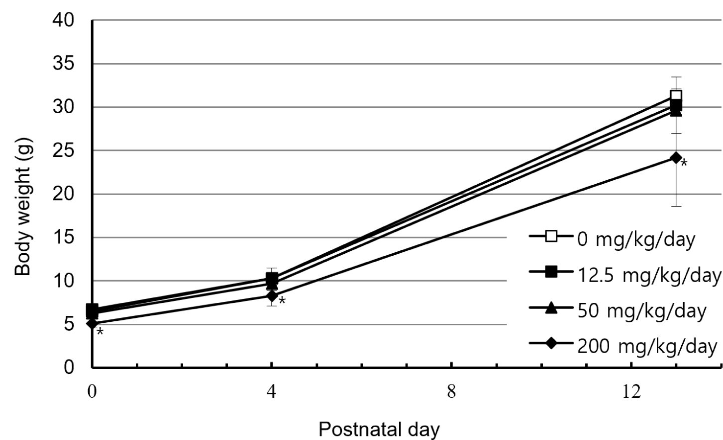
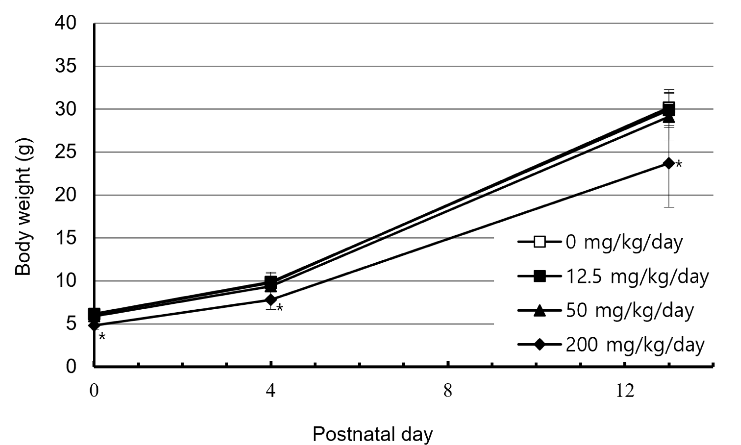
No significant changes in the levels of serum T4 and TSH were observed in any of the parental animals or offspring across all dose groups delivery (Tables 8 and 9).
A statistically significant reduction in epididymal weight was observed in the male rats in the group that received 3-hydroxy-2-naphthoic acid at the dose of 200 mg/kg/day compared with the control group. However, no changes in the weight of other organs were observed (Tables 10 and 11).
In the group that received 3-hydroxy-2-naphthoic acid at the dose of 200 mg/kg/day, one dead female rat exhibited a forestomach polyp and bilateral adrenal enlargement. Polyps in the forestomach were observed in three male and six female rats, whereas one female rat additionally exhibited redness in the forestomach (Table 12). A small right seminal vesicle was observed in two male rats in the control group. No abnormalities were observed in the pups of the control group and the groups that received 3-hydroxy-2-naphthoic acid at the doses of 12.5, 50, and 200 mg/kg/day.
Histopathological examination revealed epithelial hyperplasia and hyperkeratosis of the forestomach in both male and female rats in the groups that received 3-hydroxy-2-naphthoic acid at the doses of 50 and 200 mg/kg/day. Forestomach erosion was observed in the male rats at the dose of 50 mg/kg/day and in both sexes at 200 mg/kg/day. Furthermore, submucosal infiltration of inflammatory cells was identified in one male rat at the dose of 50 mg/kg/day. Decreased luminal fluid was observed in male rats with small seminal vesicle in the control group. No effects of 3-hydroxy-2-naphthoic acid were observed in the male rat testes and epididymides or the female rat ovaries. However, adrenal necrosis was observed in the dead female rat that received 3-hydroxy-2-naphthoic acid at a dose of 200 mg/kg/day, accompanied by erosion, epithelial hyperplasia, and hyperkeratosis of the forestomach (Table 13).
DISCUSSION
This study was conducted to obtain preliminary information on the potential effects of the test substance 3-hydroxy-2-naphthoic acid on reproductive function, maternal behavior, and offspring development from conception to PD 13. Three different doses of 3-hydroxy-2-naphthoic acid (12.5, 50, and 200 mg/kg/day) were used and each dose was administered to 24 animals (12 males and 12 females). The vehicle DMSO was administered to the animals in the control group. Throughout the observation period, multiple endpoints were assessed, including clinical signs, body weight, food consumption, estrous cycle, reproductive performance, offspring development, thyroid hormone levels, organ weights, gross necropsy findings, and histopathology. One female rat in the group receiving 3-hydroxy-2-naphthoic acid at 200 mg/kg/day died during late gestation after showing clinical signs including salivation, decreased locomotor activity, emaciation, absence of feces, and prone posture. The death was attributed to severe adrenal necrosis, which corresponded to the adrenal enlargement observed at necropsy. However, this finding was not considered toxicologically significant because spontaneous adrenal necrosis has been reported previously [7], and it occurred in only one animal in the high-dose group. Therefore, the death was considered incidental rather than test substance-related. Intermittent salivation was observed in male rats at the dose of 50 and 200 mg/kg/day while intermittent salivation was observed throughout the pre-mating, gestation, and lactation periods in female rats at the dose of 200 mg/kg/day. As salivation occurred intermittently without accompanying adverse clinical or pathological findings, it did not indicate systemic toxicity and was not toxicologically considerable; it was merely a reflex response to the taste or irritant properties of the test substance rather than direct toxicity [8]. Body weight reductions in male rats during DD 8–42 and in female rats during GD 7–20 and LD 0–13 were considered test substance-related because they were consistently observed and showed dose-related trends [9]. Similarly, decreased body weights were observed in male and female pups in the 200 mg/kg/day group on PND 0, 4, and 13. The reduced pup body weights may have been associated, at least in part, with maternal toxicity, including maternal body weight reduction during gestation and lactation, which could have adversely affected fetal growth and maternal nursing capacity or milk production. However, a direct effect of the test substance on the offspring cannot be excluded. Because toxicokinetic data, including measurements of the test substance or its metabolites in maternal milk, maternal plasma, or pup plasma, were not obtained in this study, potential direct exposure of pups through placental transfer or lactation could not be evaluated. A transient increase in food intake was observed in all female rats treated with any doses of the test substance on day 14 of the premating period. Since it was not accompanied by weight gain, it was of no toxicological significance. In contrast, decreased food consumption on LD 13 in the female rats in the group that received 3-hydroxy-2-naphthoic acid at the dose of 200 mg/kg/day was associated with gastric lesions, which contributed to loss in body weight. The test substance did not show any adverse effects on estrous cycling, mating performance, fertility indices, gestation parameters, or delivery outcomes. Similarly, no test substance-related effects on live birth index, postnatal survival, litter size, external morphology, sex ratio, anogenital distance, or nipple retention were observed in the offspring. One dam in the group that received 3-hydroxy-2-naphthoic acid at the dose of 200 mg/kg/day experienced total litter loss on the day of delivery. As it occurred in a single animal without accompanying maternal toxicity or repeated findings within the group, the finding was considered incidental. No effects on thyroid hormone levels were observed in male parental animals or offspring. The decrease in epididymal weight observed in males at the dose of 200 mg/kg/day was not supported by corresponding histopathological changes and lacked dose-dependency; thus, it was not toxicologically significant. Histopathological examination revealed epithelial hyperplasia and hyperkeratosis in the forestomachs of both sexes at the doses of 50 and 200 mg/kg/day. Erosion was observed in the forestomachs of male rats with the test substance at the dose of 50 mg/kg/day and in both sexes at the dose of 200 mg/kg/day. Additionally, submucosal infiltration of inflammatory cells was observed in one male at the dose of 50 mg/kg/day. These findings were related to the test substance because they were consistent with the commonly observed forestomach irritation triggered by repeated exposure to irritant compounds [10] and with the gross lesions. The reduced size of the right seminal vesicle observed at necropsy was caused by decreased luminal fluid; however, the finding was a spontaneous or incidental change since it was observed only in the control group. In the offspring, no gross or histopathological findings were attributed to the test substance at any doses.
The no-observed-adverse-effect level (NOAEL) for parental animals was determined to be 12.5 mg/kg/day based primarily on treatment-related clinical signs, reduced body-weight gain, and forestomach lesions observed at 50 or 200 mg/kg/day. In offspring, the NOAEL was determined to be 50 mg/kg/day based on the decreased body weights observed at 200 mg/kg/day. This OECD TG 421 study provides a limited toxicity profiles of 3-hydroxy-2-naphthoic acid suitable for short-term reproductive and developmental toxicity screening. The previous OECD TG 415 study reported several sporadic malformations and scattered visceral findings in a small number of litters, for which a clear relationship to treatment could not be established [3]. In the present study, no external abnormalities or clear treatment-related changes in anogenital distance, nipple retention, or postnatal viability were observed. Reduced pup body weights were observed at 200 mg/kg/day concurrently with decreased maternal body weights, suggesting that maternal toxicity may have contributed to the impaired offspring growth. However, because OECD TG 421 is a screening study with a relatively limited exposure period and number of developmental endpoints, the possibility of direct developmental toxicity cannot be definitively excluded. Histopathological changes in the forestomach, including epithelial hyperplasia, hyperkeratosis, erosion, and inflammation, were considered indicative of local irritation following oral administration. Overall, the present study provides screening-level information on the potential reproductive and early developmental toxicity of 3-hydroxy-2-naphthoic acid and may contribute to initial hazard identification and the determination of whether further comprehensive toxicity testing is warranted.


