
 PDF Download
 PubReader
 Export Citation
 Email the Author
 Share Facebook
 Share Twitter
 Cited by 0 Article
 Metrics (View:43)







INTRODUCTION
The embryonic ventral midbrain is a spatially and temporally organized developmental system within the mammalian central nervous system. Multiple neuronal populations are generated, migrate, differentiate, and undergo early maturation in this relatively restricted embryonic territory, within partially overlapping developmental windows, and thereby initiate the assembly of circuits underlying motor control, reward processing, cognition, and motivated behavior [1–3]. Dopaminergic (DA) and GABAergic neurons are the major populations among them, forming the substantia nigra and ventral tegmental area (VTA) and contributing to the organization and regulation of nigrostriatal and mesocorticolimbic circuits [1–5]. Because their joint assembly establishes the developmental foundation of ventral midbrain circuitry, how DA and GABAergic neurons are specified, migrate, differentiate, and mature during embryogenesis remains a central question in developmental neuroscience [4–7].
Considerable progress has been made in defining the molecular mechanisms that regulate DA and GABAergic neuron development. Work on DA neurons has identified key extracellular patterning signals, including Shh, FGF8, and Wnt signaling, together with interconnected transcriptional networks involving FOXA2, LMX1A/B, OTX2, EN1/2, NURR1, and PITX3 [2, 8–15], while work on ventral midbrain GABAergic neurons has characterized their progenitor origins, transcriptional specification, migratory routes, and subtype diversification [4, 6, 7, 16]. These populations have generally been investigated as separate developmental lineages, however, so considerably less is known about how their developmental programs become coordinated within the shared tissue environment of the embryonic ventral midbrain.
We propose the dynamic multicellular developmental niche as a framework for examining how DA and GABAergic lineages develop within a shared environment comprising radial glia, extracellular matrix (ECM), vascular structures, neighboring progenitors, and extracellular signals [5, 7, 17–22]. Throughout this review, developmental coordination is reserved for experimentally demonstrable effects of an environmental component or cell population on the timing, migration, positioning, differentiation, or early maturation of another population; coexistence alone does not constitute coordination.
Unlike a general developmental microenvironment, a neurogenic or neurovascular niche, or a radial glial scaffold, this framework considers DA and GABAergic lineages jointly across anatomical position, developmental time, and multiple cell types. It is an analytical framework rather than a newly delimited anatomical compartment, and every proposed relationship must specify its source, target, stage, location, and phenotype.
The framework is falsifiable. Stage- and cell-type-specific perturbation should produce a defined phenotype, and restoring the relevant source, signal, or target response should rescue it; failure to do so should narrow or reject the proposed relationship.
Single-cell studies have revealed heterogeneity among developing ventral midbrain neurons and radial glia [19–21, 23, 24], and spatial and multimodal approaches now relate cellular identity to native tissue architecture [22, 25–31]. Lineage tracing [31–35], CRISPR-based perturbation [36, 37], and human ventral midbrain organoids [38–50] together provide complementary means of testing candidate mechanisms and assessing their potential human relevance.
This review integrates spatial organization, temporal overlap, and the shared embryonic environment; evaluates candidate mechanisms by evidence type and anatomical relevance; and outlines an experimental roadmap for testing neuronal assembly in the ventral midbrain.
This focused narrative review was updated through August 2026 using PubMed, Web of Science, and Google Scholar. Search combinations included “ventral midbrain” or “mesencephalon” with “dopaminergic,” “GABAergic,” “development,” “neurogenesis,” “migration,” “radial glia,” “extracellular matrix,” “vascular,” “Netrin-1,” “CXCL12,” “Reelin,” “cell adhesion,” “single-cell,” “spatial transcriptomics,” and “organoid.” English-language primary studies and authoritative reviews were included when they addressed embryonic ventral midbrain lineage origin, migration, positioning, environmental regulation, or methods capable of testing cross-population mechanisms. Studies from other central nervous system regions were retained only as indirect mechanistic context and are identified as such. This was a hypothesis-oriented narrative review rather than a systematic review or meta-analysis.
SPATIAL ORGANIZATION OF DOPAMINERGIC AND GABAERGIC NEURON DEVELOPMENT IN THE EMBRYONIC VENTRAL MIDBRAIN
The embryonic ventral midbrain provides the anatomical framework within which DA and GABAergic neurons are generated, migrate, become positioned, and begin to assemble ventral midbrain circuits. It is not a homogeneous neuroepithelium but a set of progenitor domains with distinct transcriptional identities, signaling environments, and developmental potentials [2, 4, 6, 16, 19]. These spatially organized domains establish the initial positions and lineage potentials of neuronal progenitors and define the anatomical relationships within which the different populations develop. As Fig. 1 illustrates, DA and GABAergic lineages arise from distinct progenitor territories and progressively come to occupy adjacent and partially overlapping regions of a shared embryonic environment.
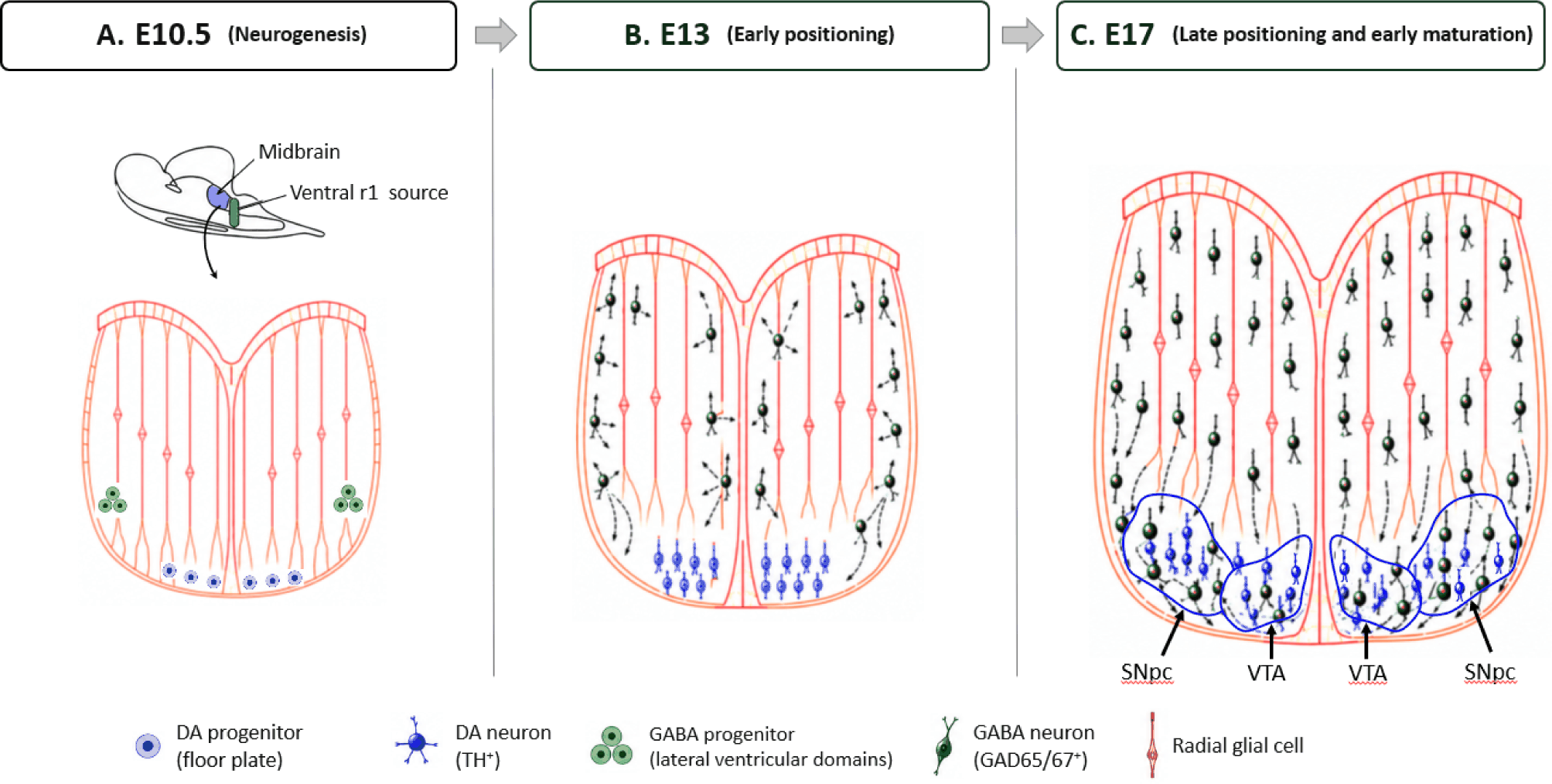
Early ventral midbrain patterning is established by the coordinated actions of Shh, FGF8, and Wnt1 signaling together with regional transcription factors such as OTX2, which collectively define the floor plate, basal plate, and adjacent progenitor territories [2, 8–12]. These domains generate multiple neuronal populations whose developmental programs proceed within partially overlapping temporal windows in neighboring embryonic regions. The embryonic ventral midbrain can therefore be regarded as a spatially organized developmental landscape in which regional patterning lays the foundation for subsequent neuronal diversification.
Despite their distinct transcriptional identities, the floor plate and basal plate remain anatomically contiguous. This arrangement places progenitors and newly generated neurons within a shared tissue context shaped by radial glia, ECM components, vascular structures, and extracellular signals [17–19]. Spatial organization thus supplies not only positional information but also the structural context in which developmental coordination may subsequently occur.
Midbrain DA neurons originate predominantly from floor plate-derived progenitors in the ventral mesencephalon. A conserved transcriptional network involving FOXA2, LMX1A/B, OTX2, EN1/2, NURR1, and PITX3 controls their specification and differentiation under the influence of Shh, FGF8, and Wnt signaling [2, 8–15, 51]. After cell-cycle exit, immature DA neurons first migrate radially from the ventricular zone toward the mantle zone and then undergo tangential redistribution that contributes to the formation of the substantia nigra pars compacta (SNpc) and VTA [2, 52, 53].
Single-cell transcriptomic and lineage-tracing analyses have further identified transcriptionally and functionally distinct radial glial subtypes in the developing ventral midbrain, which contribute differentially to DA lineage progression and to the signaling and ECM environment that supports DA neuron development [19–21, 54]. DA neuron development therefore depends on both lineage-intrinsic programs and the surrounding multicellular environment [55].
GABAergic progenitors arise from ventrolateral and neighboring mesencephalic and diencephalic ventricular domains and follow multiple radial and tangential migratory routes toward the substantia nigra pars reticulata (SNpr), VTA, and adjacent ventral and intermediate territories [4, 6, 7, 16, 56–59]. Molecularly distinct from DA progenitors though they are, their descendants develop and migrate in close anatomical proximity to the emerging DA system. By late embryogenesis, GABAergic neurons are enriched in the SNpr and adjacent ventral and intermediate territories, while smaller subsets lie within or along the borders of the SNpc and VTA, producing partial interdigitation without complete anatomical mixing [4, 6, 7, 16, 56–59].
Recent molecular profiling and lineage analyses have revealed substantial heterogeneity among developing ventral midbrain GABAergic neurons, including transcriptionally distinct subpopulations that originate from different progenitor domains and follow diverse migratory routes [6, 60]. GABAergic development therefore involves multiple spatial origins and migratory trajectories rather than a single uniform developmental program.
Although DA and GABAergic neurons originate from distinct progenitor domains, their developmental trajectories progressively bring them into adjacent and partially overlapping ventral midbrain territories. Spatial convergence here denotes the establishment of neighboring and partially shared territories rather than complete anatomical mixing of the two populations. Within these regions, developing neurons encounter a tissue environment in which radial glia, ECM, vascular structures, and extracellular guidance signals are simultaneously remodeled [7, 17–19].
This spatial convergence provides the anatomical foundation for the central concept of this review: a shared developmental environment functioning as a dynamic multicellular developmental niche. Spatial organization alone, however, cannot fully explain coordinated neuronal development, since DA and GABAergic neurons also undergo neurogenesis, migration, differentiation, and early maturation within partially overlapping developmental windows. The following section therefore examines how temporal coordination complements spatial organization within an integrated framework for ventral midbrain development.
TEMPORAL OVERLAP DURING EMBRYONIC VENTRAL MIDBRAIN DEVELOPMENT
Spatial organization defines where DA and GABAergic neurons originate and become positioned during embryogenesis (Fig. 1); equally important is when these populations undergo specification, differentiation, migration, and early maturation [2, 4–7]. These processes are not discrete events but unfold within partially overlapping temporal windows, during which radial glia, ECM, vascular structures, and extracellular signaling systems are themselves changing [17–19, 61–63].
Embryonic ventral midbrain development is therefore best viewed as a continuous, overlapping process rather than a sequence of independent cellular events. Fig. 2 summarizes the overlapping periods of predominant activity from early regional patterning through perinatal development, while indicating that circuit assembly and maturation continue postnatally; the intervals shown represent periods of predominant biological activity rather than rigid developmental boundaries. Temporal overlap among neuronal differentiation, migration, tissue remodeling, and early circuit assembly creates windows within which coordinated regulation may occur [2, 4–7, 17–19].
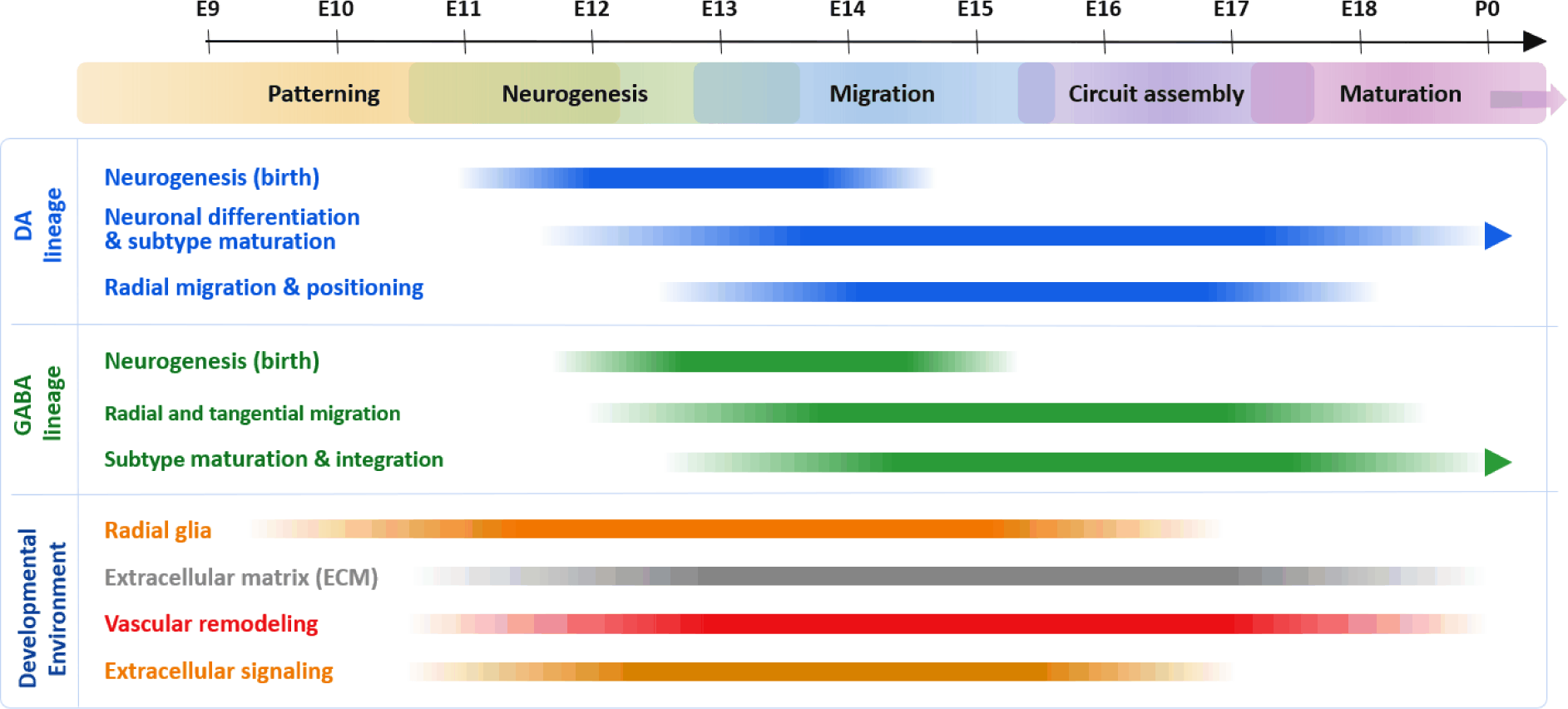
The temporal sequence of ventral midbrain development begins with regional patterning during early embryogenesis, when the coordinated actions of Shh, FGF8, and Wnt1 signaling together with regional transcription factors such as OTX2 establish molecularly distinct progenitor domains within the ventral mesencephalon and define the developmental competence of the future DA and GABAergic lineages [2, 8–12].
Midbrain DA neurogenesis begins in mice at approximately embryonic day (E) 10.5 within floor plate-derived progenitors. Newly generated DA neurons exit the cell cycle, differentiate, and migrate radially toward the ventromedial mantle zone, progressively acquiring DA identity through sequential and interconnected transcriptional programs [9–15, 19, 51, 64, 65]. This early phase establishes the initial ventromedial organization of the developing DA population, which subsequently undergoes the spatial redistribution that contributes to formation of the SNpc and VTA [2, 52, 53, 64, 66].
As summarized in the subsection “Spatial origin of GABAergic neurons, mouse GABAergic neurogenesis and radial or tangential migration overlap DA differentiation from approximately E10.5 through late embryogenesis, with major movement toward the SNpr occurring during mid-to-late embryonic stages [4, 6, 7, 16, 56, 57, 60].
Fig. 2 therefore illustrates partial temporal synchrony between DA and GABAergic development rather than complete simultaneity. DA differentiation and spatial redistribution overlap with ongoing GABAergic neurogenesis and migration, creating an extended interval during which the two populations coexist at different stages of differentiation and maturation [4–7, 52, 53].
During mid-embryogenesis, immature DA neurons continue to differentiate, extend neurites, and redistribute while GABAergic neurons undergo radial and tangential migration [5–7, 52, 53]. Radial glial subtypes are simultaneously performing neurogenic, signaling, and ECM-associated functions, ECM composition is changing, vascular networks are expanding, and guidance systems remain active [7, 17–19, 52, 53, 61–64, 67].
These concurrent processes define intervals in which interactions among neuronal and non-neuronal components are biologically possible. Temporal coincidence identifies opportunities for experimental testing, but it does not demonstrate causal interaction.
The embryonic tissue environment changes alongside neuronal development: In the schematic temporal framework used in Fig. 2, radial-glial lineage and signaling functions are emphasized from approximately E9.5 to E17 [9, 19–21, 64], ECM production and remodeling from approximately E10.5 to P0 [17, 61, 68], vascular expansion and remodeling from approximately E10.5 to P0 [18, 62, 63, 69–71], and extracellular guidance systems from approximately E10.5 to E17 [7, 52, 53, 67–70]. These intervals represent approximate evidence-supported windows of relevance rather than absolute onset or termination points.
Guidance systems are likewise stage- and lineage-dependent. Netrin-1 regulates ventral midbrain GABAergic migration and substantia nigra development, CXCL12/CXCR4 and Reelin regulate distinct phases of DA migration, and adhesion mechanisms provide additional context [7, 52, 53, 67, 72–74]. Fig. 2 summarizes this concurrent tissue remodeling rather than a neuronal timeline alone.
Fig. 2 emphasizes overlapping developmental windows rather than a rigid chronology. DA differentiation and redistribution and GABAergic migration coincide with radial glial activity, ECM remodeling, vascular expansion, and extracellular guidance [5–7, 17–19, 52, 53, 61–63, 67]. Together with the spatial relationships in Fig. 1, this temporal overlap defines when shared influences are possible without establishing a mechanism. The following section therefore defines the shared developmental environment in which such mechanisms can be tested (Fig. 3).
THE SHARED DEVELOPMENTAL ENVIRONMENT: A DYNAMIC MULTICELLULAR DEVELOPMENTAL NICHE
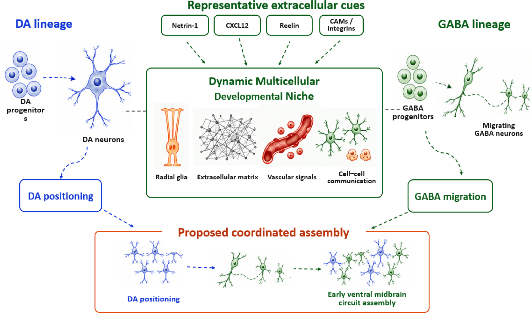
Figs. 1 and 2 define where DA and GABAergic neurons become juxtaposed and when their developmental programs overlap [2, 4–7]. These conditions permit exposure to common cellular, molecular, and extracellular influences without themselves establishing coordination. Both lineages develop within a remodeling environment containing radial glia, ECM, vascular structures, and extracellular signals [7, 17–19, 61–63, 67, 72].
We use the dynamic multicellular developmental niche to integrate developing neurons, radial glia, progenitors, ECM, endothelial cells, and extracellular signals across these overlapping windows [6, 7, 17–19, 61–63, 67, 72]. The term denotes neither a discrete anatomical compartment nor a single pathway, but a testable context in which lineage-specific programs may be modified by cellular, extracellular, and tissue-level regulation.
The shared developmental environment denotes the molecular, cellular, extracellular, and tissue context in which the distinct DA and GABAergic lineages develop. Although these lineages arise from distinct progenitor domains [2, 4, 6, 7, 16], they develop amid radial glia [9, 19–21, 64], ECM [17, 61, 68, 72], vascular networks [18, 62, 63, 69–71], and extracellular guidance cues [7, 52, 53, 67, 72–77].
This environment changes across embryogenesis and may influence migration, positioning, differentiation, and maturation. Any mechanistic claim therefore requires a defined source, responding population, anatomical location, developmental interval, and measurable outcome.
Floor-plate radial glia-like cells generate mesencephalic DA neurons, and single-cell and lineage studies have identified functionally distinct ventral midbrain radial glial subtypes [9, 19–21, 64]. Radial glia-like cell type 1 (Rgl1) contributes directly to the DA lineage, whereas Rgl3 shows signaling- and ECM-associated programs and provides factors that support DA neuron development [19].
Radial glia therefore connect progenitor production with tissue organization, extracellular signaling, and ECM regulation. Their proposed structural support for neuronal movement, and the many transcriptomically predicted interactions attributed to them, nonetheless await direct functional testing [17, 19, 61, 64, 72].
The ECM is a dynamic regulator of adhesion, tissue mechanics, extracellular diffusion, and the presentation of growth factors and guidance cues [17, 61, 72]. Laminins, fibronectin, hyaluronan, proteoglycans, and matrix factors produced by radial glial and vascular-associated populations may therefore shape the conditions encountered by differentiating DA and migrating GABAergic neurons [19, 68].
Its ventral midbrain dynamics and causal effects remain incompletely characterized. Current evidence supports the ECM as a context for cellular and signaling interactions rather than as an independently established coordinator of both lineages.
Developing vascular networks provide metabolic support and can influence neural cells through secreted factors, surface interactions, and vascular-associated ECM [18, 62, 63, 69–71]. Because direct evidence for coordinated DA and GABAergic regulation is limited, the vascular compartment is presented here as a plausible but unvalidated niche component.
Extracellular cues carry unequal evidentiary support. Netrin-1 links defined GABAergic migration with substantia nigra organization, whereas Reelin and CXCL12/CXCR4 primarily regulate DA migration, and adhesion and integrin mechanisms remain more context-dependent [7, 19, 52, 53, 67, 72, 73].
Within the proposed niche, neuronal populations, radial glia, ECM, vascular elements, and extracellular cues contribute at differing levels of experimental support to progenitor behavior, migration, positioning, survival, and tissue maturation [6, 7, 17–19, 52, 53, 61–64, 67, 68]. The following section evaluates these candidate relationships by evidence type and anatomical relevance.
CANDIDATE MOLECULAR AND CELLULAR MECHANISMS RELEVANT TO DEVELOPMENTAL INTEGRATION
The preceding sections established the spatial, temporal, and conceptual foundations of coordinated DA and GABAergic development in the embryonic ventral midbrain. This section evaluates candidate mechanisms according to whether they rest on direct cross-lineage evidence, direct lineage-specific evidence, functional evidence for a shared environmental component, or primarily descriptive and computational support. The available evidence ranges from mutant, explant, transplantation, and genetic guidance studies to transcriptomic associations, spatial colocalization, and computational predictions [5–7, 22, 28–30, 78–81].
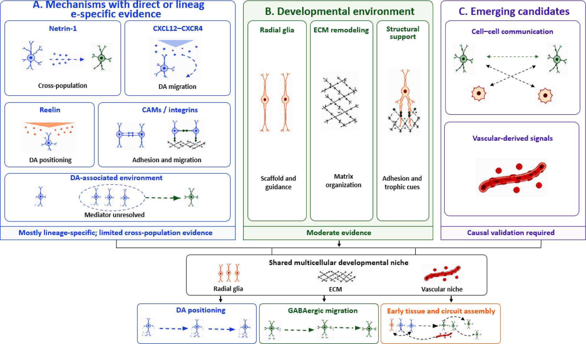
Evidence strength is accordingly kept separate from presumed biological importance. We graded each relationship using five operational criteria: (1) anatomical relevance to the embryonic ventral midbrain, (2) specification of source and responding population, (3) developmental-stage resolution, (4) direct perturbation of the proposed component with a measurable phenotype, and (5) rescue or replication in a complementary model. Descriptive temporal or spatial correspondence and computational ligand–receptor inference were classified as low or emerging evidence; direct lineage-specific loss-of-function or gain-of-function evidence was classified as moderate; and source- and target-resolved perturbation with rescue or convergent validation was classified as moderate–high for an environmental effect. Cross-population confidence was assigned separately and required that manipulating one population or its defined product altered a phenotype in the other population. Fig. 4 summarizes this provisional hierarchy, and Table 1 lists model, study design, evidence basis, confidence, and limitations for each mechanism.
The most direct evidence that the developing DA system can influence GABAergic development comes from the Pitx3-deficient aphakia mouse [5]. Disruption of the normal DA neuronal architecture in this model was accompanied by reduced accumulation and abnormal positioning of GABAergic neurons in the ventral mesencephalon, with no detectable reduction in GABAergic neurogenesis—observations that link the pre-existing DA-associated ventral midbrain environment to the subsequent migration of GABAergic neurons.
Functional explant and transplantation experiments strengthened this interpretation. Wild-type basal plate-derived cells migrated toward wild-type ventral midbrain explants but showed little attraction toward aphakia ventral midbrain tissue, and replacing the mutant tissue with wild-type ventral midbrain restored the migration and integration of GAD65-positive cells [5]. These findings together support a chemoattractive or otherwise permissive activity associated with normally developing ventral midbrain DA territories.
Pitx3 deficiency nonetheless has pleiotropic effects on DA differentiation, migration, survival, and regional tissue organization, so these experiments do not establish dopamine itself, or any DA neuron-derived molecule, as the sole instructive signal. Neither the responsible source nor the molecular mediator has been resolved, and the evidence is most accurately read as DA neuron-associated regulation of GABAergic migration rather than as a fully defined direct DA-to-GABA signaling pathway.
This study nonetheless provides an important benchmark for the present framework, since it combines cross-population phenotyping with functional tissue assays and partial environmental rescue. Distinguishing DA neuron-autonomous signals from indirect effects mediated through radial glia, ECM, axonal inputs, or other components of the ventral midbrain environment will require cell-type-specific perturbation and molecular rescue experiments.
Extracellular guidance pathways provide direct evidence for defined migratory and positioning events in the embryonic ventral midbrain [6, 7, 52, 53], but their evidentiary interpretation differs by pathway. Netrin-1 links the migration of defined substantia nigra-associated GABAergic populations with DA substantia nigra organization, whereas CXCL12/CXCR4 and Reelin are established primarily as regulators of DA neuronal migration and positioning. Evidence for an integrated network coordinating both lineages remains incomplete.
Netrin-1 currently offers the strongest available evidence linking GABAergic migration with DA substantia nigra organization. In mouse embryos, forebrain-derived Netrin-1 delivered by axons guides SIX3-positive anterior SNpr GABAergic precursors into the ventral substantia nigra. Loss of axonal Netrin-1 or disruption of DSCAM in these migrating GABAergic cells reduces their ventral accumulation; DCC was tested but was not required for this GABAergic migratory response [7]. The resulting failure of GABAergic positioning permits abnormal ventral dispersion of DA neurons, indicating an indirect organizational effect of GABAergic migration on the DA compartment rather than a demonstrated GABA-derived molecular signal to DA neurons. By contrast, DCC-dependent Netrin-1 migration reported for interpeduncular-nucleus populations involves a distinct target population and should not be conflated with the DSCAM-dependent SNpr mechanism [82].
CXCL12/CXCR4 and Reelin regulate distinct aspects of DA neuron migration and positioning. CXCL12/CXCR4 signaling influences migratory behavior and contributes to the appropriate distribution of DA neurons, whereas Reelin promotes the rapid, laterally directed migration required for correct organization of the SNpc [52, 53, 74]. These studies establish extracellular signaling as an important regulator of context-dependent DA neuron positioning, but direct evidence that either pathway simultaneously regulates GABAergic development in the ventral midbrain remains limited.
CXCL12/CXCR4 signaling also controls tangential migration and regional distribution of GABAergic interneurons in other developing CNS regions [75, 83–85]. Such observations support its broader relevance to GABAergic migration without establishing an equivalent function in the ventral midbrain, so region- and cell-type-specific perturbation will be required to determine whether CXCL12/CXCR4 participates in ventral midbrain GABAergic migration or in DA–GABA developmental coordination.
Additional systems, including integrins and cell-adhesion molecules, may influence neuronal migration, axonal extension, adhesion, and tissue organization [19, 67, 72, 73]. Evidence from other CNS regions suggests that these molecules establish permissive or restrictive environments for neuronal movement, yet their direct contributions to coordinated DA and GABAergic development remain incompletely defined, and their activities may depend on cellular source, receptor distribution, ECM composition, and local tissue architecture [17–19, 61, 64, 72].
When considered as source–ligand–receptor/target–stage–phenotype chains, these mechanisms show different levels of resolution. In Pitx3-deficient aphakia mouse embryos, disruption of the DA-associated ventral midbrain environment reduces accumulation and alters positioning of GAD65-positive GABAergic neurons without detectably reducing their neurogenesis; wild-type ventral midbrain tissue restores migration and integration, but the responsible cellular source and mediator remain unresolved [5]. For Netrin-1, a forebrain-derived ligand transported by axons acts during embryonic substantia nigra assembly on SIX3-positive anterior SNpr GABAergic precursors. Genetic loss of the axonal Netrin-1 source and analysis of Dscam-mutant embryos demonstrate a DSCAM-dependent migration defect, whereas DCC is not required for this GABAergic response [7]. DCC-dependent migration in the developing interpeduncular nucleus concerns Pax7-, Nkx6.1-, Irx2-, Otp-, and Otx2-defined populations and is separate evidence [82]. CXCL12 acts through CXCR4 on migrating DA neurons during the early radial phase to regulate trajectory and final distribution; comparable ventral midbrain GABAergic targeting remains unproven [52, 74, 75, 83–85]. Reelin engages ApoER2/VLDLR–Dab1 signaling during later lateral DA migration, and pathway disruption impairs SNpc organization; a direct effect on ventral midbrain GABAergic migration has not been established [52, 53]. CAM and integrin mechanisms remain plausible but are supported mainly by other CNS regions [19, 67, 72, 73].
At the intracellular level, these receptors converge on signaling modules that regulate adhesion and cytoskeletal dynamics. Netrin receptor complexes can engage Src/focal-adhesion signaling and Rho-family GTPases; CXCR4, a Gαi-coupled receptor, can activate PI3K–AKT, ERK, and Rho-family pathways; and Reelin binding to ApoER2/VLDLR induces Src-family kinase-dependent Dab1 phosphorylation with downstream adhesion and cytoskeletal responses [7, 52, 53, 74, 76]. These cascades provide plausible molecular links from extracellular cues to migration, but their complete source-, receptor-, and cell-type-specific operation has not been demonstrated in both DA and GABAergic populations of the embryonic ventral midbrain.
Extracellular guidance should therefore be regarded as a group of partially overlapping pathways rather than as an experimentally established, integrated network. Netrin-1 currently provides the strongest direct evidence connecting GABAergic migration with DA substantia nigra development, whereas CXCL12 and Reelin provide well-supported mechanisms regulating DA neuron migration and positioning [7, 52, 53]. How these pathways interact within the shared developmental environment, and whether they jointly coordinate the development of both populations, remains a major unresolved question.
Radial glia and ECM remodeling provide the structural and molecular contexts within which neuronal development and extracellular guidance occur [17–19, 61, 64, 72]. Lineage tracing, transcriptomic profiling, and selected functional studies support these functions, but direct evidence that they coordinate DA and GABAergic development remains comparatively limited; they are therefore treated here as moderately supported organizers of the developmental environment.
As described in the subsection “Cellular components of the shared environment,” ventral midbrain radial glia include lineage-producing and signaling/ECM-associated subtypes, providing a heterogeneous cellular framework for DA development [9, 19–21, 64].
The elongated morphology of radial glia may provide structural substrates for neuronal movement from ventricular progenitor regions toward the mantle zone. This potential migratory function should be distinguished, however, from their experimentally established progenitor and signaling roles, since direct evidence that radial glial processes guide both DA and GABAergic neurons in the ventral midbrain is currently insufficient.
Radial glial subtypes express extracellular guidance molecules, adhesion proteins, secreted factors, and ECM-associated genes that may influence neighboring cells independently of their neurogenic functions [19–21]. Selected Rgl3-associated ECM molecules enhance the survival and yield of human stem cell-derived DA neurons, which demonstrates that radial glial products can modify neuronal development [19, 68]. Many ligand–receptor relationships inferred from radial glial transcriptomes nonetheless remain computational predictions awaiting validation in vivo.
The ECM constitutes another dynamic component of the developmental environment. Matrix remodeling can modify cell adhesion, tissue mechanics, extracellular diffusion, and the local presentation of growth factors and guidance signals [17, 61, 72], so laminins, fibronectin, hyaluronan, proteoglycans, and related molecules may shape the biochemical and biomechanical conditions encountered by differentiating DA neurons and migrating GABAergic neurons [68]. The spatiotemporal dynamics of ECM remodeling in the embryonic ventral midbrain nevertheless remain poorly characterized.
ECM components can bind, retain, concentrate, or restrict diffusible molecules and thereby influence the distribution and activity of Netrin-1, CXCL12, Reelin, and other extracellular signals [61, 72]. Radial glia and the ECM may also regulate one another, since radial glial subtypes produce matrix components and remodeling factors while responding to matrix-derived biochemical and mechanical signals [17, 19, 61, 64, 72]. This reciprocal relationship could create a dynamic structural environment integrating neurogenesis, neuronal migration, and extracellular signaling.
Existing evidence therefore supports radial glial and ECM functions in DA lineage production, extracellular signaling, neuronal support, and tissue organization. The principal knowledge gap is whether cell-type-specific radial glial activity or ECM remodeling causally coordinates DA and GABAergic development within the same embryonic tissue.
Single-cell, spatial, and computational analyses predict extensive communication among neurons, radial glia, progenitors, endothelial cells, pericytes, microglia, and other supporting populations in the developing ventral midbrain [19–22, 28–30, 78, 79]. These approaches have identified candidate signaling sources, receptors, ligands, and cellular interactions that conventional lineage-specific analyses did not reveal, though most predicted interactions have yet to be functionally validated in vivo.
Computational methods such as CellChat and CellPhoneDB infer potential communication networks from the coordinated expression of ligands, receptors, and associated molecular components [78, 79]. Valuable though these tools are for hypothesis generation and prioritization, transcript coexpression does not establish ligand secretion, receptor activation, spatial proximity, or causal developmental function, so computationally inferred interactions must be regarded as candidates rather than confirmed signaling mechanisms.
Studies of ventral midbrain radial glia illustrate both the value and the limitations of this approach. Ligand–receptor inference identified Rgl3 as a potential signaling source and predicted extracellular guidance, trophic, adhesion-related, and ECM-mediated interactions [19], and functional experiments subsequently showed that selected Rgl3-associated ECM molecules enhance DA neuron survival and yield in vitro. These findings support part of the predicted signaling environment, but most inferred interactions, and their potential effects on GABAergic development, remain untested.
Spatial transcriptomics strengthens such analyses by relating gene expression to native tissue architecture [22, 25, 26, 28–30]. It can establish whether ligand-producing and receptor-expressing populations occupy anatomically compatible territories and can reveal relationships among cellular identity, ECM organization, and tissue structure. Spatial colocalization alone cannot demonstrate functional communication, however, and targeted perturbation remains necessary to establish causality.
The embryonic vascular system is another potential component of the developmental niche. Studies across the developing CNS indicate that endothelial and vascular-associated cells can influence neural progenitors and neurons through VEGF-family, PDGF, angiopoietin, cytokine or chemokine, cell-surface, and vascular-ECM signals, as well as by altering oxygen and nutrient availability [18, 62, 63, 69–71]. These examples are cited as broader-CNS candidates, since direct evidence for their roles in the joint organization of DA and GABAergic development in the embryonic ventral midbrain is limited.
Direct evidence that vascular-derived signaling specifically coordinates DA and GABAergic development in the ventral midbrain is likewise limited, because most neurovascular studies have addressed general vascularization, neurogenesis, progenitor regulation, or neuronal survival rather than interactions between these two populations. Microglia and other immune-lineage cells may also contribute cytokine or trophic signals, but this possibility remains predominantly transcriptomic or extrapolated in the present context. Vascular and immune signaling should therefore be treated as biologically plausible but emerging components of the proposed niche.
Classifying cell–cell communication and vascular signaling as emerging mechanisms reflects the predominance of transcriptomic, computational, spatial, and descriptive evidence rather than any judgment of their biological significance. Establishing their developmental functions will require cell-type- and stage-specific perturbation combined with quantitative analysis of both DA and GABAergic populations.
Current evidence supports relationships validated to different degrees. The Pitx3/aphakia experiments demonstrate an influence of the DA-associated environment on GABAergic migration [5]. The Netrin-1 study demonstrates DSCAM-dependent positioning of anterior SNpr GABAergic cells and a consequent constraint on DA substantia nigra organization [7], but it does not identify a GABA-derived signal acting directly on DA neurons. Thus, reciprocal molecular signaling between the two neuronal populations has not been established. CXCL12/CXCR4 and Reelin are established mainly for DA migration and positioning; radial glia and ECM remodeling carry moderate environmental support; and cell–cell communication and vascular-derived signaling remain emerging candidates. The hierarchy in Fig. 4 and Table 1 reflects evidentiary strength rather than presumed biological importance.
Most studies examine a single lineage, pathway, or developmental event, and many candidate interactions remain descriptive or computational [22, 25, 26, 28–30, 78, 79]. The key gaps are whether a given mechanism affects both populations within the same tissue and whether its source, target, location, developmental window, and causal outcome can be resolved.
Future work should combine spatial and multimodal profiling with lineage tracing, genome engineering, live imaging, quantitative phenotyping, and organoids, using negative or lineage-restricted results to narrow the proposed interactions.
Fig. 5 organizes the testing of candidate mechanisms into four interconnected phases: discovery, hypothesis prioritization, experimental validation, and systems-level integration. This flexible roadmap is intended to translate descriptive observations into provisional mechanistic models without presupposing coordinated regulation.
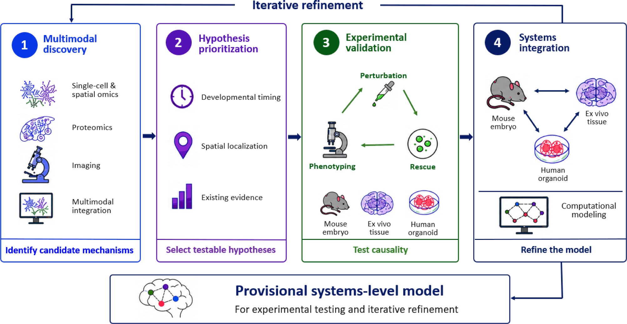
Candidate mechanisms should first be identified through integrated single-cell and spatial transcriptomics, multimodal molecular profiling, and quantitative imaging. Single-cell analyses can resolve transcriptionally distinct neuronal and radial glial populations, whereas spatial approaches relate these cellular states to their native anatomical environment [20–31, 45]. Computational analysis of ligand–receptor expression can further identify potential signaling relationships among neurons, radial glia, vascular-associated cells, and other components of the embryonic niche [78–81].
Discovery studies should integrate molecular identity with spatial localization and developmental timing. A candidate interaction becomes more biologically plausible when the predicted signal-producing and responding populations occupy neighboring territories during the developmental interval in which the relevant migratory or differentiation event occurs. Transcriptomic association and spatial proximity nonetheless remain hypothesis-generating evidence rather than proof of functional communication.
Because high-throughput approaches generate numerous candidate regulators, hypotheses should be prioritized by cell-type specificity, spatial proximity, temporal correspondence, evolutionary conservation, reproducibility across datasets, and the strength of existing functional evidence. Mechanisms supported by loss-of-function, gain-of-function, or rescue studies should generally take precedence over relationships inferred solely from gene-expression data.
Directly demonstrated mechanisms provide useful benchmarks for prioritization. These include the influence of the DA-associated environment on embryonic midbrain GABAergic migration and the contribution of Netrin-1-dependent GABAergic migration to substantia nigra development [5, 7]. Each selected hypothesis should specify the predicted signal source, responding population, developmental interval, anatomical location, and measurable developmental outcome.
Prioritized candidates should undergo functional perturbation using conditional genetic models, CRISPR/Cas-mediated genome engineering, RNA interference, pharmacological manipulation, or spatially and temporally restricted approaches [36, 37]. The resulting phenotypes should then be evaluated by quantitative imaging, spatial molecular profiling, and lineage tracing in order to distinguish effects on progenitor specification, neuronal differentiation, migration, positioning, survival, and tissue organization [22, 25, 26, 28–37].
Cell-type-specific and temporally controlled manipulations matter particularly here, since broad perturbations may affect neuronal development indirectly through changes in regional patterning, vascularization, or tissue integrity. Perturbation experiments should be complemented wherever feasible by rescue studies restoring gene function or signaling activity. Evidence for coordination will be strongest when manipulating one neuronal lineage or a shared niche component produces a reproducible effect on the development of another lineage, with the signal source, target population, and developmental interval experimentally resolved [5, 7].
Human pluripotent stem cell-derived ventral midbrain organoids provide complementary systems for assessing cross-system correspondence and potential conservation [38–50], though organoid concordance alone establishes neither causality nor conservation. A conserved phenotype should ideally show comparable regional and cell-type identities, developmental timing, source–target expression patterns, phenotype direction, and responses to orthologous perturbation and rescue in at least one native-tissue or ex vivo model.
Current organoids do not independently reproduce native embryonic anatomy, complete GABAergic migratory routes, vascular and meningeal organization, or long-range tissue interactions. Defined ECM scaffolds, endothelial co-culture or vascularization strategies, perfusion or bioreactor systems, and regionally fused assembloids may improve selected niche features [38, 39, 42–44], but each addition introduces model-specific variables. Organoid findings should therefore be interpreted alongside causal evidence from mouse embryos and ex vivo preparations.
Systems-level integration should combine molecular, spatial, temporal, and phenotypic evidence across mouse embryos, ex vivo tissues, human organoids, and developmental datasets [20, 21, 23–25, 28–31, 38–50]. Confidence should rise from expression and spatial correspondence through causal perturbation to replication across complementary systems, while computational models return experimentally supported predictions to hypothesis prioritization [28–30, 36, 37, 78–81].
FUTURE PERSPECTIVES
Future progress will depend on predictive models that connect lineage-specific mechanisms with the spatial, temporal, and multicellular organization of embryonic tissues. Predictive models should connect progenitor identity, position, developmental timing, migration, and tissue architecture while preserving the distinction between demonstrated and inferred relationships [2, 4–7, 19]. Their value will be measured by whether they prospectively identify mechanisms and phenotypes that withstand targeted experimental testing [36, 37, 78–81].
Regarding new technologies and system integration, single-cell and spatial transcriptomics, lineage tracing, CRISPR perturbation, and quantitative imaging should be integrated across embryos, ex vivo tissues, and human ventral midbrain organoids to connect molecular identity with cellular behavior and assess cross-system correspondence [20–50, 86].
Because radial glia, ECM, vascular signals, guidance molecules, and adhesion mechanisms operate broadly in neural development, the framework may guide studies in other regions [17, 18, 61, 67, 72, 73, 87–90]. It should nevertheless be treated as an experimental perspective, not a universally conserved program, and retained only where region-specific predictions withstand causal testing.
CONCLUSIONS
DA and GABAergic neurons follow lineage-specific programs yet develop within a partially shared embryonic ventral midbrain environment. The Pitx3/aphakia evidence supports a DA-associated environmental influence on GABAergic migration, while Netrin-1/DSCAM-dependent positioning of anterior SNpr GABAergic cells constrains DA substantia nigra organization without establishing a direct GABA-derived molecular signal to DA neurons. Reciprocal molecular signaling between the two neuronal populations therefore remains unproven. CXCL12/CXCR4 and Reelin are established mainly as DA-lineage regulators, and radial glia, ECM remodeling, vascular signals, and multicellular communication carry more limited or emerging support for DA–GABA coordination. The dynamic multicellular developmental niche remains a provisional framework for source-, target-, stage-, location-, and phenotype-specific hypotheses that can be confirmed, narrowed, or rejected through perturbation, quantitative phenotyping, rescue, and comparison across animal and human models.


