


INTRODUCTION
Progressive multifocal leukoencephalopathy (PML) is a demyelinating disease of the central nervous system (CNS) caused by oligodendroglial cells infection by the John Cunningham virus (JC) strain of papovavirus [1]. PML usually develops in immunosuppressed adults with predisposing disorders, such as lymphoproliferative diseases, chronic infections, chronic inflammatory diseases, and iatrogenic immunosuppression. PML reflects viral reactivation, and increased incidence has been caused by the rise of the acquired immune deficiency syndrome (AIDS) epidemic [1]. JC virus infects only humans, and PML targets the white matter, particularly oligodendrocytes in subcortical and deep cerebral white matter, the cerebellum, and the brainstem, and rarely involves the spinal cord. Patients show progressive symptoms reflecting the distribution of the underlying demyelination. To date, the radiographic findings and clinical features consistent with PML, coupled with the confirmation of JC virus infection by cerebrospinal fluid (CSF) polymerase chain reaction (PCR), are sufficient for the unequivocal diagnosis of PML in immunodeficiency [2-4]. However, it is difficult to suspect PML without patient history or clinical evidence of immunodeficiency. Here, we describe a case of PML diagnosed by brain biopsy without medical history.
CASE REPORT
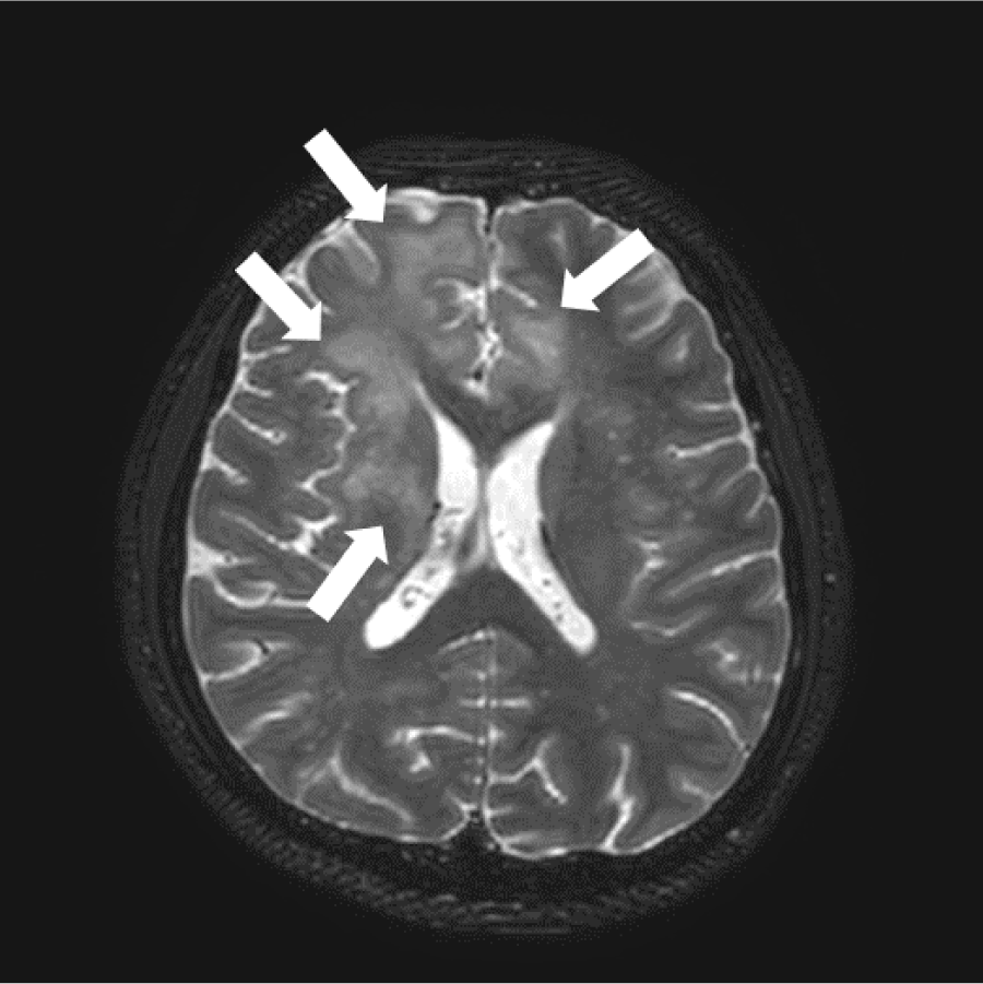
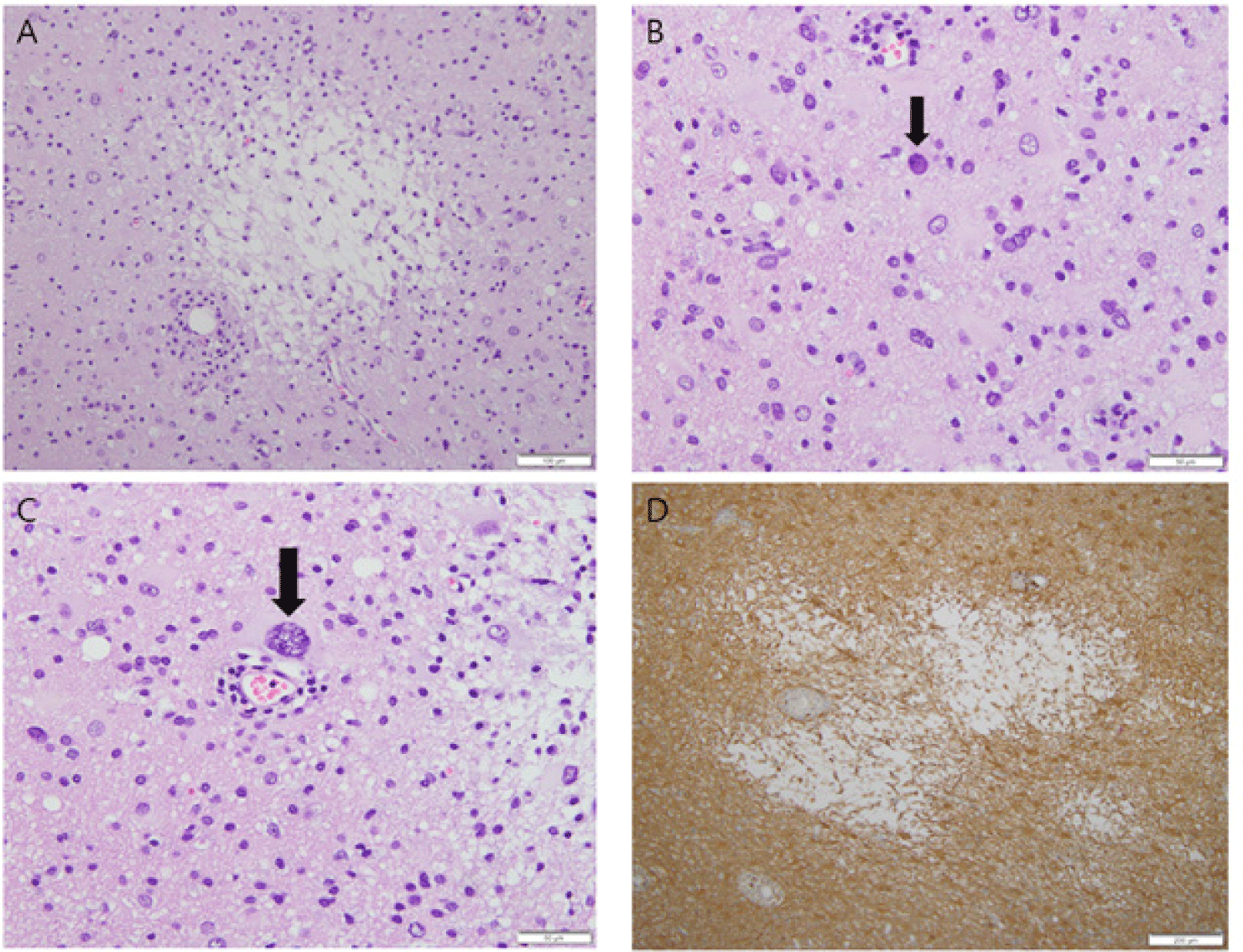
A 32-year-old man presented with intermittent headache in the left parietal region for a month without any antecedent events and an uneventful medical history. He has no personality changes or cognitive abnormalities. The pain was described as waxing and waning, and was characterized by stabbing, 20 minute headaches. Severity was 5–6 (mild to moderate) in the Numerical Rating Scale score, and there were no aggravating factors. Brain magnetic resonance imaging (MRI) demonstrated multifocal ill-defined confluent T2 high signal intensity lesions in the frontal lobes, right parietal lobe, right basal ganglia, and medulla (Fig. 1). Based on clinical and image features, the differential diagnoses included demyelinating lesion and neoplasms. To distinguish white matter demyelinating diseases from tumorous lesions, a surgical biopsy was performed. One burr hole was made just anterior to the coronal suture and approximately 4 cm left of the midline. One external ventricular drain catheter was inserted into the left posterior horn of the lateral ventricle. Under neuronavigation guidance, the patient underwent stereotactic biopsy. Aspiration revealed a yellowish CSF. Microscopy revealed multiple foci of loss of myelin and oligodendroglial cells with a variable inflammatory infiltrate, especially macrophages (Fig. 2A). Significant perivascular lymphocytic and histiocytic infiltration was identified. Oligodendroglial cell had enlarged nuclei and plum-colored inclusions (ground glass-appearance) with chromatin margination (Fig. 2B). This inclusion was observed along the periphery of the lesion. Atypical astrocytes mimicking neoplastic conditions had large or multiple nuclei (Fig. 2C). Immunohistochemical staining revealed myelin loss and a few residual glial cells (Fig. 2D). Pathological diagnosis was “multifocal demyelinating and degenerative white matter, consistent with PML”.
After brain biopsy, human immunodeficiency virus (HIV) infection was confirmed by serum chemiluminescent immunoassay (CLIA) and JC virus infection was detected by CSF PCR. Although there were no respiratory symptoms, Pneumocystis jirovecii was identified by sputum PCR. A highly active antiretroviral therapy was initiated. The patient’s postoperative course was uneventful, and the headache disappeared. The patient was discharged without any complications. This study was approved by the institutional review board (CBNUH IRB, Cheongju, No. 2022-01-012), and informed consent was obtained from the patient.
DISCUSSION
PML is a fatal demyelinating disease of the CNS sed by the JC virus, which infects oligodendrocytes. Demyelinating lesions are a consequence of virus-induced oligodendrocyte necrosis. Patients present with focal neurologic defects according to the location of the demyelinating sites. These defects include headache, motor dysfunction, visual loss, and cognitive abnormalities. PML occurs almost exclusively in immunosuppressed individuals. HIV infection often coexists with PML. After the AIDS pandemic, PML became characterized as a disease of younger people who are affected by AIDS, as is the case of our patient. He visited the hospital because of intermittent headaches and no previous medical history. Hyper-intense signal abnormalities of the white matter on T2-weighted MRI suggested demyelinating disease or other tumors. A brain biopsy was performed before any other serological tests. The microscopic findings were consistent with those of PML. These pathologic and clinical findings led to serum CLIA to confirm HIV infection, as the clinical headache and pathological PML were the first manifestations in the patient. In the diagnosis of PML, the detection of JC virus DNA in CSF by PCR is more useful than brain biopsy. JC virus infection can be diagnosed by identification of the virions in oligodendrocytic nuclei by electron microscopy examination, recognition of JC virus proteins by immunohistochemistry of the infected cells, and as well as in situ hybridization of the viral genome. However, it is unlikely that these tests for JC virus would be considered without establishing an immunosuppressed status, such as HIV infection.
Therefore, it is very important for pathologists to be aware of the histological features of PML [5, 6]. JC virus induces PML as well as other brain disorders, such as granule cell neuronopathy of the cerebellum and fulminant JC virus encephalopathy [2]. This virus can be even found in the brains of healthy individuals. The demonstration of the presence of this virus is insufficient to establish a diagnosis of PML; this should be accompanied by the clinicopathological features.