INTRODUCTION
Natural relics excavated from historic sites provide important data for understanding the lifestyle and cultural level of humans at that time. Animal bone relics can reveal the type, distribution, and animal geography of animals hunted or raised at the time and can be used as valuable data for future research [1–7].
Reports on Korea’s ancient livestock include the historical study of the horse husbandry in Korea [8], systematic studies on the Korean native horse [9], studies on the origin of Korean native goats [10], and study on the veterinary science in the Three Kingdoms period [11].
Research on animal bone relics discovered at historical sites includes animal bones from the Chondrichthyes, Osteichthyes, Amphibia, Aves, and Mammalia classes excavated from the Sugari shell mound in Gimhae [12]. Another report is on Pisces, Aves, and Mammalia natural relics from the Yongwon Shell Mound site in Jinhae [13], and another is on bone relics from the Maldeung ruins in Baengnyeong Island [14]. Among the animal bone relics in Jeju Island, animal bones excavated from the Gwakji archaeological site [4, 15], the Jongdal-ri shell mound site [3], and relics excavated from the Gwenae Cave site in Gimnyeong-ri have been reported [5].
This study examined Cervidae bones identified among those excavated from a well presumed to be from the Three Kingdoms period in Gasan-ri, Ibanseong-myeon, Jinju, excavated by the Gyeongnam Cultural Heritage Research Institute. The size and characteristics of Cervidae bones from that period were observed by gross anatomy.
MATERILS AND METHODS
The animal bones in this study were Cervidae bone artifacts excavated from a well in Gasan-ri, Ibanseong-myeon, Jinju [16]. The excavated Cervidae bones were classified according to the body part and compared to existing animal skeleton specimens stored in the anatomy specimen room of the college of veterinary medicine at gyeongsang national university to consider the species, age, and size of animals similar to the Cervidae bone artifacts. A comparative analysis was conducted on the animal skeletal samples.
The excavated animal bones were classified by anatomic region according to Schmid’s method [17] and divided into skull, vertebral axial skeletons, forelimb bones, and hindlimb bones. The measurements of the Cervidae bone artifacts were made with a Mitutoyo caliper (0.01 mm) according to Driesch’s method [18].
RESULTS
Animal bones were excavated from a well in Gasan-ri, Ibanseong-myeon, Jinju, about 30 meters east of the Three Kingdoms period ruins. According to the classification method of Schmid [17] and Driesch [18], the teeth arrangement for Cervidae animal bones is 0/3 1/1 3/3 3/3. Among the animal bones from the well in Gasan-ri, Ibanseong-myeon, Jinju, those with deer-like tooth structures were classified as Cervidae. Among the 447 excavated animal bones, 102 (22.82%) were classified as Cervidae, and the weight of the Cervidae bones (453.79 g) accounted for 46.53% of the total weight of the identified bones (975.30 g).
The classification of the excavated Cervidae bones according to body area identified 19 (18.63%) skull bones including the maxilla and mandible, 14 (13.73%) vertebral axial skeletons, 28 ribs and sternum (28.4%), 16 (15.69%) forelimb bones, and 19 (18.63%) hindlimb bones. The metatarsal bone and the phalanges were indistinguishable in six cases, and many Cervidae skeletons were intact.
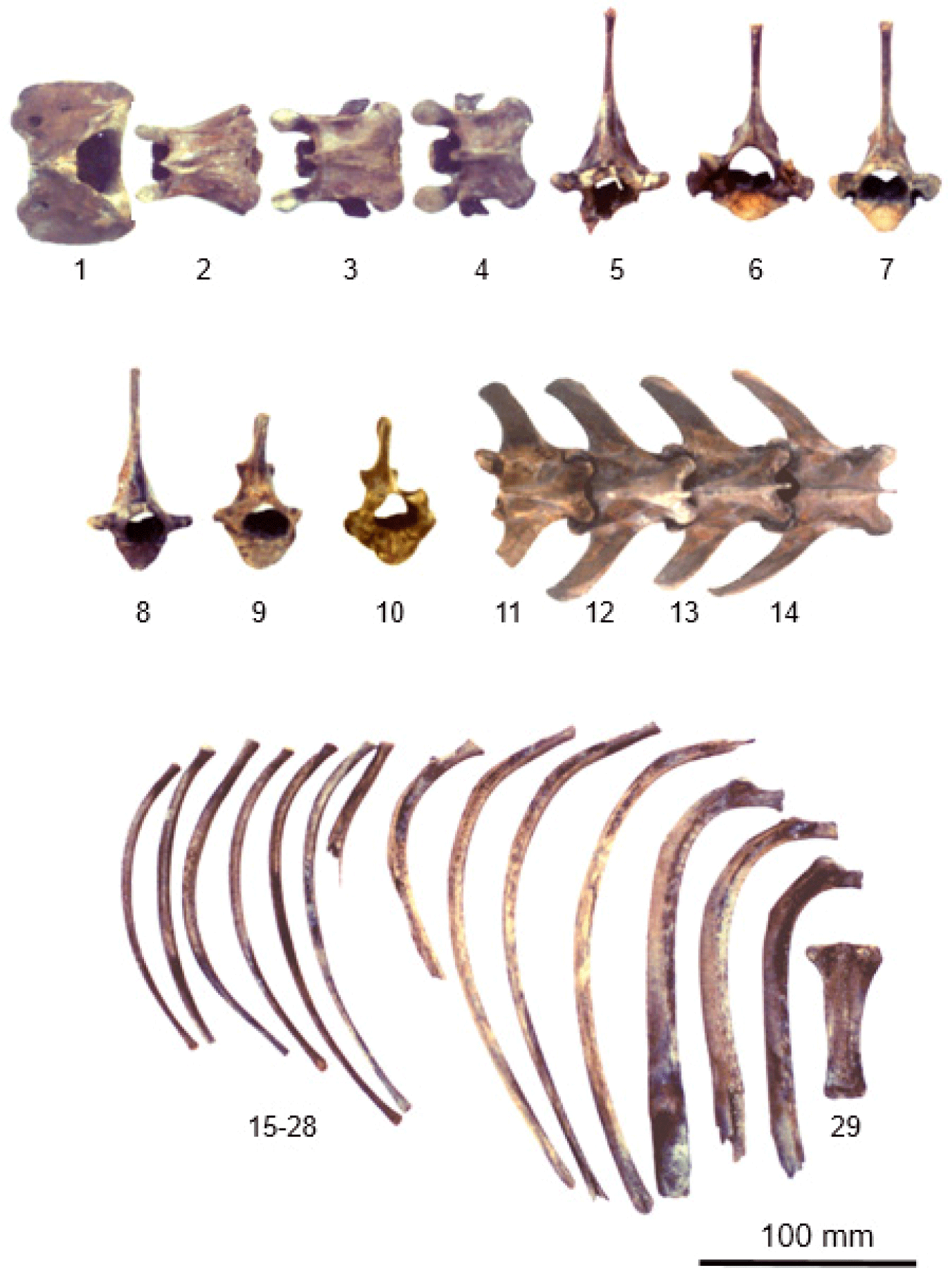
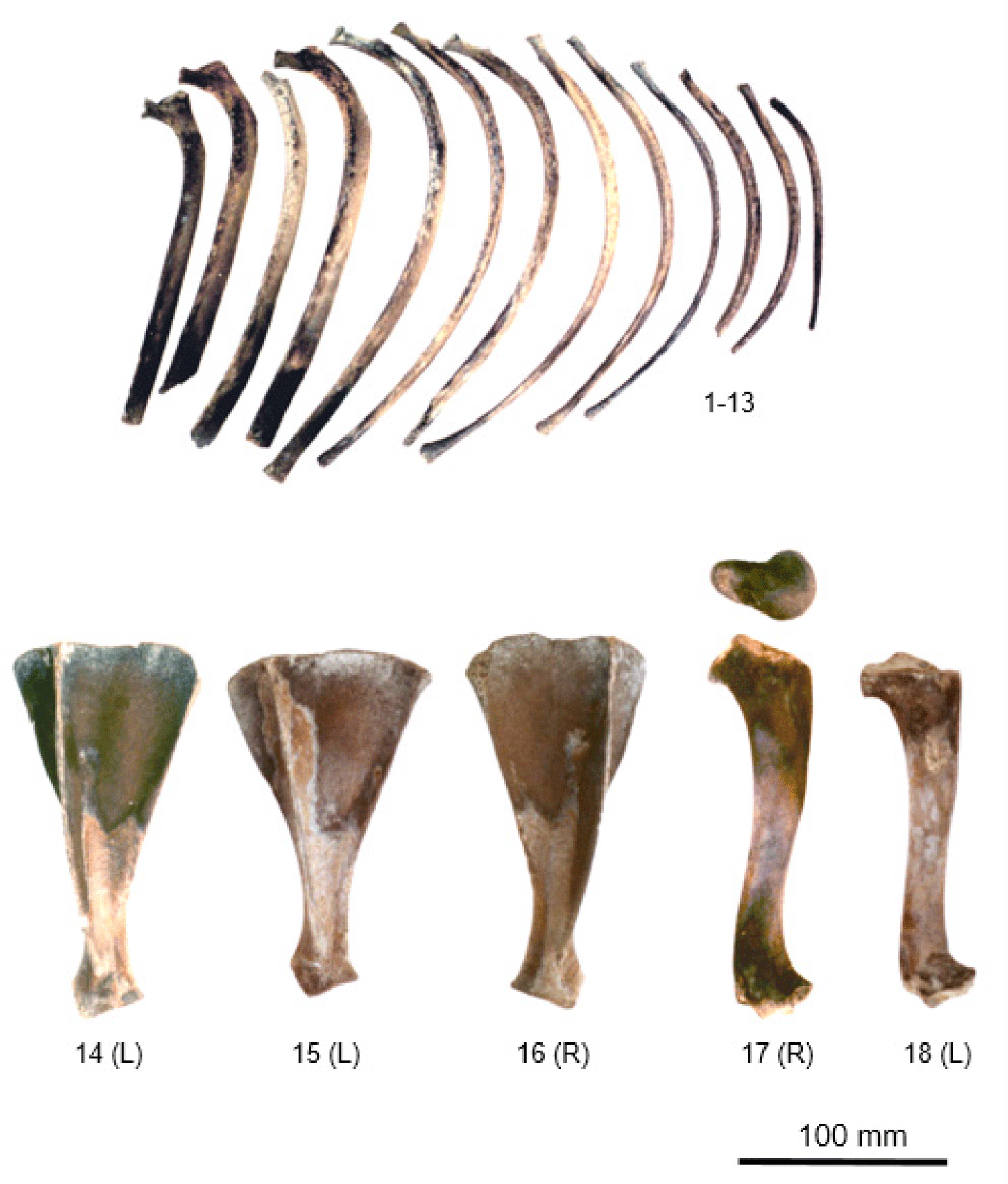
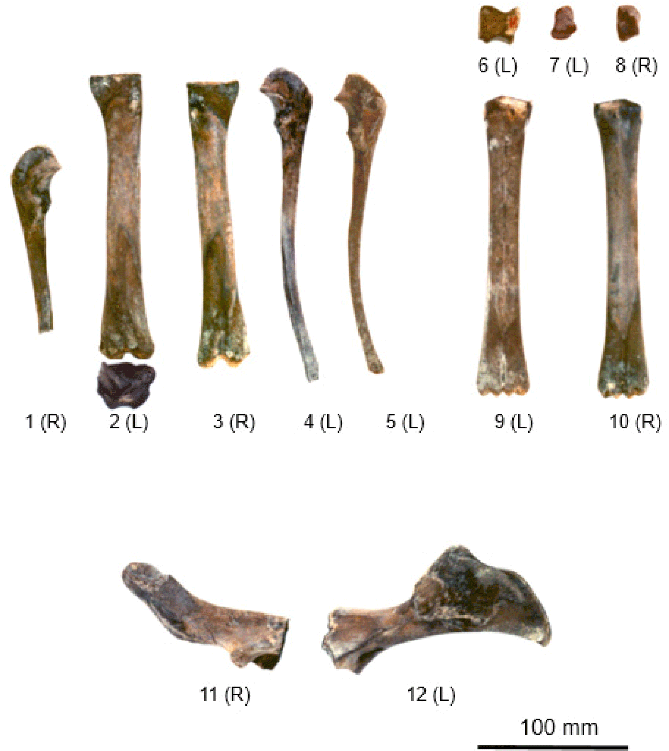
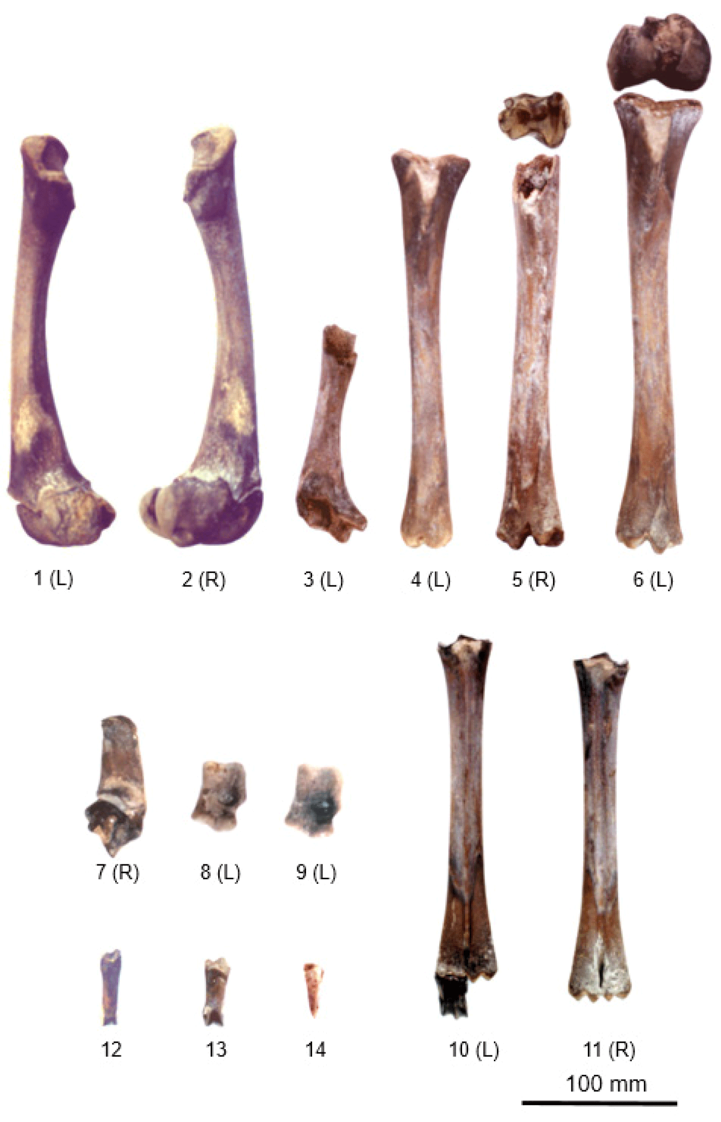
The animal bones classified as Cervidae were from two animals. The skulls were severely damaged, and the horns were missing, making it difficult to clearly distinguish between roe deer and deer. Additionally, three canine teeth were observed in the maxillary bone. Considering that the canine teeth on the maxillary bone were short and small, these specimens were presumed not to be water deer (Hydropotes inermis argyropus). The canine teeth were also not long, so it was difficult to classify them as roe deer (Capreolus capreolus) or deer (Cervus sp). Therefore, they were classified as Cervidae. The Cervidae bones were divided into skull bones, vertebrae, ribs, sternum, hip bones, forelimb bones, and hindlimb bones (Figs. 1, 2, 3, 4, and 5).
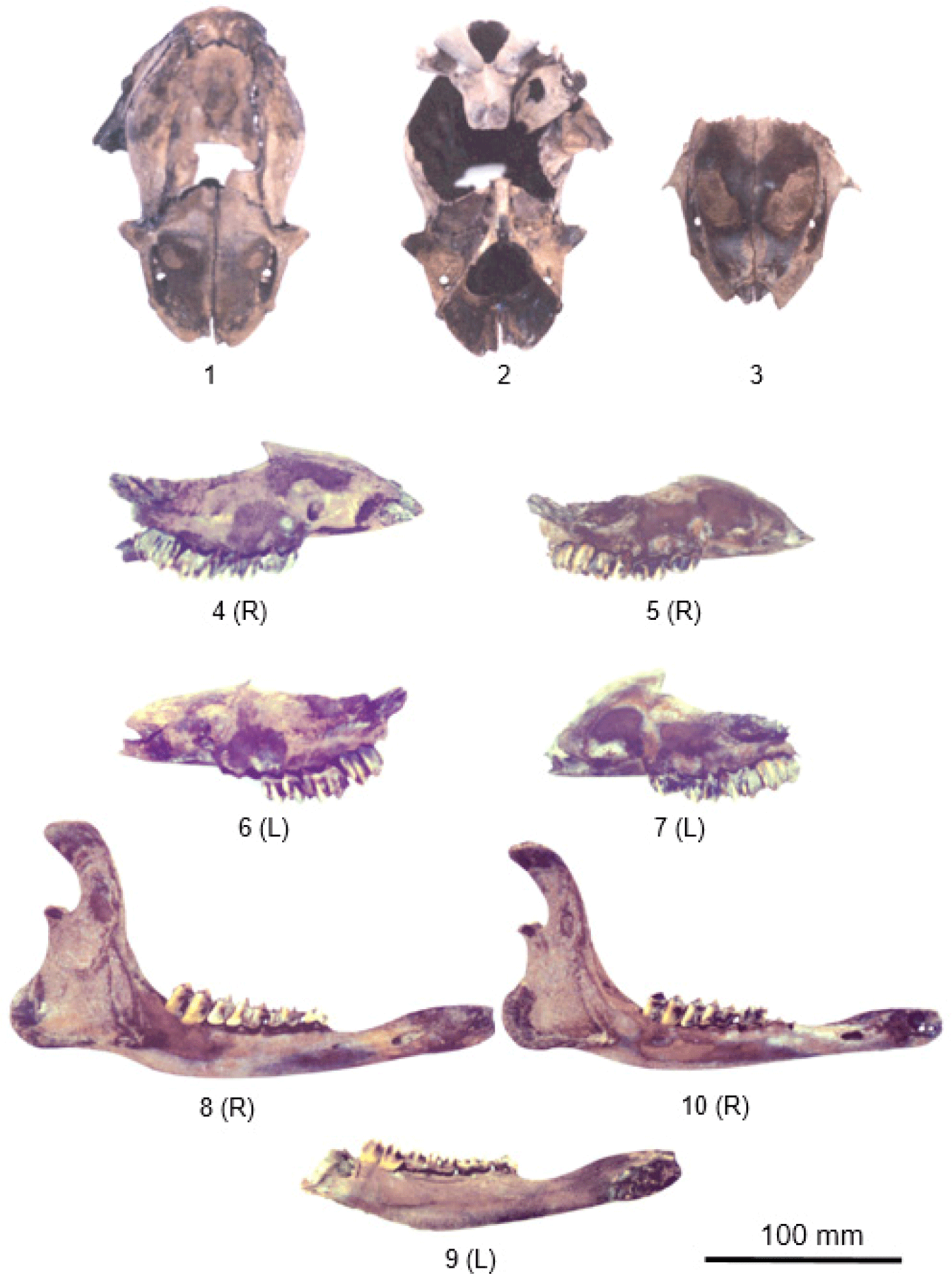
The type and quantity of each body region of the two identified sets of Cervidae bones are shown in Table 1. These features were compared with those of existing water deer and deer skeletons stored in the animal specimens of the College of Veterinary Medicine at Gyeongsang National University. The length of the premolar row in each Cervidae skeleton, which is the standard for head size, was 25.65 mm and 24.58 mm, respectively, which was longer than that of existing 1- to 1.5-year-old water deer but shorter than that of deer. Additionally, the length from the angle (angle of the mandible) of each mandible was 113.37 and 102.48 mm, length from the condyle was 108.47 and 101.19 mm, length of the premolar row was 26.23 and 24.03 mm, aboral height of the vertical ramus was 33.75 and 29.74 mm, middle height of the vertical ramus was 31.44 and 28.27 mm, oral height of the vertical ramus was 54.45 and 49.07 mm, respectively, which was longer than that of existing 1- to 1.5-year-old water deer and shorter than that of deer.
Nine bone fragments were recovered from one individual’s skull, including one bone fragment each from the left and right maxillae, while further portions of the temporal bone, sphenoid bone, and incisor bone were damaged or missing. The skull of the other individual was only partially restored by combining three bone fragments. There was one left and right maxillary bone fragment each, and skull bone behind the border between the frontal bone and the parietal bone. The parietal bone near the posterior parietal point (bregma), which is the boundary between the frontal bone and the parietal bone, had a hole believed to have been caused by a blow and was severely broken into several skull fragments (Fig. 1[1–3]).
There were four maxillary bones (2 on the left, 2 on the right; same below; Fig. 1[4–7]), and the canine teeth were not long. The two skeletons had three mandibular fragments (1 in one skeleton and 2 in the other), and because the first molars had erupted, the skeletons were estimated to be from individuals 5–6 months old (Fig. 1[8–10]). A total of 29 teeth were preserved, 18 in the maxilla and 11 in the mandible, with no incisor preserved.
A total of 14 vertebrae bones were identified: four cervical vertebrae, six thoracic vertebrae, and four lumbar vertebrae, and the atlas and axis were included among the cervical vertebrae (Fig. 2[1–14]). Twenty-seven ribs (13 left, 14 right) were preserved (Figs. 2[15–28] and 3[1–13]). The maximum length of the head of the Cervidae ribs was 5.83 (5.08–6.63) mm, which was much shorter than the maximum length of the tubercle of the ribs, which was 8.05 (5.62–9.13) mm. Only one xiphoid process in the sternum was preserved (Fig. 2[29]).
Among the forelimb bones, there were three scapulae (2 left, 1 right; Fig. 3[14–16]) and two humerus bones (1 left, 1 right), which were located on the left side of the small individual. In the humerus, both the proximal epiphysis and the distal epiphysis were separated and missing (Fig. 3[17, 18]). There were two radial bones (1 left, 1 right; Fig. 4[2, 3]) and three ulnar bones (2 left, 1 right; Fig. 4[1, 4, 5]). A total of three carpal bones were preserved, including one forearm on the ulnar side of the left front foot and one for each of the left and right intermediate carpal bones (Fig. 4[6–8]). There were two metacarpal bones (1 left, 1 right), one each for the left and right front metacarpals, consisting of the third and fourth metacarpal bones (Fig. 4[9, 10]), and the greatest length of metacarpal bones was 133.74 mm (Table 1).
In the bones of the hindlimb, only one right ischium and one left ilium were preserved as hip bones (Fig. 4[11, 12]). Three femurs (2 left, 1 right; Fig. 5[1–3]) and three fibulae (2 left, 1 right) were also preserved (Fig. 5[4–6]). Three tarsal bones were preserved: one calcaneus (1 right; Fig. 5[7]) and two taluses (2 left; Fig. 5[8, 9]). Two hind metatarsal bones were present (1 left, 1 right), and the third and fourth hind metatarsal bones were fused (Fig. 5[10, 11]). Three phalanges were present, one each for the proximal phalanx, middle phalanx, and distal phalanx (Fig. 5[12–14]). The greatest length of the proximal phalanx was 133.74 mm, the breadth of the distal end was 8.94 mm, and the length of the dorsal surface of the distal phalanx was 21.37 mm (Table 1).
DISCUSSION
Pit dwellings, high-rise buildings, and wells were identified at the Gasan-ri site ruins in Jinju. Judging from the excavated artifacts, the age of the ruins was estimated to be the 8th century or earlier [16]. Among all animal bones excavated in this study, Cervidae bones accounted for 22.82% (weight ratio: 46.53%) of the skeletons found. The proportion of deer skeletal bones in the Jongdal-ri shell mound site in Jeju was 53.3% [1], and the proportion of deer skeletal bones in the Gwakji shell mound in Jeju was 36.4% [4]. The proportion of deer skeletal bones in the Gwenaegi ruins in Gimnyeong-ri Jeju was 11% [5]. The remains of terrestrial mammals excavated from the Yongwon shell mound in Jinhae included those of wild animals, such as deer, wild boars, roe deer, otters, and raccoons, as well as those of domestic animals, such as dogs, cows, and deer. Among them, deer bones accounted for the largest proportion at 61.2%. Therefore, it is estimated that the proportion of Cervidae was very high during the Three Kingdoms period.
Animal bone fragments excavated from the Jongdalli shell mound site in Jeju Island were reported to be 61.75% skull bones, 6.24% vertebrae, 12.97% forelimb bones, and 14.81% hindlimb bones [1]. Among the 515 animal bones identified at the Gonaeri site in Jeju, skulls (50%) were the most common [2]. Nishinakagawa et al. reported that skull bones (27.3%, 22%), vertebral axial skeletons (0%, 28%), forelimb bones (20.8%, 22%), and hindlimb bones (25.7%, 28%) were evenly excavated [6, 7]. Shin et al. reported that skull bones accounted for the majority at 91% [5]. However, most studies did not accurately classify the excavated proportions by animal. In this study, the excavated Cervidae bones included 18.63% skull bones, 13.73% vertebral axial skeletons, 28.4% ribs and sternum, 15.69% forelimb bones, and 18.63% hindlimb bones. The ratio of each excavated animal part appeared to vary depending on the circumstances and surrounding environment of the era in which the animal remains were excavated.
Previous studies reported that diseases were searched in the bones of excavated ruins [19] and found that bone-related diseases existed in ancient times. Just as in modern times, tumors were observed in the radial bones and lumbar vertebrae of relic wild boars [7]. In this investigation, the Cervidae bones were from 5 to 6-month-old individuals, and no lesions or tumors were observed on the bones.
The skull of a Cervidae excavated from the well in Gasan-ri, Ibanseong-myeon, Jinju was damaged in the left parietal area from the crown of the frontal bone and parietal bone toward the occipital bone along the midline. The destruction of the skull of the 5 to 6-month-old Cervidae in this study was by artificial blows presumed to have been done for the purpose of using the brain. However, the preserved cartilage of the excavated animals’ ribs, humerus, radius, ulna, femur, and fibula suggests that the animals were not used for food. Additionally, we could not rule out the possibility that the discovery of animal remains inside the well represented either an object disposed of while using the well or an object disposed of from some kind of ritualistic act.
The well relics were located about 30 m east of a residence, and animal bone relics were excavated only from the lowest level, along with jars and earthenware, wooden tools, plant remains, and animal remains, such as Cervidae. Among the accompanying relics, jars, and earthenware were presumed to have been thrown into the well after being used in ancestral rites, as the well is used not only for drinking water but also as an important ritual space [16]. In particular, it was not clear whether the animal remains were disposed of during well use rather than being used for human consumption or whether they were placed there for some kind of ceremonial purpose and disposed of in the well.
In conclusion, the results of anatomical investigation of Cervidae bone artifacts excavated from a well site in Gasan-ri, Ibanseong-myeon, Jinju-si are as follows. A total of 447 animal bones were excavated, of which 102 (22.82%) were classified as Cervidae bones. The weight of the Cervidae bone was 453.79 g, accounting for 46.53% of the total weight of the identified bone (975.30 g). The Cervidae bones were identified as belonging to two animals with an estimated age of 5 to 6 months. Cervidae bones were divided into skull bones, vertebrae, ribs, sternum, hip bones, forelimb bones, and hindlimb bones. The 102 Cervidae bones include 19 skull bones (18.63%), 14 axial vertebrae (13.72%), 28 ribs and sternum (28.43%), 16 forelimb bones (15.69%), and 19 hindlimb bones (18.63%). and the remaining six were difficult to distinguish. A parietal bone fracture was observed near the bregma of the skull and was presumed to have been caused by an artificial blow. This study can be used as basic data to estimate the types of animals and human culture at the time through Cervidae bones believed to be relics from the Three Kingdoms period.


