Original Article

Effect of inhaled 1,2-dichlorobenzene on cytochrome P450s and lipid peroxidation in B6C3F1 mice

Eun-Sang Cho1,*https://orcid.org/0000-0001-7297-0063, Yong-Soon Kim1https://orcid.org/0000-0001-9875-3833, Hye-yeon Choi1https://orcid.org/0009-0003-0636-7874, Kyung-Taek Rim1https://orcid.org/0000-0001-6443-6362
Author Information & Copyright
1Inhalation Toxicity Research Center, Occupational Safety and Health Research Institute, Korea Occupational Safety and Health Agency (KOSHA), Daejeon 34122, Korea
*Corresponding author: Eun-Sang Cho, Inhalation Toxicity Research Center, Occupational Safety and Health Research Institute, Korea Occupational Safety and Health Agency (KOSHA), Daejeon 34122, Korea, Tel: +82-42-869-8513, E-mail: escho@kosha.or.kr

© Research Institute of Veterinary Medicine, Chungbuk National University.

Received: Jul 26, 2023; Revised: Aug 17, 2023; Accepted: Aug 18, 2023

Abstract

1,2-Dichlorobenzene (1,2-DCB) is in various industries as solvents in herbicides, pesticides, and wax, and as degreasers or in the production of dyes. Studies on the hazards and risks of 1,2-DCB showed that this substance can cause skin corrosion, skin irritability, respiratory irritability, and certain target organ toxicity. Industrial workers are exposed to 1,2-DCB by inhalation or skin exposure and there is a lack of information on human hazards even though they can be exposed to organic compounds such as benzene or other DCB complexes rather than a single substance. In this study, we investigated the specific organ toxicity of 1,2-DCB and sex differences using whole-body inhalation in laboratory mice. Male and female mice were exposed to 0–120 ppm of the test substance for 13 weeks. After euthanization, the organs were collected, histopathological assessments and immunohistochemistry (IHC) were performed, and lipid peroxidation was analyzed. Macro and microscopic lesions were observed in the livers of male mice exposed to the test substance, and microscopic alterations were observed in the nasal cavities of male and female mice. IHC analysis of the liver confirmed a greater increase in cytochrome P450 induction in males than in female mice, and malondialdehyde and 4-hydroxynonenal were increased in both sexes by 1,2-DCB inhalation. Based on the relevant literature and experimental results, 1,2-DCB is believed to cause specific organ toxicity in the livers of male mice and the nasal cavities of both sexes of mice, which is supposed to be related to sex differences in cytochrome P450 induction and changes in lipid and oxidative products associated with the early metabolites of the test substance.

Keywords: 1,2-dichlorobenzene; inhalation toxicity; liver toxicity; B6C3F1; cytochrome P-450 enzyme system

INTRODUCTION

1,2-Dichlorobenzene (CAS No. 95-50-1, o-dichlorobenzene or o-DCB) is a colorless, halogenated aromatic liquid compound known as a dichlorobenzene isomer, which includes 1,3-dichlorobenzene (meta-dichlorobenzene) and 1,4-dichlorobenzene (para-dichlorobenzene). This compound is primarily used as a base for herbicides, pesticides, solvent in wax, resin, tar, rubber, oil, and paint [1, 2].

This substance is classified as an acute toxic agent 4, eye irritant 2, and skin irritant 2, and has specific target organ toxicity (single) according to the globally harmonized system of classification and labeling of chemicals health hazards categories, and about 10,000–100,000 tonnes per year are manufactured and/or imported in the European Economic Area [3]. The American Conference of Governmental Industrial Hygienists (ACGIH) recommended an exposure limit of 25 ppm as the time-weighted and 50 ppm as the short-term exposure limit. The Occupational Safety and Health Administration (OSHA) and National Institute for Occupational Safety and Health (NIOSH) recommended 50 ppm as the permissible exposure limit-ceiling or recommended exposure limit-ceiling, respectively [4]. 1,2-Dichlorobenzene showed a high level of liver toxicity, causing mortality in an acute oral study and animal inhalation studies using rats and mice [5, 6].

Several researchers also studied differences in liver microsomal metabolism between humans and mice through pharmacokinetic model studies, but further studies are needed because these studies are insufficient to use as evidence for risk assessment [2, 7]. We identified unexpected differences in hepatotoxicity between male and female animals through a subacute inhalation toxicity study, which encouraged us to study the risk of 1,2-dichlorobenzene exposure to occupational workers. While there are some toxicologic data from animal studies on this substance, our study investigated subchronic inhalation toxicity using laboratory mice, which is the main route of exposure to workers, and confirmed differences in specific organ toxicity in male and female mice.

MATERIALS AND METHODS

Animals

Six-week-old male and female specific-pathogen-free B6C3F1 mice were purchased (Japan SLC, Shizuoka, Japan) and acclimatized for nine days in polycarbonate cages. We followed the methods of Cho et al. 2020 for the inhalation toxicity study [8]. Exposures were conducted in whole-body inhalation chambers with attached temperature and humidity control devices (Model No. WITC-06M, HCT, Icheon, Korea). All animals were maintained in individual wire mesh cages with three chambers, one for each concentration of the test substance and one control chamber with HEPA-filtered clean air. The ambient temperature and relative humidity of the chamber were 22 ± 3°C and 50 ± 20%, respectively, with a 12:12 hour light/dark cycle with 150~300 lux of illumination. The mice received rodent chow (2918C, Envigo RMS, Indianapolis, IN, USA) and tap water ad libitum. We also provided wooden chew sticks to each mouse as an environmental enrichment item. All animal care and inhalation studies were performed under Good Laboratory Practice (GLP) conditions with modified OECD guidelines (TG413) [9]. The animal study protocol was approved by the Committee on Animal Research Committee of the Occupational Safety and Health Research Institute (Approval No. IACUC-1816).

Chemicals and inhalation exposure

1,2-Dichlorobenzene (99.0% pure, Lot No.: 010917,012618; CAS No.:95-50-1) was purchased from Samchun Pure Chemicals (Pyeongtaek, Korea). The Environmental conditions were monitored every 30 min (Model No. ITC manager, HCT). We generated the concentration of 1,2-DCB in each chamber using a liquid vapor generator (LVG-04-A, HCT) at least three times during each exposure day and also analyzed the concentration of the test substance using gas chromatography (TRACE1310, Thermo Fisher Scientific, Waltham, MA, USA). The maximum concentration (120 ppm) of 1,2-DCB used in this study was selected based on results from a preliminary subacute inhalation study.

Experimental design

The subchronic inhalation toxicity test was performed following the protocol described by OECD guidelines (TG413), with modifications. A total of 40 mice were randomly assigned to four groups for each sex (n = 10): control (filtered air), T1 (30 ppm), T2 (60 ppm), and T3 (120 ppm). All mice were exposed to 1,2-DCB or filtered air for six hours/day, five days/week for 13 weeks. Body weight data were collected twice a week for six weeks and then once a week until 13 weeks. Individual food consumption were also measured once a week, and clinical observations were recorded twice a day during all exposure periods.

Sample collection

At the end of the study, all animals were anesthetized with isoflurane (I-Fran liquid, Hana Pharm, Seongnam, Korea) and euthanized by exsanguination from the abdominal aorta and vein after blood sampling from the caudal vena cava. The blood samples were collected in EDTA2K-containing sample tubes or serum separation tubes, then analyzed for hematology or serum biochemical tests after centrifugation at 1,800 × g for 10 min to obtain serum. At the necropsy of all animals, organs, including the adrenal glands, brain, epididymides, heart, kidneys, liver, lung, ovaries, spleen, testes, thymus, and uterus, were examined and weighed. The following organs and tissues were also examined, collected, and fixed in 10% neutral buffered formalin or Davison’s fixative (only for the testes and eyes/optic nerves) for histopathologic evaluation: aorta, bone marrow, cecum, colon, duodenum, esophagus, eyes/optic nerves, femur, gallbladder, Harderian glands, ileum, jejunum, larynx, lymph nodes (tracheobronchial and mesenteric), mammary gland, nasopharyngeal tissue, pancreas, parathyroids, pituitary, prostate, rectum, salivary glands (submandibular, sublingual, and parotid), sciatic nerve, seminal vesicle, skeletal muscle, skin, spinal cord, sternum, stifle joint, stomach, teeth, thyroid, tongue, urinary bladder, and vagina. Part of the liver tissue was also resected before fixation and kept at –80°C until relevant examination.

Hematology and blood biochemical analyses

Hematologic and biochemical blood examinations were performed using a hematology analyzer (ADIVA 2120i, Siemens Diagnostics, Tarrytown, NY, USA) or blood chemistry analyzer (TBA-120FR, Toshiba, Tokyo, Japan) for the following parameters: white blood cell counts with differential counts (neutrophils, lymphocytes, monocytes, eosinophils, and basophils), red blood cell count, hemoglobin, hematocrit, mean corpuscular volume, mean corpuscular hemoglobin, mean corpuscular hemoglobin concentration, platelet count, and reticulocytes; and biochemical parameters, including alanine aminotransferase (ALT), aspartate aminotransferase (AST), alkaline phosphatase (ALP), total protein, albumin, globulin, triglycerides (TG), total cholesterol, total bilirubin, blood urea nitrogen, creatinine, sodium, chloride, potassium, calcium, and inorganic phosphorus.

Histopathology, immunohistochemistry, and image analysis

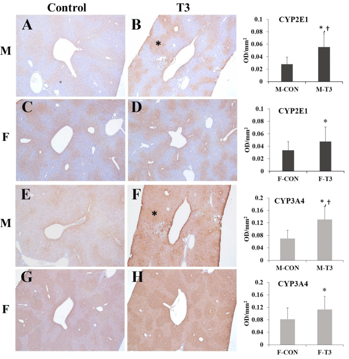
Fixed organs and tissues from the controls and T3 group and the liver and nasal cavities of the T1 and T2 groups were routinely processed for paraffin embedding. Four μm thick tissue was prepared and stained with hematoxylin and eosin (Automatic Stainer, DAKO, Glostrup, Denmark). Five-um thick liver tissue sections were prepared and stained with a trichrome staining kit (Ventana Benchmark Special Stains, Roche, Indianapolis, IN, USA) according to the manufacturer’s instructions. Immunostaining was conducted using EnVisionTM Detection System kit according to the manufacturer’s instructions (Peroxidase/DAB+, Rabbit.Mouse, K5007, DAKO). Briefly, 4-um thick paraffin-embedded liver sections were prepared and deparaffinized, hydrated, and blocked for endogenous peroxidase. Then, heat-induced epitope retrieval was conducted using a pressure cooker with citrate buffer. Cytochrome P450 2E1 and 3A4 antibodies are used for IHC. The microscopic features of the liver were examined and photographed under a light microscope (DM3000, Leica, Wetzlar, Germany). The intensity of immunostaining for CYP2E1 and 3A4 in the central vein area was analyzed in representative images of the serial sections at 20 ×, with five fields for each animal, using the ImageJ program (NIH, Bethesda, MD, USA).

Determination of lipid peroxidation

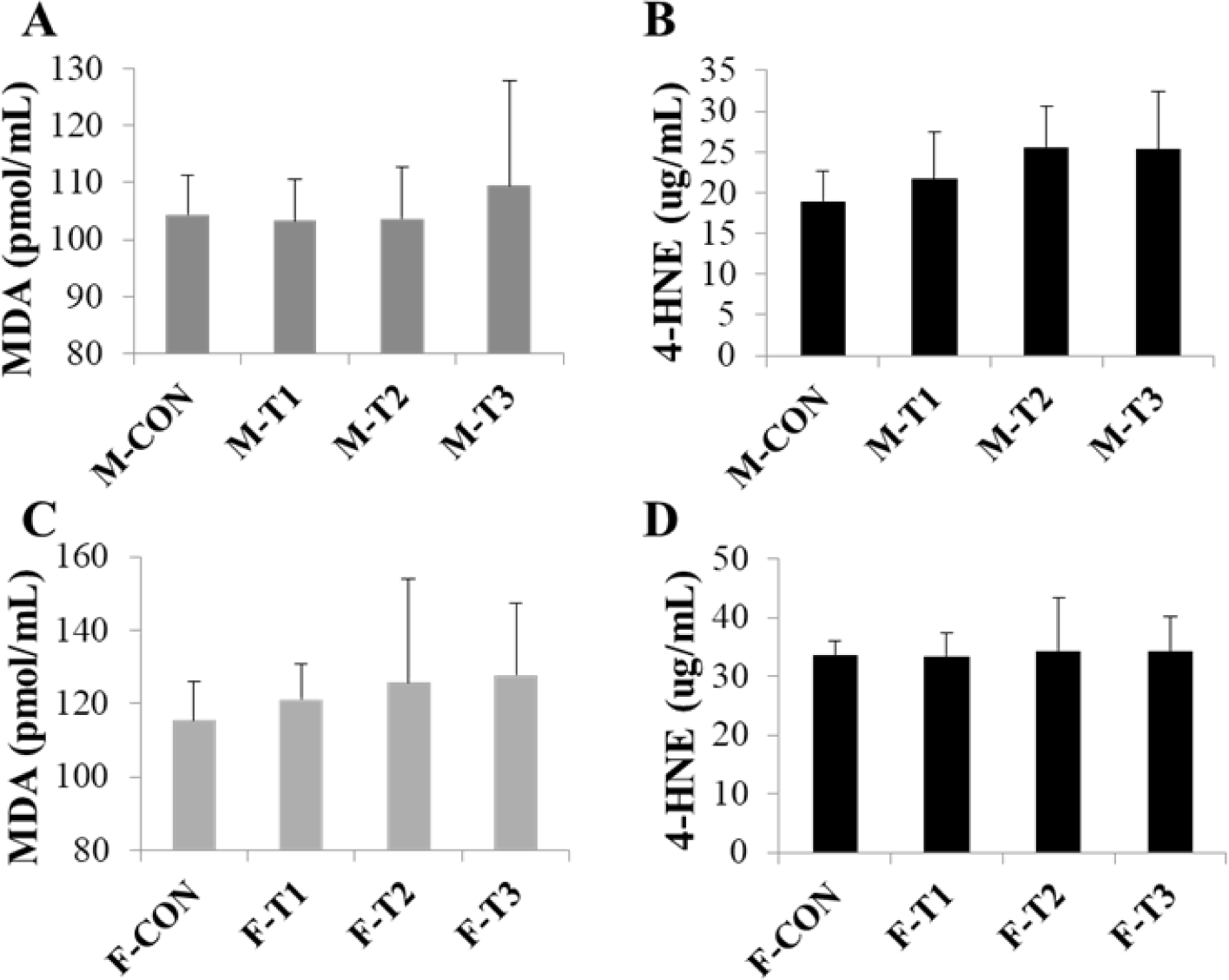
Lipid peroxidation levels were measured in liver homogenates as levels of malondialdehyde (MDA) and 4-hydroxynonenal (4-HNE), the parameters of oxidative stress. Each assay was conducted using kits (OxiSelectTM MDA Adduct Competitive ELISA kit or OxiSelectTM HNE Adduct Competitive ELISA kit, Cell Biolabs, San Diego, CA, USA) following the manufacturer’s instructions.

Statistical analysis

The data are expressed as the mean ± S.D. Statistical analyses were performed using the Pristima® system (Version 2.0; Xybion, Princeton, NJ, USA) to analyze parameters, including body weight, food consumption, organ weight, and hematological and serum biochemical data. The data were analyzed using Levene’s test for homogeneity of variance and one-way analysis of variance with Dunnett’s multiple test or Kruskal-Wallis with Dunn rank sum test for comparisons between the control and inhaled test substance groups. Statistical analysis of the images and lipid peroxidation were performed using the Student’s t-test (Microsoft Excel, Redmond, WA, USA). A value of p<0.05 or p<0.01 were considered statistically significant.

RESULTS

Monitoring of inhalation exposure

The actual concentrations of 1,2-DCB in the chambers during the exposure period were measured. The mean (± S.D.) concentrations were 30.04 (± 1.06), 59.95 (± 1.97), and 122.83 (± 13.67) ppm for nominal concentrations of 30, 60, and 120 ppm, confirming the tested concentrations of 1,2-DCB.

In-life parameters

During the 90-day study period, there were no animal deaths and adverse clinical signs or decreases in food consumption related with test substance (Data not shown). The mean body weight differences between the control and dosing groups are shown in Table 1. At the end of the study, there were significant differences or tendencies toward decreased body weights in the T1, T2, and T3 groups of male and female mice compared to the control group.

Table 1. Summary of body weights of mice inhaling 1,2-dichlorobenzene for 90 days
Terminal body weights (g) Groups (ppm)
Control T1 (30) T2 (60) T3 (120)
N 10 10 10 10
Male 33.635 ± 2.1759 30.349 ± 1.4930** 30.781 ± 1.4484** 31.835 ± 1.1452*
Female 26.492 ± 1.0597 24.533 ± 2.2826* 24.868 ± 1.1955* 25.817 ± 0.9752

Data are expressed as the mean ± S.D.

* Dunnett’s LSD test significant at the 0.05 level.

** Dunnett’s LSD test significant at the 0.01 level.

N, number of animals examined.

Download Excel Table
Hematology and blood chemistry

The hematology and blood biochemistry data are shown in Tables 2, 3, 4, and 5. In the blood biochemistry data, there was a significant increase in TG in T3 males and ALP in T1, T2, and T3 males compared to control mice. Other changes in hematology or blood biochemistry value were not considered to be test substance-related.

Table 2. Summary of hematological data in male mice inhaling 1,2-dichlorobenzene for 90 days
Parameters Groups (ppm)
Control T1 (30) T2 (60) T3 (120)
N 10 10 10 10
WBC (× 103/μL) 3.122 ± 1.5171 2.643 ± 0.9141 2.957 ± 0.9259 3.599 ± 1.3634
RBC (× 106/μL) 9.560 ± 0.3722 9.518 ± 0.3725 9.642 ± 0.3463 9.478 ± 0.4123
HGB (g/dL) 13.93 ± 0.455 14.00 ± 0.419 14.16 ± 0.341 13.84 ± 0.386
HCT (%) 45.61 ± 1.609 45.91 ± 1.725 46.30 ± 1.656 45.11 ± 1.591
MCV (fL) 47.72 ± 0.590 48.26 ± 0.519 48.01 ± 0.407 47.62 ± 0.808
MCH (pg) 15.55 ± 0.212 14.71 ± 0.251 14.71 ± 0.256 14.60 ± 0.267
MCHC (g/dL) 30.55 ± 0.242 30.49 ± 0.348 30.62 ± 0.551 30.67 ± 0.356
PLT (× 103/μL) 1,131.1 ± 39.66 1,113.3 ± 52.67 1,134.4 ± 47.63 1,100.1 ± 77.51
NEU% (%) 15.65 ± 3.379 21.41 ± 2.569* 18.95 ± 5.077 16.62 ± 5.870
LYM% (%) 79.06 ± 3.005 72.49 ± 2.731** 75.46 ± 5.418 77.10 ± 3.628
MON% (%) 2.12 ± 0.777 1.66 ± 0.548 1.44 ± 0.440 3.10 ± 5.627
EOS% (%) 2.58 ± 0.689 3.91 ± 1.021* 3.60 ± 1.146 2.29 ± 1.010
BAS% (%) 0.15 ± 0.097 0.17 ± 0.116 0.15 ± 0.118 0.18 ± 0.092
NEUA (× 103/μL) 0.462 ± 0.1536 0.576 ± 0.2305 0.554 ± 0.1977 0.611 ± 0.3060
LYMA (× 103/μL) 2.493 ± 1.3017 1.907 ± 0.6429 2.239 ± 0.7400 2.787 ± 1.0984
MONA (× 103/μL) 0.063 ± 0.0250 0.044 ± 0.0284 0.044 ± 0.0232 0.095 ± 0.1507
EOSA (× 103/μL) 0.083 ± 0.0485 0.101 ± 0.0360 0.102 ± 0.0397 0.077 ± 0.0333
BASA (× 103/μL) 0.005 ± 0.0071 0.003 ± 0.0048 0.004 ± 0.0052 0.005 ± 0.0053
RET% (%) 2.558 ± 0.0832 2.395 ± 0.1602 2.282 ± 0.1528** 2.151 ± 0.1819**
RETA (× 109/μL) 244.44 ± 9.848 228.17 ± 19.467 220.20 ± 18.955** 203.55 ± 15.800**

Data are expressed as the mean ± S.D.

* Dunnett’s LSD test significant at the 0.05 level.

** Dunnett’s LSD test significant at the 0.01 level.

** N, number of animals examined; WBC, white blood cell count; RBC, red blood cell count; HGB, hemoglobin; HCT, hematocrit; MCV, mean corpuscular volume; MCH, mean corpuscular hemoglobin; MCHC, mean corpuscular hemoglobin concentration; PLT, platelets; NEU%, neutrophil relative; LYM%, lymphocyte relative; MON%, monocyte relative; EOS%, eosinophil relative; BAS%, basophil relative; NEUA, neutrophil absolute; LYMA, lymphocyte absolute; MONA, monocyte absolute; EOSA, eosinophil absolute; BASA, basophil absolute; RET%, reticulocyte relative; RETA, reticulocyte absolute.

Download Excel Table
Table 3. Summary of hematological data in female mice inhaling 1,2-dichlorobenzene for 90 days
Parameters Groups (ppm)
Control T1 (30) T2 (60) T3 (120)
N 10 10 10 10
WBC (× 103/μL) 2.837 ± 1.3475 2.105 ± 0.3558 2.177 ± 1.0874 2.140 ± 0.8163
RBC (× 106/μL) 9.517 ± 0.3773 9.390 ± 0.2698 9.382 ± 0.4270 9.287 ± 0.1579
HGB (g/dL) 14.08 ± 0.573 13.81 ± 0.335 13.71 ± 0.570 13.81 ± 0.183
HCT (%) 46.10 ± 1.672 45.70 ± 1.205 45.25 ± 1.909 45.49 ± 1.080
MCV (fL) 48.45 ± 0.698 48.68 ± 0.609 48.27 ± 0.540 48.98 ± 0.833
MCH (pg) 14.81 ± 0.238 14.73 ± 0.195 14.62 ± 0.249 14.86 ± 0.113
MCHC (g/dL) 30.56 ± 0.363 30.24 ± 0.386 30.34 ± 0.440 30.37 ± 0.433
PLT (× 103/μL) 997.0 ± 100.82 1,015.8 ± 62.54 1,034.6 ± 70.34 1,093.4 ± 39.21
NEU% (%) 19.50 ± 3.292 25.52 ± 5.433 26.26 ± 5.617* 26.48 ± 7.171*
LYM% (%) 74.69 ± 3.265 68.71 ± 5.733* 67.22 ± 4.699** 67.57 ± 6.497*
MON% (%) 1.53 ± 0.611 1.87 ± 0.796 1.86 ± 0.568 1.47 ± 0.474
EOS% (%) 3.54 ± 2.049 3.20 ± 1.665 3.98 ± 1.767 3.88 ± 1.648
BAS% (%) 0.19 ± 0.120 0.18 ± 0.092 0.20 ± 0.194 0.22 ± 0.109
NEUA (× 103/μL) 0.546 ± 0.2581 0.523 ± 0.0519 0.578 ± 0.3265 0.554 ± 0.2187
LYMA (× 103/μL) 2.141 ± 1.0728 1.460 ± 0.3464 1.463 ± 0.7226 1.454 ± 0.6142
MONA (× 103/μL) 0.044 ± 0.0255 0.039 ± 0.0129 0.043 ± 0.0320 0.032 ± 0.0211
EOSA (× 103/μL) 0.089 ± 0.0526 0.066 ± 0.0317 0.079 ± 0.0331 0.081 ± 0.0437
BASA (× 103/μL) 0.005 ± 0.0053 0.003 ± 0.0048 0.002 ± 0.0042 0.003 ± 0.0050
RET% (%) 2.719 ± 0.4397 2.112 ± 0.4531** 2.245 ± 0.3738* 1.961 ± 0.3264**
RETA (× 109/μL) 258.49 ± 41.231 198.94 ± 44.295** 211.02 ± 38.54* 182.30 ± 31.022**

Data are expressed mean ± S.D.

* Dunnett’s LSD test significant at the 0.05 level.

** Dunnett’s LSD test significant at the 0.01 level.

N, number of animals examined; WBC, white blood cell count; RBC, red blood cell count; HGB, hemoglobin; HCT, hematocrit; MCV, mean corpuscular volume; MCH, mean corpuscular hemoglobin; MCHC, mean corpuscular hemoglobin concentration; PLT, platelet; NEU%, neutrophil relative; LYM%, lymphocyte relative; MON%, monocyte relative; EOS%, eosinophil relative; BAS%, basophil relative; NEUA, neutrophil absolute; LYMA, lymphocyte absolute; MONA, monocyte absolute; EOSA, eosinophil absolute; BASA, basophil absolute; RET%, reticulocyte relative; RETA, reticulocyte absolute.

Download Excel Table
Table 4. Summary of blood biochemistry data in male mice inhaling 1,2-dichlorobenzene for 90 days
Parameters Groups (ppm)
Control T1 (30) T2 (60) T3 (120)
N 10 10 10 10
Na (mmol/L) 147.30 ± 0.941 148.99 ± 1.098 148.46 ± 2.134 147.75 ± 2.249
K (mmol/L) 5.000 ± 0.1927 5.478 ± 0.3193 5.450 ± 0.5836 5.430 ± 0.4165
Cl (mmol/L) 113.01 ± 2.213 115.44 ± 1.806 114.75 ± 2.158 114.88 ± 1.635
TP (g/dL) 3.61 ± 0.120 3.65 ± 0.135 3.61 ± 0.137 3.57 ± 0.116
ALB (g/dL) 2.97 ± 0.082 3.00 ± 0.082 2.98 ± 0.114 2.93 ± 0.082
CREA (mg/dL) 0.298 ± 0.0336 0.283 ± 0.0316 0.282 ± 0.0249 0.283 ± 0.0356
BUN (mg/dL) 31.40 ± 7.358 24.86 ± 8.383 30.48 ± 10.482 35.80 ± 10.832
GLU (mg/dL) 248.57 ± 21.505 231.95 ± 23.266 236.96 ± 21.199 218.16 ± 28.281
Ca (mg/dL) 8.32 ± 0.162 8.38 ± 0.193 8.35 ± 0.227 8.33 ± 0.231
IP (mg/dL) 7.49 ± 1.074 7.71 ± 1.411 7.60 ± 1.190 7.29 ± 1.311
TBIL (mg/dL) 0.171 ± 0.0360 0.192 ± 0.0343 0.191 ± 0.0341 0.165 ± 0.0314
TCHO (mg/dL) 149.17 ± 11.516 142.49 ± 13.219 147.82 ± 10.776 149.98 ± 7.924
TG (mg/dL) 60.66 ± 17.763 57.06 ± 18.624 65.21 ± 13.763 78.57 ± 12.725*
AST (IU/L) 44.03 ± 6.319 45.91 ± 9.374 46.06 ± 6.730 48.30 ± 5.456
ALT (IU/L) 27.11 ± 5.184 29.28 ± 8.397 29.55 ± 5.921 33.18 ± 5.319
ALP (IU/L) 166.51 ± 12.384 192.05 ± 10.168 192.89 ± 11.440** 205.40 ± 18.207**
A/G ratio 4.66 ± 0.306 4.68 ± 0.620 4.77 ± 0.538 4.60 ± 0.323

Data are expressed as the mean ± S.D.

* Dunnett’s LSD test significant at the 0.05 level.

** Dunnett’s LSD test significant at the 0.01 level.

N, number of animals examined; Na, sodium; K, potassium; Cl, chloride; TP, total protein; ALB, albumin; CREA, creatinine; BUN, blood urea nitrogen; GLU, glucose; Ca, calcium; IP, inorganic phosphorus; TBIL, total bilirubin; TCHO, total cholesterol; TG, triglycerides; AST, aspartate aminotransferase; ALT, alanine aminotransferase; ALP, alkaline phosphatase; A/G ratio, albumin/globulin ratio.

Download Excel Table
Table 5. Summary of blood biochemistry data in female mice inhaling 1,2-dichlorobenzene for 90 days
Parameters Groups (ppm)
Control T1 (30) T2 (60) T3 (120)
N 10 10 10 10
Na (mmol/L) 136.47 ± 1.125 137.08 ± 0.809 137.38 ± 1.023 137.67 ± 0.686*
K (mmol/L) 4.070 ± 0.1947 4.230 ± 0.4498 4.210 ± 0.2807 4.230 ± 0.2263
Cl (mmol/L) 105.46 ± 0.757 104.46 ± 1.410 104.74 ± 0.797 105.80 ± 0.871
TP (g/dL) 3.48 ± 0.114 3.42 ± 0.092 3.39 ± 0.099 3.46 ± 0.126
ALB (g/dL) 3.10 ± 0.115 3.11 ± 0.099 3.07 ± 0.082 3.15 ± 0.108
CREA (mg/dL) 0.279 ± 0.0292 0.299 ± 0.0321 0.291 ± 0.0233 0.291 ± 0.0242
BUN (mg/dL) 23.44 ± 4.944 28.26 ± 0.0321 26.96 ± 13.219 21.39 ± 3.911
GLU (mg/dL) 230.85 ± 39.892 240.55 ± 16.181 226.35 ± 38.649 251.25 ± 19.196
Ca (mg/dL) 8.20 ± 0.183 8.40 ± 0.189 8.37 ± 0.134 8.36 ± 0.171
IP (mg/dL) 8.55 ± 1.782 7.68 ± 1.782 7.70 ± 1.547 7.33 ± 1.067
TBIL (mg/dL) 0.148 ± 0.0290 0.176 ± 0.0280 0.150 ± 0.0163 0.157 ± 0.0241
TCHO (mg/dL) 112.13 ± 5.175 119.06 ± 12.402 114.82 ± 5.910 114.57 ± 8.379
TG (mg/dL) 42.56 ± 20.670 42.34 ± 11.628 33.33 ± 9.648 41.22 ± 8.273
AST (IU/L) 53.19 ± 17.043 50.08 ± 15.005 47.50 ± 11.557 49.24 ± 11.098
ALT (IU/L) 26.54 ± 10.621 26.29 ± 5.926 26.48 ± 9.776 30.65 ± 9.706
ALP (IU/L) 265.82 ± 23.915 290.98 ± 27.062 284.87 ± 33.157 275.20 ± 16.195
A/G ratio 8.51 ± 1.839 10.38 ± 2.200 9.98 ± 2.198 10.50 ± 2.315

Data are expressed as the mean ± S.D.

* Dunnett’s LSD test significant at the 0.05 level.

N, number of animals examined; Na, sodium; K, potassium; Cl, chloride; TP, total protein; ALB, albumin; CREA, creatinine; BUN, blood urea nitrogen; GLU, glucose; Ca, calcium; IP, inorganic phosphorus; TBIL, total bilirubin; TCHO, total cholesterol; TG, triglycerides; AST, aspartate aminotransferase; ALT, alanine aminotransferase; ALP, alkaline phosphatase; A/G ratio, albumin/globulin ratio.

Download Excel Table
Organ weights

The absolute and relative (% for terminal body weight) organ weights are shown in Tables 6 and 7. There were some differences in organ weights between the controls and dosing groups, but these were not considered test substance-related adverse changes.

Table 6. Summary of absolute organ weight data in mice inhaling 1,2-dichlorobenzene for 90 days
Organs (g) Groups (ppm)
Control T1 (30) T2 (60) T3 (120)
N (Male) 10 10 10 10
Adrenal glands 0.0050 ± 0.00105 0.0061 ± 0.00251 0.0048 ± 0.00220 0.0047 ± 0.00177
Brain 0.4518 ± 0.01729 0.4544 ± 0.01381 0.4475 ± 0.01627 0.4505 ± 0.01417
Epididymides 0.0905 ± 0.00930 0.0937 ± 0.00808 0.0926 ± 0.00788 0.0957 ± 0.00865
Heart 0.1294 ± 0.00786 0.1196 ± 0.00928*,*** 0.1232 ± 0.00629 0.1192 ± 0.00365*,***
Kidneys 0.4504 ± 0.02906 0.4219 ± 0.02759 0.4326 ± 0.01724 0.4368 ± 0.01692
Liver 1.3084 ± 0.17467 1.1258 ± 0.13487* 1.1532 ± 0.07995* 1.3326 ± 0.13144
Lung 0.0579 ± 0.00812 0.0600 ± 0.00657 0.0558 ± 0.00394 0.0570 ± 0.00645
Spleen 0.0708 ± 0.00678 0.0549 ± 0.00513** 0.0533 ± 0.00495** 0.0600 ± 0.00514**
Testes 0.2158 ± 0.01219 0.2205 ± 0.00870 0.2150 ± 0.01504 0.2179 ± 0.00894
Thymus 0.0297 ± 0.00263 0.0287 ± 0.00568 0.0284 ± 0.00227 0.0315 ± 0.00587
N (Female) 10 10 10 10
Adrenal glands 0.0108 ± 0.00169 0.0107 ± 0.00200 0.0097 ± 0.00134 0.0096 ± 0.00217
Brain 0.4860 ± 0.00909 0.4690 ± 0.00808** 0.4597 ± 0.00913** 0.4681 ± 0.01169**
Heart 0.1106 ± 0.00546 0.1032 ± 0.00773* 0.1022 ± 0.00498* 0.1064 ± 0.00580
Kidneys 0.3021 ± 0.01301 0.2944 ± 0.01700 0.2975 ± 0.02314 0.3063 ± 0.01994
Liver 1.0430 ± 0.10080 1.0265 ± 0.12723 1.0183 ± 0.12106 1.1194 ± 0.07258
Lung 0.0620 ± 0.01175 0.0594 ± 0.00386 0.0561 ± 0.00681 0.0563 ± 0.00422
Ovaries 0.0144 ± 0.00196 0.0119 ± 0.00197 0.0166 ± 0.01124 0.0125 ± 0.00207
Spleen 0.0910 ± 0.00819 0.0661 ± 0.00930** 0.0697 ± 0.00683** 0.0766 ± 0.00881**
Thymus 0.0359 ± 0.00695 0.0329 ± 0.007465 0.0317 ± 0.00347 0.0350 ± 0.00514
Uterus 0.1561 ± 0.03960 0.1511 ± 0.004185 0.1401 ± 0.04118 0.1499 ± 0.04428

Data are expressed as the mean ± S.D.

* Dunnett LSD test significant at the 0.05 level.

** Dunnett LSD test significant at the 0.01 level.

*** Dunn rank sum test significant at the 0.05 level.

N, number of animals examined.

Download Excel Table
Table 7. Summary of relative organ weight data in mice inhaling 1,2-dichlorobenzene for 90 days
Organs (%) Groups (ppm)
Control T1 (30) T2 (60) T3 (120)
N (Male) 10 10 10 10
Adrenal glands 0.0154 ± 0.00325 0.0206 ± 0.00844 0.0159 ± 0.00715 0.0151 ± 0.00576
Brain 1.3817 ± 0.08392 1.5508 ± 0.10332** 1.4932 ± 0.07216* 1.4471 ± 0.07831
Epididymides 0.2767 ± 0.03049 0.3190 ± 0.02259** 0.3085 ± 0.02236* 0.3082 ± 0.03361*
Heart 0.3954 ± 0.02492 0.4073 ± 0.02729 0.4114 ± 0.02993 0.3829 ± 0.02065
Kidneys 1.3764 ± 0.09646 1.4373 ± 0.08849 1.4433 ± 0.06828 1.4020 ± 0.05840
Liver 3.9860 ± 0.43985 3.8307 ± 0.39814 3.8511 ± 0.33944 4.2722 ± 0.37117
Lung 0.1770 ± 0.02601 0.2043 ± 0.02037* 0.1860 ± 0.01117 0.1828 ± 0.02012
Spleen 0.2164 ± 0.02127 0.1869 ± 0.01562** 0.1778 ± 0.01622** 0.1926 ± 0.01693*
Testes 0.6607 ± 0.05937 0.7518 ± 0.04072** 0.7170 ± 0.04892* 0.6998 ± 0.03970
Thymus 0.0908 ± 0.00883 0.0977 ± 0.01869 0.0947 ± 0.00732 0.1009 ± 0.01809
N (Female) 10 10 10 10
Adrenal glands 0.0429 ± 0.00644 0.0430 ± 0.00756 0.0397 ± 0.00577 0.0376 ± 0.00870
Brain 1.9318 ± 0.06903 1.881 ± 0.06855 1.8806 ± 0.089614 1.8327 ± 0.05827*
Heart 0.4397 ± 0.02632 0.4150 ± 0.02610 0.4182 ± 0.02896 0.4163 ± 0.01732
Kidneys 1.2002 ± 0.04937 1.1843 ± 0.06135 1.2138 ± 0.05535 1.1980 ± 0.05764
Liver 4.1389 ± 0.33508 4.1313 ± 0.52865 4.1577 ± 0.44517 4.3813 ± 0.27266
Lung 0.2467 ± 0.04883 0.2391 ± 0.01623 0.2298 ± 0.03116 0.2203 ± 0.01530
Ovaries 0.0572 ± 0.00735 0.0479 ± 0.00782 0.0681 ± 0.04969 0.0125 ± 0.00207
Spleen 0.3611 ± 0.02628 0.2654 ± 0.03248** 0.2852 ± 0.03233** 0.2994 ± 0.02909**
Thymus 0.1423 ± 0.02518 0.1325 ± 0.03218 0.1293 ± 0.01131 0.1368 ± 0.01881
Uterus 0.6210 ± 0.16142 0.6055 ± 0.16109 0.5712 ± 0.15539 0.5870 ± 0.17213

Data are expressed as the mean ± S.D.

* Dunnett’s LSD test significant at the 0.05 level.

** Dunnett’s LSD test significant at the 0.01 level.

N, number of animals examined.

Download Excel Table
Gross and histopathological findings

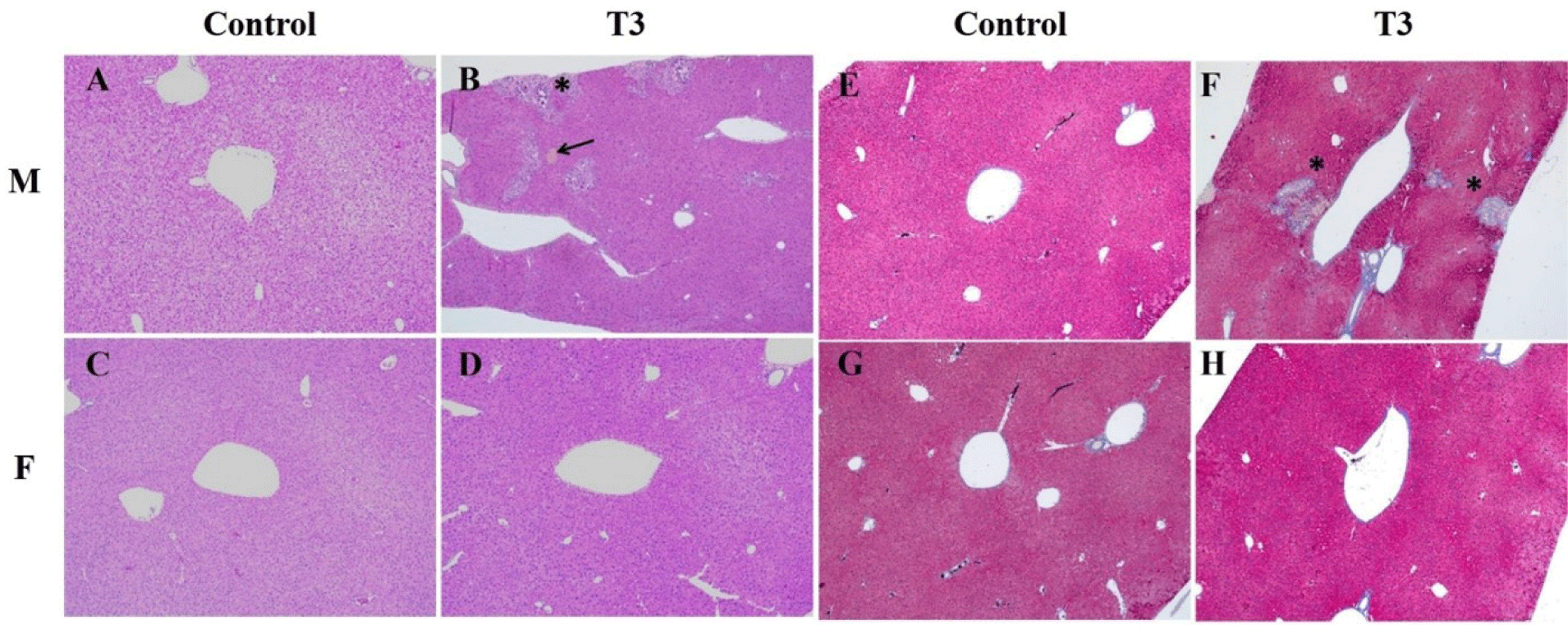
Macroscopic findings, including liver spots, were observed in male T3 mice (Table 8), which were correlated with mineralization on histopathological findings. The microscopic findings in the liver and nasal cavities of male and female mice are summarized in Table 9. Dystrophic mineralized deposits, necrosis, and pigmentation of the liver were observed in male T3 mice, and Masson’s trichrome stain revealed a fibrotic region surrounding the mineralization and necrosis (Fig. 1). Eosinophilic globules, basal cell hyperplasia, and respiratory metaplasia of the nasal cavity were also observed in 1,2-DCB-inhaled male and female mice (Table 9). Other macro and microscopic findings were within the range of normal background alterations seen in animals of this strain and age and were not related to the test substance.

Table 8. Summary of gross findings in mice inhaling 1,2-dichlorobenzene for 90 days
Variable Male Female
Control T1 (30 ppm) T2 (60 ppm) T3 (120 ppm) Control T1 (30 ppm) T2 (60 ppm) T3 (120 ppm)
N 10 10 10 10 10 10 10 10
Organ
 Liver
  Focus 0 0 0 0 0 0 0 0
  Increased size 0 0 0 0 0 1 0 0
  Spot 0 0 0 4 0 0 0 0
 Ovary
  Discoloration 0 0 0 0 0 0 1 0
 Seminal vesicle
  Cyst 1 0 0 0
 Skin
  Hair loss 4 3 4 1 4 1 1 5
 Thymus
  Decreased size 0 0 0 0 0 1 0 0

N, number of animals examined.

Download Excel Table
Table 9. Summary of major histopathological findings in mice inhaling 1,2-dichlorobenzene for 90 days
Variable Male Female
Control T1 (30 ppm) T2 (60 ppm) T3 (120 ppm) Control T1 (30 ppm) T2 (60 ppm) T3 (120 ppm)
N 10 10 10 10 10 10 10 10
Organ
 Liver
  Mineralization, dystrophic 1a 0 0 0 3 0 0 0 0
2a 0 0 0 3 0 0 0 0
3a 0 0 0 3 0 0 0 0
Total 0 0 0 9 0 0 0 0
  Necrosis 1a 0 0 0 1 0 0 0 0
Total 0 0 0 1 0 0 0 0
  Pigmentation 1a 0 0 0 8 0 0 0 0
2a 0 0 0 1 0 0 0 0
Total 0 0 0 9 0 0 0 0
  Infiltration, mononuclear cell 1a 2 0 0 2 4 1 0 1
Total 2 0 0 2 4 1 0 1
 Nasal cavity
  Atrophy 1a 0 0 0 0 0 2 1 2
2a 0 0 0 1 0 2 3 1
3a 0 0 0 0 0 1 0 2
4a 0 0 0 1 0 0 0 0
Total 0 0 0 2 0 5 4 5
  Dilatation, glands 2a 0 0 0 0 0 1 4 6
3a 0 0 0 0 0 4 6 0
Total 0 0 0 0 0 5 10 6
  Eosinophilic globules 1a 0 7 2 2
2a 0 3 3 5 0 4 1 0
3a 0 0 3 1 0 5 4 8
4a 0 0 2 2 0 1 5 1
Total 0 10 10 10 0 10 10 9
  Hyperplasia, basal cell 1a 0 1 3 3 0 3 3 6
2a 0 2 2 3 0 4 3 3
3a 0 4 5 3 0 2 2 0
4a 0 3 0 1 0 1 0 0
Total 0 10 10 10 0 10 8 9
  Respiratory metaplasia 1a 0 0 6 0 0 1 0 0
2a 0 3 2 1 0 3 5 0
3a 0 5 2 0 0 4 4 0
4a 0 2 0 0 0 2 1 0
Total 0 10 10 1 0 10 10 0

a Severity: 1, minimal; 2, mild; 3, moderate; 4, marked.

N, number of animals examined.

Download Excel Table
jbtr-24-3-67-g1
Fig. 1. Histopathological findings of the liver in control and T3 (120 ppm) mice that inhaled 1,2-dichlorobenzene (M: male; F: female). Asterisks indicate mineralization with fibrotic regions in the liver, and the arrow indicates pigment deposits with pigment-laden macrophages. (A–D) Hematoxylin and eosin staining, × 20; (E–F) Masson’s trichrome staining, × 20.
Download Original Figure
Immunohistochemical analysis of CYP proteins

The immunohistochemical analysis of CYP protein expression is shown in Fig. 2. An obvious expression of CYP2E1 and CYP3A4 was detected in the centrilobular area of the livers of male and female mice in the T3 group compared to the control group (p<0.05). In addition, CYP450 expression was increased in male T3 mice compared to female T3 mice (p<0.05).

jbtr-24-3-67-g2
Fig. 2. Immunohistochemical analysis of the liver sections of control and T3 (120 ppm) mice that inhaled 1,2-dichlorobenzene (M: male; F: female; CON: control). Asterisks (B, F) indicate the expression of CYP2E1 or CYP3A4 in the centrilobular area of the liver. (A–D) CYP2E1, (E–F) CYP3A4. The mean area revealed a higher expression of CYPs in T3 mice compared to both sexes of control mice and in males compared to females. (A–H) Immunostaining, × 20. *,† Student's t-test, significant compared to the controls or females, p<0.05.
Download Original Figure
Assessment of lipid peroxidation levels

MDA and 4-HNE levels, indicating lipid peroxidation products, were analyzed in liver tissue (Fig. 3). A slightly increasing trend was observed in MDA in the T3 group of male and female mice, and an increased trend in 4-HNE was observed in male T3 group mice, although not statistically significant.

jbtr-24-3-67-g3
Fig. 3. Levels of lipid peroxidation markers in the liver tissue of male (A, B) and female (C, D) mice that inhaled 1,2-dichlorobenzene. The data are expressed as the mean ± S.D. MDA, malondialdehyde; 4-HNE, 4-hydroxynonenal; M, male; T1, 30 ppm; T2, 60 ppm; T3, 120 ppm; CON, control; F, female.
Download Original Figure

DISCUSSION

There are few restricted data concerning the effects of 1,2-DCB or mixture containing 1,2-DCB such as irritation to the eyes and respiratory passages, hematopoietic disorder, headache, nausea and malaise in workers [3, 10]. Data on health hazards of 1,2-DCB were mostly identified as results of animal or in vitro experiments and there were no reports of on volunteers [10]. In the toxicity of 1,2-DCB, a subchronic oral study using Sprague-Dawley rats reported histopathologic lesions in the liver, such as centrilobular degeneration and centrilobular hypertrophy in both sexes at high doses, single-cell necrosis in males, and necrosis in females. In the clinical biochemistry results, an increase in ALT in middle-dose rats was not accepted because there was no histopathologic evidence, dose-dependency, or change in other enzymes that indicated liver damage [11]. Similar to the results of the above study, in another oral study using F344 rats, hepatotoxicity was identified, including centrilobular degeneration, single-cell necrosis, and necrosis of the liver in high-dose rats of both sexes. Body weight decreases in high-dose males, and relative liver weight increases in middle and high-dose males and females were also noted for determination of no-observed-adverse-effect-level (NOAEL) [6, 12]. Chronic and carcinogenic toxicity studies of 1,2-DCB oral exposure in F344 rats reported the occurrence of tumors, including adrenal gland pheochromocytomas and interstitial-cell tumors of the testis in males, but these have not been recognized as carcinogenic test substances [6, 12]. In mouse oral toxicity studies, the high-dose administration of 1,2-DCB induced body weight decreases, liver lesions, such as centrilobular necrosis, single-cell necrosis, hepatocellular degeneration, mineral deposits in the heart, and lymphoid depletion in the thymus and spleen in both sexes. Pigmentation, hepatocellular degeneration, and single-cell necrosis of the liver were observed in middle-dose males [6, 12]. Chronic and carcinogenic studies revealed non-neoplastic kidney lesions considered to be test substance-related in male mice and malignant histiocytic lymphomas and alveolar/bronchiolar carcinomas, which were considered not to be related to the test substance [6, 12]. A few reports using the inhalation test route in animals, including rats, guinea pigs, and rabbits, reported body weight loss, spleen weight decreases, and lesions in the liver and kidneys. An inhalation study using Swiss male mice exposed to 1,2-DCB (64 and 163 ppm) reported the induction of upper respiratory damage, including damaged olfactory epithelium, which can regenerate [13].

In our preliminary study conducted to determine test concentrations, dosing with 150 ppm led to the death of the male mice (2 of 5) or liver lesions, including hepatic necrosis, accompanied by increases in biochemical enzymes, such as AST and ALT, in the surviving mice. Since liver toxicity was not confirmed in females, differences in enzymatic expression and lipid peroxidation in the liver between the sexes were confirmed in an additional 90-day inhalation study. In the subchronic study, exposure to 1,2-DCB (30, 60, 120 ppm) induced slight decreases in the body weight of the mice and liver damage only in the male T3 group without serum biochemical changes or damage of the upper respiratory tract, especially nasal cavity in both sexes of all dosing mice. In addition, fibrous tissue proliferated around the areas of mineralization, and some necrotic areas were identified in the livers.

Liver injury and regeneration induced by chemicals have been well-recognized through many studies. In the short term, the liver can recover normal structure by proliferating or reconstructing the remaining cells. However, prolonged damage causes liver fibrosis, which can cause cirrhosis or liver cancer [14]. In the results of our study using B6C3F1 mice, 1,2-DCB is believed to have caused repeated tissue damage, and individual animals adapted to the initial toxicity after primary metabolism occurred in the liver and recovered. Previous studies showed that the test substance was quickly absorbed and oxidized by CYP450 in the early stages of metabolism to form an intermediate epoxide, which is known to be associated with hepatotoxicity [15]. Some researchers also studied how alterations in CYP450 or glutathione affected hepatotoxicity in animals [16]. Moreover, in the literature on sex differences in the metabolism of 1,2-DCB in rats and mice, the liver microsomes of female rats metabolized 1,2-DCB faster than males, but in contrast, this process was more efficient in male mice than in females. Additionally, the metabolism of the test substance is known to be faster in mice than in rats [17]. CYP450s, key enzymes in the liver metabolism of drugs and chemicals, can produce reactive oxygen species, causing oxidative damage, lipid peroxidation, protein modification, and mitochondrial damage [1820]. The IHC analysis showed that CYP2E1 and CYP3A4 were expressed around the central vein of the liver in T3 mice, and both males and females showed a significant increase in expression compared to the control group. In addition, the expression of CYPs was significantly increased in males compared to females. In this regard, the analysis of MDA and 4-HNE in the liver confirmed increased changes in male and/or female T3 mice and sex differences.

An evaluation of CYP450 enzymatic activity is important for identifying inhibition or induction characteristics in drug and chemical metabolism, and major isoforms, including 1A2, 2E1, 2C9, 2C19, and 3A have been identified in previous studies [21, 22]. Although there is a difference between humans and animal species, the similarities between human and mouse hepatic-metabolizing characteristics were shown in recent studies and could strengthen our research findings [23]. Thus, despite the need for further studies on the relevance of 1,2-DCB to human, the mechanistic approaches to the toxicity identified in vivo would be useful because health hazards data on human associated with 1,2-DCB could not exclude the effects of accidental exposure or mixture form.

In summary, we conducted 90-day inhalation toxicity studies using male and female B6C3F1 mice to evaluate the toxicity of 1,2-DCB under inhalation exposure conditions in accordance with GLP and OECD guidelines. The present study showed that inhaled 1,2-DCB induced upper respiratory damage in both sexes of mice and hepatotoxicity only in males and that differences in liver toxicity were associated with lipid peroxidation due to CYP450 alterations.

Conflict of Interest

No potential conflict of interest relevant to this article was reported.

Acknowledgements

This study was supported by the Korea Occupational Safety and Health Agency, Ministry of Employment and Labor, Korea, and a Grant-in-Aid for chemical hazard assessment, 2019. The authors sincerely thank Minha Kim, Junghee Han, Taeyeon Shim, and Dongseok Seo, colleagues in the Inhalation Toxicity Research Center, OSHRI, for their help. Also some of results of the study was accounced as a poster presentation through 2022 Korean Association for Laboratory Animal Science (KALAS) International Symposium.

Ethics Approval

The animal study protocol was approved by the Committee on Animal Research Committee of the Occupational Safety and Health Research Institute (Approval No. IACUC-1816).

References

1.

Office of Environmental Health Hazard Assessment [OEHHA]. Summary of the public health goal for 1,2-dichlorobenzene (1,2-DCB) [Internet]. 1997 [cited 2019 Nov 20]. Available from: https://oehha.ca.gov/water/public-health-goal/summary-public-health-goal-12-dichlorobenzene-12-dcb

2.

Office of Environmental Health Hazard Assessment [OEHHA]. Update of the PHG for 1,2-dichlorobenzene [Internet]. 2009 [cited 2019 Nov 20]. Available from: https://oehha.ca.gov/water/report/12-dichlorobenzene

3.

European Chemical Agency [ECHA]. 1,2-Dichlorobenzene_registration dossier [Internet]. 2023 [cited 2023 Jul 24]. Available from: https://echa.europa.eu/registration-dossier/-/registered-dossier/13734/2/1

4.

Occupational Safety and Health Administration [OSHA]. o-Dichlorobenzene, OSHA occupational chemical database [Internet]. 2021 [cited 2023 Jul 24]. Available from: https://www.osha.gov/chemicaldata/755

5.

Umemura T, Saito M, Takagi A, Kurokawa Y. Isomer-specific acute toxicity and cell proliferation in livers of B6C3F1 mice exposed to dichlorobenzene. Toxicol Appl Pharmacol 1996;137:268-274.

6.

Organisation for Economic Cooperation and Development [OECD]. SIDS initial assessment report: 1,2-dichlorobenzene [Internet]. 2001 [cited 2023 Jul 24]. Available from: https://hpvchemicals.oecd.org/ui/handler.axd?id=40127CF8-F9A5-4437-BC08-0E1199DF65C7

7.

Hissink AM, Van Ommen B, Krüse J, Van Bladeren PJ. A physiologically based pharmacokinetic (PB-PK) model for 1,2-dichlorobenzene linked to two possible parameters of toxicity. Toxicol Appl Pharmacol 1997;145:301-310.

8.

Cho ES, Lee YH, Han JH, Kim S, Park K, Jo J, Lee SB. Evaluation of 1-propanol toxicity in B6C3F1 mice via repeated inhalation over 28 and 90 days. J Toxicol 2020;2020:9172569.

9.

Organisation for Economic Cooperation and Development [OECD]. Test no. 413: subchronic inhalation toxicity: 90-day study, OECD guidelines for the testing of chemicals, section 4 [Internet]. 2018 [cited 2023 Jul 24]. Available from: https://www.oecd.org/env/test-no-413-subchronic-inhalation-toxicity-90-day-study-9789264070806-en.htm

10.

Environmental Protection Agency [EPA]. Proposed designation of o-dichlorobenzene (CASRN 95-50-1) as a high-priority substance for risk evaluation [Internet]. 2019 [cited 2023 Jul 24]. Available from: https://www.epa.gov/sites/default/files/2019-08/documents/o-dichlorobenzene_95-50-1_high-priority_proposeddesignation_082319.pdf

11.

Robinson M, Bercz JP, Ringhand HP, Condie LW, Parnell MJ. Ten- and ninety-day toxicity studies of 1,2–dichlorobenzene administered by oral gavage to Sprague-Dawley rats. Drug Chem Toxicol 1991;14:83-112.

12.

National Toxicology Program [NTP]. Toxicology and carcinogenesis studies of 1,2-dichlorobenzene (o-dichlorobenzene) (CAS no. 95-50-1) in F344/N rats and B6C3F1 mice (gavage studies). Washington: United States Department of Health and Human Services; 1985.

13.

Zissu D. Histopathological changes in the respiratory tract of mice exposed to ten families of airborne chemicals. J Appl Toxicol 1995;15:207-213.

14.

Tanaka M, Miyajima A. Liver regeneration and fibrosis after inflammation. Inflamm Regen 2016;36:19.

15.

Stine ER, Gunawardhana L, Sipes IG. The acute hepatotoxicity of the isomers of dichlorobenzene in Fischer-344 and Sprague-Dawley rats: isomer-specific and strain-specific differential toxicity. Toxicol Appl Pharmacol 1991;109:472–481.

16.

Valentovic MA, Ball JG, Anestis D, Madan E. Modification of P450 activity and its effect on 1,2-dichlorobenzene toxicity in Fischer 344 rats. Toxicology 1993;79:169–180.

17.

Nedelcheva V, Gut I, Souček P, Frantı́k E. Cytochrome P450 catalyzed oxidation of monochlorobenzene, 1,2- and 1,4-dichlorobenzene in rat, mouse, and human liver microsomes. Chem Biol Interact 1998;115:53–70.

18.

Cederbaum AI, Wu D, Mari M, Bai J. CYP2E1-dependent toxicity and oxidative stress in HepG2 cells. Free Radic Biol Med 2001;31:1539-1543.

19.

Renton KW. Alteration of drug biotransformation and elimination during infection and inflammation. Pharmacol Ther 2001;92:147-163.

20.

Younis HS, Hoglen NC, Kuester RK, Gunawardhana L, Sipes IG. 1,2-Dichlorobenzene-mediated hepatocellular oxidative stress in Fischer-344 and Sprague–Dawley rats. Toxicol Appl Pharmacol 2000;163:141-148.

21.

Zhou SF, Liu JP, Chowbay B. Polymorphism of human cytochrome P450 enzymes and its clinical impact. Drug Metab Rev 2009;41:89-295.

22.

Kathirvel E, Chen P, Morgan K, French SW, Morgan TR. Oxidative stress and regulation of anti-oxidant enzymes in cytochrome P4502E1 transgenic mouse model of non-alcoholic fatty liver. J Gastroenterol Hepatol 2010;25:1136-1143.

23.

Turpeinen M, Ghiciuc C, Opritoui M, Tursas L, Pelkonen O, Pasanen M. Predictive value of animal models for human cytochrome P450 (CYP)-mediated metabolism: a comparative study in vitro. Xenobiotica 2007;37:1367-1377.