INTRODUCTION
Corneal ulcers are commonly diagnosed in dogs, with deep ulcers characterized by > 50% stromal loss [1]. Furthermore, descemetocele is a deeper corneal lesion in which the corneal epithelium and stroma are completely destroyed, leaving a lesion lined only by Descemet’s membrane, which is only 3–12-μm thick, and the corneal endothelium thus easily ruptures [2]. Common presentations include lacrimation, ocular discharge and pain, blepharospasm, photophobia, conjunctival hyperemia, and corneal edema [3]. Lesions are variable, but the distribution of blood vessels around the corneal defect is the most common characteristic. The diagnosis of a corneal ulcer is made based on these clinical signs and the retention of topically applied fluorescein dye by the corneal stroma. In addition, corneal ulcer depth should be estimated using magnification, focal illumination using a slit-beam, topical fluorescence to distinguish the stroma and Descemet’s membrane, and optical coherence tomography.
As deep corneal ulcers can progress to perforation and lead to potential vision loss and anterior synechia formation, surgical correction of deep corneal ulcerations is required to provide tectonic support, maintain ocular integrity, and limit corneal opacification [1].
Corneo-conjunctival transposition (CCT) utilizes autologous cornea and conjunctiva advanced into the ulcer bed to treat deep corneal wounds. This surgical technique has been used successfully in conjunction with keratectomy for corneal sequestra in cats and allows for a clearer and thicker visual axis than conjunctival pedicle grafts [4]. Although a number of cases utilizing CCT have been reported in dogs, to the best of our knowledge, there are no reports of bidirectional CCT in dogs.
CASE
A 12-year-old spayed female Maltese dog, weighing 2.4 kg, presented to Chungbuk National University Veterinary Teaching Hospital in Korea after having been diagnosed with superficial corneal ulcer of right eye (OD) initially at a local animal hospital, but two days was later referred to the emergency visiting with a suspected descemetocele. The chief complaint was acute blepharospasm of the OD that started a week prior to presentation, with eye discharge. At presentation, there was a deep wound on approximately half of the cornea, and the central part of the ulcer was very thin and protruded in the form of a blister (Fig. 1). A descemetocele was present in the center of the thinned cornea, and there was a small perforation in the lower left part. As a result of intraocular bleeding due to perforation, hemorrhage was observed in the central part of the corneal wound. Vascularization in the periphery of the cornea and edema of the entire cornea were observed. Before surgery, the intraocular pressure (IOP) was 5 and 13 mmHg in the OD and left eye (OS), respectively, measured using TonoVet (iCare, Vantaa, Finland). The menace response, dazzle reflex, and pupillary light reflex were all negative in the OD and positive in the OS. Fluorescence was stained only in the center of the corneal ulcer site because the peripheral wound site was epithelialized. Corneal edema and shallow anterior chamber due to loss of aqueous humor by corneal perforation inhibited light from entering the anterior part of the eye.

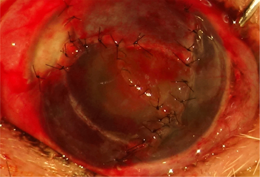
The dog underwent emergency surgery 1 day after the initial consultation. Corneal edema and vascularization progressed to the vicinity of the ulcer lesion. The dog was pre-medicated with butorphanol (0.2 mg/kg, IV), and anesthesia was induced with midazolam (0.2 mg/kg, IV) and propofol (4 mg/kg, IV). The patient was intubated and maintained on oxygen and isoflurane. The periocular region and OD were washed with a 0.5% povidone-iodine solution. Debridement was performed using a diamond burr around the descemetocele. Ophthalmic viscoelastic (Kata Hylon, Ajupharm, Seoul, Korea) was applied, and the corneal perforation site was sutured with 8–0 Vicryl (Ethicon, Raritan, NJ, USA) once. Subsequently, the aqueous humor did not leak from the sutured site. With a crescent knife at 12 o'clock on the dorsal side, the cornea and conjunctiva were picked in a trapezoidal shape, 1 mm larger than the lesion with 1/2 to 2/3 thickness. Bidirectional CCT grafts were applied since the lesion area was large, and central vision was thus secured (Fig. 2). The first CCT at 12 o’ clock and the second CCT, performed at 7 o'clock on the ventral side, were sutured with simple interrupted and continuous patterns using ophthalmic 8–0 nylon (USIOL, Lexington, KY, USA) to avoid blocking the central field of view. Cefazoline (50 mg/0.1 mL) was administered via subconjunctival injection. Tissue plasminogen activator (25 ug/0.1 mL) was injected into the anterior chamber using an insulin syringe to prevent intracameral fibrin formation. Temporary tarsorrhaphy was applied using 5–0 nylon and a stent on the eyelids.

The patient was hospitalized for 3 days. During hospitalization, ampicillin-sulbactam (22 mg/kg, IV, BID), meloxicam (0.1 mg/kg, SC, SID), tramadol (4 mg/kg, IV, BID), famotidine (0.5 mg/kg, IV, BID), and topical eye drops including atropine sulfate (1% isopto-atropine, Alcon, Geneva, Switzerland), tobramycin sulfate (Ocuracin, Samil, Seoul, Korea), moxifloxacin sulfate (Vigamox, Novartis, Basel, Switzerland), and bromfenac sulfate (Bronuck, Taejun, Seoul, Korea) were administered.
During the follow-up period, the patient’s IOP was stable between 10 and 20 mmHg in the OD. Vascularization was observed in the entire lesion of the CCT graft 2 weeks after surgery (Fig. 3). Afterwards, the blood vessels gradually regressed, and after 8 weeks, all blood vessels in the central cornea disappeared. Although the cornea was in better condition than before the operation, it did not recover to a completely transparent state due to scar formation. At 36 weeks after surgery, the prognosis was similar, and complications such as glaucoma, uveitis, re-perforation, and ulcer did not occur. After surgery, the dazzle reflex was restored, but clinical visual acuity did not improve because of traumatic cataract and corneal scar formation. Postoperatively, mild kerato-conjunctivitis sicca developed and was managed with cyclosporine eye ointment.

DISCUSSION
Deep corneal ulcers, corneal perforations, and descemetoceles are frequently presented in veterinary ophthalmology practice, and prompt treatment is required to prevent vision loss and ensure anatomical preservation of the globe [5–7]. Surgical treatments for deep corneal ulcers include direct corneal sutures, conjunctival graft transplantation, and biomaterials such as porcine small intestinal submucosa (SIS) and bovine amniotic membrane. These approaches can be applied to increase the stability of corneal structures [7, 8]. Furthermore, the use of autologous lamellar corneal transposition procedures in dogs has previously been described to repair deep corneal wound and small size perforation [9, 10]. Among them, CCT is one of the most advanced surgical strategies that utilizes autologous adjacent healthy cornea with limbus and conjunctiva advanced into the ulcer bed to treat deep corneal ulcers. The goal of bidirectional CCT is to provide tectonic support and preserve relatively clear optical vision at the center of the cornea. Commonly used surgical techniques, such as conjunctival pedicle grafting, result in postoperative corneal opacification, which may be visually limiting. CCT is thicker than the conjunctival flap and contains a limbus with abundant stem cells. Therefore, compared to other methods, such as the conjunctival flap and SIS, it can induce thick and rapid regeneration of the stroma in deep corneal wounds. The limitations of this study include the absence of advanced ophthalmic examinations, such as optical coherence tomography, and the lack of complete recovery of sight due to scar and traumatic cataract formation.
This case report describes the first successful case of bidirectional CCT in a dog with a large and deep corneal defect. Bidirectional CCT could be an effective strategy for patients with large and deep corneal wounds to improve vision and structural support in the eye as an alternative to corneal transplantation.


