Original Article

Preventive effect of polydatin on diabetes-related hypofunction of salivary gland in streptozotocin-induced diabetic rats

Hyung Rae Kim1https://orcid.org/0000-0003-0239-3170, Junghyun Kim1,*https://orcid.org/0000-0002-9003-2158
Author Information & Copyright
1Department of Oral Pathology, School of Dentistry, Jeonbuk National University, Jeonju 54896, Korea
*Corresponding author: Junghyun Kim, Department of Oral Pathology, School of Dentistry, Jeonbuk National University, Jeonju 54896, Korea, Tel: +82-63-270-4032, E-mail: dvmhyun@jbnu.ac.kr

© Research Institute of Veterinary Medicine, Chungbuk National University.

Received: Nov 12, 2021; Revised: Dec 01, 2021; Accepted: Dec 02, 2021

Abstract

Declined salivary gland function is commonly observed in patients with diabetes. Advanced glycation end products (AGEs) are believed to contribute to the pathogenesis of diabetes-induces hypofunction of the salivary glands. Polydatin (resveratrol-3-O-β-mono-D-glucoside) is a polyphenol that can be easily accessed from peanut, grape, and red wines. Although polydatin is known to have anti-glycation, anti-oxidation, and anti-inflammation effects, its effect in the salivary gland is not known. In the present study, we evaluated the AGEs burden in the salivary gland in streptozotocin (STZ)-induced diabetic rats and the protective effect of polydatin on diabetes-related salivary hypofunction. Polydatin (50 and 100 mg/kg/day) was orally administered in the STZ-induced diabetic rats for 4 weeks. The results showed that the salivary flow rate of the STZ-induced diabetic rats was reduced compared with that in the normoglycemic control rats. The circulating AGEs in serum and secreted AGEs in saliva increased in the STZ-induced diabetic rats. The reactive oxygen specises (ROS) were highly generated in the salivary gland in these diabetic rats. The salivary gland from the diabetic rats showed increased acinar cell apoptosis compared to normoglycemic control mice. However, polydatin suppressed all of these diabetes-related salivary changes. Overall, polydatin could provide a beneficial option for diabetes-related salivary hypofunction.

Keywords: diabetes; glycation; xerostomia; polydatin; salivary glands

INTRODUCTION

Most patients with diabetes have a clinical symptom of diabetes-related hyposalivation [1]. The maintaining of normal function of the salivary glands is important for a healthy oral environment. The salivary gland hypofunction can induced several clinical discomforts, such as sallowing problem, xerostomia, and halitosis [2]. Moreover, this functional disorder of the salivary gland wersens the quality of life and increase the prevalance of various oral diseases, such as periodontitis, mucositis, and tooth decay [3]. The pathogenic risk factors of salivary gland hypofunction are chemical medications, several chronic diseases, radiation therapy, and aging process [4]. To control this clinical symptom, there are several treatment options, such as artificial saliva, chewing gum, malic acid and pilocarpine [5]. However, these medications only help temporary relif from symptom through an increased salivary flow rate.

Advanced glycation end products (AGEs) are non-enzymatic modifications of proteins or lipids with sugars. AGEs form in vivo in hyperglycemic environments and during aging [6, 7]. AGEs accumulate in various tissues and bind to AGE-specific receptors (RAGEs), which play an important role in the development of diabetes-related organ hypofunction [8]. Evidence shows that AGEs could cause the generation of reactive oxygen species (ROS) [9]. Cells in the human body are chronically exposed to oxidative stress during the development of diabetes, thereby causing cell injury [10]. The irreversible formation of AGEs increases the risk for the development of periodontitis, pulptitis in oral tissues [11]. RAGE is present in the salivary gland and highly expressed in the salivary gland with Sjögren’s syndrome [12]. However, it is not clear whether AGE accumulation is due to diabetes-related salivary gland hypofunction.

Polydatin (resveratrol-3-O-β-mono-D-glucoside) is a polyphenol that can be easily accessed from peanut, grape, and red wines [13]. Polydatin has potent anti-diabetic, anti-oxidant, anti-inflammation, anti-cancer, and cardiovascular protection effects [14]. However, its preventive role on diabetes-related salivary gland hypofunction has not been investigated. Thus, we examined the protective effect of polydatin on diabete-related hypofunction of the salivary glands in streptozotocin-induced diabetic rats.

MATERIALS AND METHODS

Animal experiments

Diabetes was induced by the intraperitoneal injection of streptozotocin (STZ, 60 mg/kg, Sigma-Aldrich, St. Louis, MO, USA) in 7-week-old male Sprague Dawley rats (Koatech, Pyeongtaek, Korea). Normoglycemic control rats were administered vehicle only. Normoglycemic and hyperglycemic rats (blood glucose levels > 300 mg/dL blood) were randomized into the following 4 groups of 8 rats: normoglyemic control group (NOR), diabetic group (DM), polydatin (50 and 100 mg/kg/day)-treated two groups. Polydatin (Sigma-Aldrich) was given orally once a day for 4 weeks. To measure the salivary flow rate, rats were intraperitoneally injected with pilocarpine hydrochloride (2 mg/kg, Sigma-Aldrich). Saliva samples were then collected for 15 min. At necropsy, blood samples were collected, and submandibular salivary glands were isolated. All experimental procedures were performed under the supervision of our Institutional Animal Care and Use Committee (IACUC No. 2020-24).

Quantification of advanced glycation end products (AGEs) in saliva and blood

The total protein levels in saliva and serum were examined using a Quick Start™ Bradford protein assay kit (Bio-Rad, Hercules, CA, USA). Briefly, saliva samples were centrifuged at 13,000×g for 10 minutes at 4℃ to remove food residues. Total protein concentration in saliva and serum was determined by the Bradford method, using bovine serum albumin as a standard, and the plates were read at 600 nm in a microplate reader (Tecan, Männedorf, Swiss). The AGEs levels were detected using a rat AGEs ELISA kit (MyBioSource, San Diego, CA, USA) according to the manufacturer’s instructions.

Oxidative stress assay in salivary gland

A protein extraction solution (RIPA, Elpis Biotech, Daejeon, Korea) containing a HaltTM Protease Inhibitor Cocktail (Thermo Fisher Scientific, Waltham, MA, USA) was applied to lyse the the submandibular salivary gland tissues. Afterward, a Protein Assay Dye Reagent Concentrate (Bio-Rad) was conducted to examine the extracted protein. The levels of ROS in the submandibular salivary gland tissues were examined using a Rat Reactive Oxygen Species ELISA Kit (MyBioSource) according to the manufacturer’s instructions.

Histopathological analysis

Fixing process of submandibular salivary gland tissues was conducted to evaluate the tissue structure through the following steps: fixation in paraformaldehyde solution (4%), dehydration by using gradient ethanol, incorporation with paraffin, incision into smaller parts (4 µm), and staining with hematoxylin and eosin (H & E). The tissue sections were examined under a light microscopy (BX51, Olympus, Tokyo, Japan).

Apoptosis analysis

Apoptosis was determined using an in situ cell death detection kit (Roche, Mannheim, Germany) according to the manufacturer's instructions. After paraffin removal, the cut sections were incubated in 100 μL volume of proteinase K (20 µg/mL) for 10 min at ambient temperature. Then, they were transferred into a TUNEL reaction mixture with a volume of 50 μL and placed in a dark incubator for 1 h at 37℃. A solution of Vectashield Mounting Medium with 4′,6-diamidino-2-phenylindole (Vector Laboratories, Road Burlingame, CA, USA) was exerted to stain the prepared sections. The numbers of TUNEL-positive cells were counted under a fluorescence microscope (BX51, Olympus).

Data analysis

The results were evaluated statistically using a one-way analysis of variance followed by Tukey’s multiple comparison test using GraphPad Prism 6.0 (GraphPad Software, San Diego, CA, USA).

RESULTS

Polydatin improves hyposalivation in streptozotocin (STZ)-induced diabetic rats

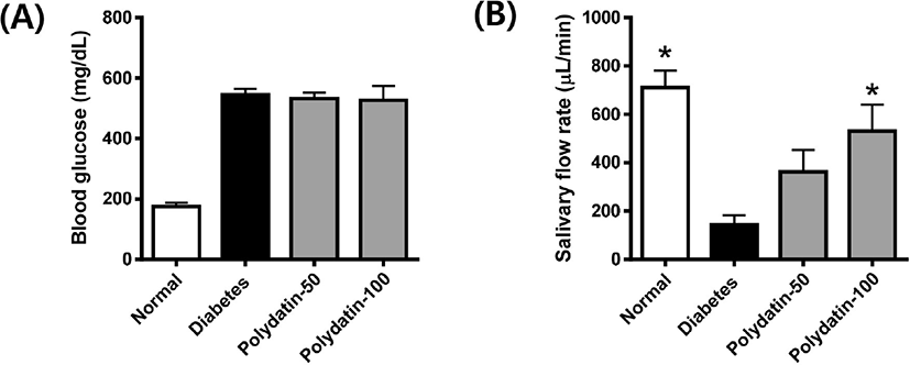
The blood glucose level in the STZ-induced diabetic rats was significanly increased compared with that in the control group and was not significantly altered by polydatin (Fig. 1A). The salivary flow rate was reduced in the STZ-induced diabetic rats, but reduced saliva secretion was significantly recovered following treatment with polydatin (Fig. 1B). These data suggest that diabetic rats had salivary hypofunction owing to decreased saliva secretion, which can be alleviated by polydatin.

jbtr-22-4-159-g1
Fig. 1. Effect of polydatin on salivary gland hypofunction in the STZ-induced diabetic rats. (A) Blood glucose levels and (B) salivary flow rate. The values in the bar graphs represent the means ± S.E. (n = 8). * p < 0.05 vs. diabetic group. STZ, streptozotocin.
Download Original Figure
Polydatin decreases the levels of circulating advanced glycation end products (AGEs) in serum and secreted advanced glycation end products (AGEs) in saliva

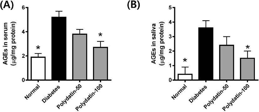
To determine whether polydatin reduced the AGEs formation in the body, we examined the levels of circulating AGEs in serum and secreted AGEs in saliva. As shown in Fig. 2, both circulating and secreted AGEs levels in the STZ-induced diabetic rats were significantly higher compared with the normoglycemic control rats. Polydatin decreased the levels of circulating and secreted AGEs compared with the DM.

jbtr-22-4-159-g2
Fig. 2. Effect of polydatin on circulating AGEs levels in serum (A) and secreted AGEs levels in saliva (B) from the salivary gland from the STZ-induced diabetic rats. The AGEs levels were determined by ELISA. The values in the bar graphs represent the means ± S.E. (n = 8). * p<0.05 vs. diabetic group. AGE, advanced glyaction end product. STZ, streptozotocin.
Download Original Figure
Polydatin decreases the oxidative stress in the salivary gland

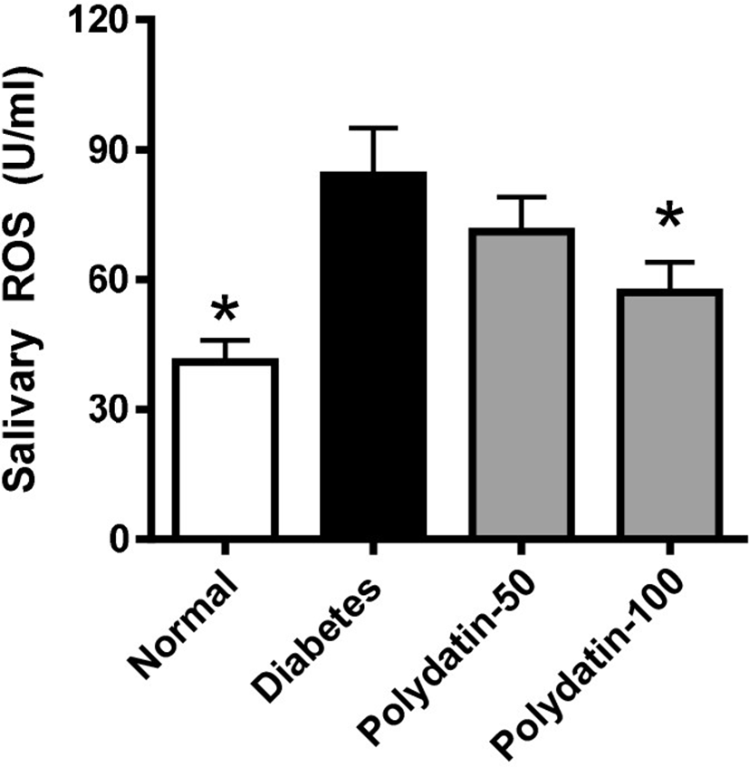
To evaluate the status of salivary oxidative stress in the STZ-induced diabetic rats, we examined the ELISA assay of ROS in the salivary gland tissues. As shown in Fig. 3, ROS generations were largely increased in the STZ-induced diabetic rats compared with the normoglycemic control rats. Polydatin significantly prevented ROS generation in the diabetic rats to a level similar to that observed in normoglycemic control rats.

jbtr-22-4-159-g3
Fig. 3. Effect of polydatin on ROS generation in the salivary gland from the STZ-induced diabetic rats. ELISA assay for ROS in the salivary gland. The values in the bar graphs represent the means ± S.E. (n = 8). * p < 0.05 vs. diabetic group. ROS, reactive oxygen specises. STZ, streptozotocin.
Download Original Figure
Polydatin inhibits apoptosis in the salivary glands of streptozotocin (STZ)-induced diabetic rats

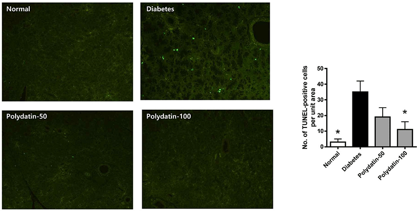
Our previous study reported that salivary acinal cells were histopathologically degenerated and damaged by apoptotic injury in the STZ-induced diabetic rats [15]. Thus, TUNEL staining was used to determine whether polydatin inhibits acinar cells die due to apoptosis. As shown in Fig. 4, apoptosis was detected by TUNEL staining and was increased in the DM and restored by polydatin. Polydatin protects salivary gland tissues by reducing apoptosis.

jbtr-22-4-159-g4
Fig. 4. Effect of polydatin on apoptosis in the salivary gland from the STZ-induced diabetic rats. Representative TUNEL staining in the salivary glands. Original magnifications: ×100. TUNEL-positive cells were determined. The values in the bar graphs represent the means ± S.E. (n = 8). * p < 0.05 vs. diabetic group. STZ, streptozotocin.
Download Original Figure

DISCUSSION

The gradual decline in salivary function is commonly observed in patients with diabetes. Patients with salivary hypofunction have a high risk factor for developing oral diseases such as tooth decay and periodontitis [16, 17]. Thus, the diabetes-related decline in salivary function should be managed to maintain a healthy oral environment. In this study, we assessed the effect of polydatin on hypofunctioning salivary glands by reducing AGEs burden.

Our study showed evidence indicating that enhanced AGEs burden occurred in the salivary gland of the STZ-induced diabtic rats. The functional alteration of the salivary gland was significantly induced in these diabetic rats compared with the normoglycemic control rats. These findings indicate that the significant AGEs burden in the salivary glands led to significant hypofunction of this tissue. This hyposalivation is in agreement with the results of Knaś et al. [18], who observed a decrease in the salivary flow rate and the enhanced oxidative stress in STZ-induced diabetic rats. Our results described the link between diabetes-related hypofunction of the salivary gland and AGEs burden. In addition, we showed that polydatin treatment exerts preventive effects on salivary glands in the diabetic rats.

The cytotoxic effects of AGEs have been shown in several studies [19]. AGEs can accumulate. Since the body does not contain any enzymes capable of AGE structural degradation, AGEs can accumulate in many tissues [20]. The interaction between AGEs and its receptor can induce the overproduction of ROS [21]. Oxidative stress is an important feature of diabetic complications [22]. ROS production was accelerated under diabetic conditions [10]. ROS frequently contribute to the tissue-damaging effects [9, 23]. Collectively, These results suggest that the diabetic conditions in the STZ-induce diabetic rats can induce salivary injury through AGEs-related oxidative stress and inflammation, which can lead to salivary gland dysfunction.

Here, we hypothesized that the reduction in AGEs burden by polydatin may contribute to the inhibition of salivary gland dysfunction. The current study clearly demonstrated that polydatin restored the levels of both circulating and secreted AGEs to near-normal levels in the the diabetic ats, in parallel with a marked increase of salivary flow rate. These findings provide evidence that polydatin has a beneficial effect on diabetes-related salivary gland hypofunction. Polydatin has the potent anti-glycation [24], and anti-oxidative activities [25, 26]. Chen et al. showed that polydatin inhibited the AGEs burden and renal injury in db/db diabetc mice [27]. In addition, polydatin enhanced tear production by attenuating oxidative stress in an animal model of dry eye disease [28].

Acinar cell loss by apoptosis can inevitably affect salivary flow rate, resulting in salivary hypofunction [29]. Moreover, high AGEs concentrations contribute to apoptotic cell death whenever they are generated in the context of the apoptotic process [30, 31]. The present study showed that diabetic conditions increased the number of TUNEL-positive cells in the salivary gland. However, we found that polydatin markedly decreased the number of TUNEL-positive cells in the STZ-induced diabetic rats. Similarly, polydatin has anti-apoptotic activity in neurodegenerative diseases [32], liver with acetaminophen-indced hepatotoxicity [33] and myocadial ischemia/reperfusion injury [34]. Thus, our findings suggest that polydatin has a potential anti-apoptotic effect in the salivary gland.

In conclusion, our study demonstrates that the AGEs burden was increased in the salivary gland of the STZ-induced diabetic rats. Polydatin has protective effects on the salivary gland of diabetic rats. These novel findings provide insight into the effects of polydatin against diabetes-related salivary hypofunction.

Conflict of Interest

No potential conflict of interest relevant to this article was reported.

Acknowledgements

This work was supported by a National Research Foundation of Korea (NRF) grant funded by the Korea government (MEST) (No. NRF-2019R1A2C1008773)

Ethics Approval

All of the experimental procedures using animals were performed under the supervision of the Institutional Animal Care and Use Committee, Jeonbuk National University Hospital, Korea (IACUC No. 2020-24).

REFERENCES

1.

López-Pintor RM, Casañas E, González-Serrano J, Serrano J, Ramírez L, de Arriba L, Hernández G. Xerostomia, hyposalivation, and salivary flow in diabetes patients. J Diabetes Res 2016;2016:4372852.

2.

Rao PKJ, Chatra L, Shenai P, Veena K, Prabhu RV, Kushraj T, Shetty P, Hameed S. Xerostomia: few dry facts about dry mouth. Arch Med Health Sci 2014;2:190-194.

3.

Ahmad MS, Bhayat A, Zafar MS, Al-Samadani KH. The impact of hyposalivation on quality of life (QoL) and oral health in the aging population of Al Madinah Al Munawarrah. Int J Environ Res Public Health 2017;14:445.

4.

Saleh J, Figueiredo MAZ, Cherubini K, Salum FG. Salivary hypofunction: an update on aetiology, diagnosis and therapeutics. Arch Oral Biol 2015;60:242-255.

5.

Visvanathan V, Nix P. Managing the patient presenting with xerostomia: a review. Int J Clin Pract 2010;64:404-407.

6.

Goh SY, Cooper ME. The role of advanced glycation end products in progression and complications of diabetes. J Clin Endocrinol Metab 2008;93:1143-1152.

7.

Tessier FJ. The Maillard reaction in the human body. The main discoveries and factors that affect glycation. Pathol Biol 2010;58:214-219.

8.

Brownlee M, Cerami A, Vlassara H. Advanced glycosylation end products in tissue and the biochemical basis of diabetic complications. N Engl J Med 1988;318:1315-1321.

9.

Lu J, Wu D, Zheng Y, Hu B, Zhang Z, Ye Q, Liu C, Shan Q, Wang Y. Ursolic acid attenuates D-galactose-induced inflammatory response in mouse prefrontal cortex through inhibiting AGEs/RAGE/NF-κB pathway activation. Cereb Cortex 2010;20:2540-2548.

10.

Xu H, Chen M, Forrester JV. Para-inflammation in the aging retina. Prog Retin Eye Res 2009;28:348-368.

11.

Ilea A, Băbţan AM, Bosca BA, Crişan M, Petrescu NB, Collino M, Sainz RM, Gerlach JQ, Câmpian RS. Advanced glycation end products (AGEs) in oral pathology. Arch Oral Biol 2018;93:22-30.

12.

Katz J, Stavropoulos F, Bhattacharyya I, Stewart C, Perez FM, Caudle RM. Receptor of advanced glycation end product (RAGE) expression in the minor salivary glands of patients with Sjögren’s syndrome: a preliminary study. Scand J Rheumatol 2004;33:174-178.

13.

Du QH, Peng C, Zhang H. Polydatin: a review of pharmacology and pharmacokinetics. Pharm Biol 2013;51:1347-1354.

14.

Wang Y, Ye J, Li J, Chen C, Huang J, Liu P, Huang H. Polydatin ameliorates lipid and glucose metabolism in type 2 diabetes mellitus by downregulating proprotein convertase subtilisin/kexin type 9 (PCSK9). Cardiovasc Diabetol 2016;15:19.

15.

Kang WS, Jung WK, Park SB, Kim HR, Kim J. Gemigliptin suppresses salivary dysfunction in streptozotocin-induced diabetic rats. Biomed Pharmacother 2021;137:111297.

16.

Tsai C, Hayes C, Taylor GW. Glycemic control of type 2 diabetes and severe periodontal disease in the US adult population. Community Dent Oral Epidemiol 2002;30:182-192.

17.

Mealey BL, Ocampo GL. Diabetes mellitus and periodontal disease. Periodontology 2000 2007;44:127-153.

18.

Knaś M, Maciejczyk M, Daniszewska I, Klimiuk A, Matczuk J, Kołodziej U, Waszkiel D, Ładny JR, Żendzian-Piotrowska M, Zalewska A. Oxidative damage to the salivary glands of rats with streptozotocin-induced diabetes-temporal study: oxidative stress and diabetic salivary glands. J Diabetes Res 2016;2016:4583742.

19.

Brownlee MD. Advanced protein glycosylation in diabetes and aging. Annu Rev Med 1995;46:223-234.

20.

Pokupec R, Kalauz M, Turk N, Turk Z. Advanced glycation endproducts in human diabetic and non-diabetic cataractous lenses. Graefes Arch Clin Exp Ophthalmol 2003;241:378-384.

21.

Yamamoto Y, Yamamoto H. Interaction of receptor for advanced glycation end products with advanced oxidation protein products induces podocyte injury. Kidney Int 2012;82:733-735.

22.

Giacco F, Brownlee M. Oxidative stress and diabetic complications. Circ Res 2010;107:1058-1070.

23.

Cuzzocrea S, McDonald MC, Filipe HM, Costantino G, Mazzon E, Santagati S, Caputi AP, Thiemermann C. Effects of tempol, a membrane-permeable radical scavenger, in a rodent model of carrageenan-induced pleurisy. Eur J Pharmacol 2000;390:209-222.

24.

Sheng Z, Ai B, Zheng L, Zheng X, Xu Z, Shen Y, Jin Z. Inhibitory activities of kaempferol, galangin, carnosic acid and polydatin against glycation and α-amylase and α-glucosidase enzymes. Int J Food Sci Technol 2018;53:755-766.

25.

Chen Y, Zhang D, Liao Z, Wang B, Gong S, Wang C, Zhang M, Wang G, Cai H, Liao FF, Xu J. Anti-oxidant polydatin (piceid) protects against substantia nigral motor degeneration in multiple rodent models of Parkinson’s disease. Mol Neurodegener 2015;10:4.

26.

Huang K, Chen C, Hao J, Huang J, Wang S, Liu P, Huang H. Polydatin promotes Nrf2-ARE anti-oxidative pathway through activating Sirt1 to resist AGEs-induced upregulation of fibronetin and transforming growth factor-β1 in rat glomerular messangial cells. Mol Cell Endocrinol 2015;399:178-189.

27.

Chen C, Huang K, Hao J, Huang J, Yang Z, Xiong F, Liu P, Huang H. Polydatin attenuates AGEs-induced upregulation of fibronectin and ICAM-1 in rat glomerular mesangial cells and db/db diabetic mice kidneys by inhibiting the activation of the SphK1-S1P signaling pathway. Mol Cell Endocrinol 2016;427:45-56.

28.

Park B, Jo K, Lee TG, Hyun SW, Kim JS, Kim CS. Polydatin inhibits NLRP3 inflammasome in dry eye disease by attenuating oxidative stress and inhibiting the NF-κB pathway. Nutrients 2019;11:2792.

29.

Stephens LC, Schultheiss TE, Price RE, Ang KK, Peters LJ. Radiation apoptosis of serous acinar cells of salivary and lacrimal glands. Cancer 1991;67:1539-1543.

30.

Mahali S, Raviprakash N, Raghavendra PB, Manna SK. Advanced glycation end products (AGEs) induce apoptosis via a novel pathway: involvement of Ca2+ mediated by interleukin-8 protein. J Biol Chem 2011;286:34903-34913.

31.

Li DX, Deng TZ, Lv J, Ke J. Advanced glycation end products (AGEs) and their receptor (RAGE) induce apoptosis of periodontal ligament fibroblasts. Braz J Med Biol Res 2014;47:1036-1043.

32.

Fakhri S, Gravandi MM, Abdian S, Akkol EK, Farzaei MH, Sobarzo-Sánchez E. The neuroprotective role of polydatin: neuropharmacological mechanisms, molecular targets, therapeutic potentials, and clinical perspective. Molecules 2021;26:5985.

33.

Liu YH, Huang QH, Wu X, Wu JZ, Liang JL, Lin GS, Xu LQ, Lai XP, Su ZR, Chen JN. Polydatin protects against acetaminophen-induced hepatotoxicity in mice via anti-oxidative and anti-apoptotic activities. Food Funct 2018;9:5891-5902.

34.

Ling Y, Chen G, Deng Y, Tang H, Ling L, Zhou X, Song X, Yang P, Liu Y, Li Z, Zhao C, Yang Y, Wang X, Kitakaze M, Liao Y, Chen A. Polydatin post-treatment alleviates myocardial ischaemia/reperfusion injury by promoting autophagic flux. Clin Sci 2016;130:1641-1653.