


Introduction
The raccoon dog is native to East Asia and has established itself widely throughout Eastern Europe [1]. These animals are omnivores with long torsos and short four legs with total lengths that can range from 50 to 68 cm and weights of 4 to 10 kg. Nyctereutes procynoides has been introduced into the entire Korean peninsula. Native East Asian raccoon dog populations have declined in recent years because of hunting, fur trade and destruction of habitats by urbanization [2]. Traffic accidents involving wildlife are also a major cause of anthropogenic mortality [3, 4]. Fractures due to traffic collisions are also common in wild animals. Limb amputation is a salvage surgical procedure relatively common in small animals and other species. This surgical method has been considered a viable option in animals to treat malignant neoplasm, severe trauma, extensive nerve injury, ischemia, necrosis of limb, severe infection, congenital deformities and severe disability [5, 6]. Orthopedic and neurological evaluation should be carefully performed before the amputation [7]. Anecdotally, most veterinarians believe that companion animals adapt quickly to ambulation after limb amputation [8]. However, animals after forelimb amputation have many difficulties in returning to normal life because they place more than 60 percent of their weight on their front legs [9, 10]. In addition, arboreal quadrupeds use their forelimbs to carry most of their weight and to hold feed. Thus, the aim of this study was to evaluate forelimb amputation and its outcomes in two raccoon dogs.
Case Report
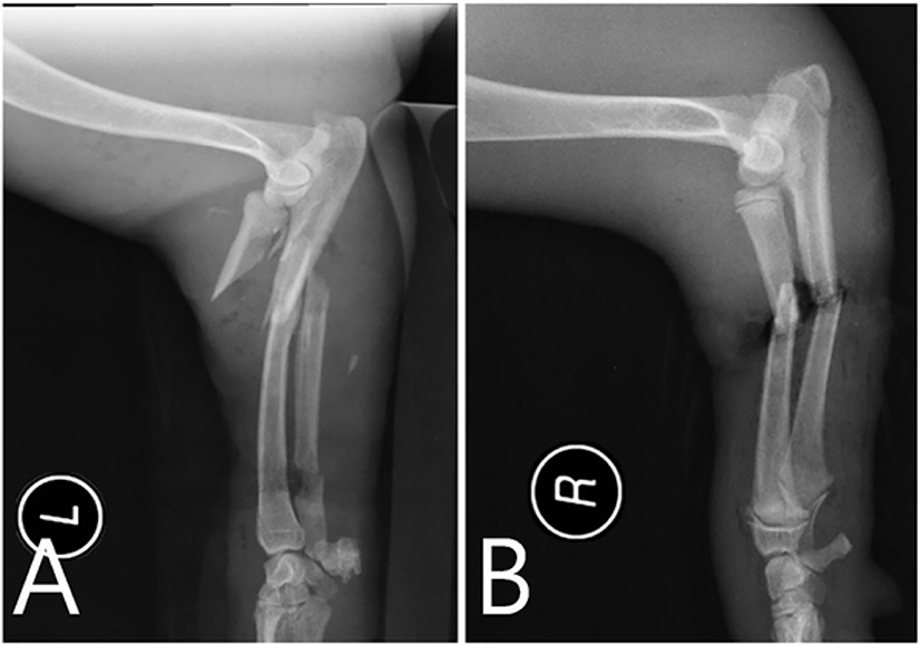
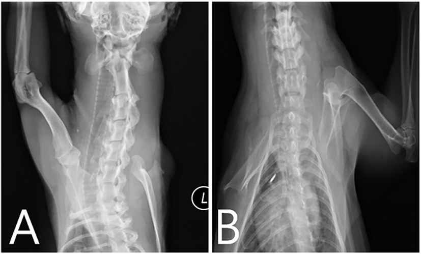
Two unknown aged male raccoon dogs of 3.2 and 4.5 kg were captured and presented to the veterinary teaching hospital for radio-ulnar fractures of unknown reasons. One had severe edema and infection in the right shoulder region and a right radio-ulnar open fracture with severe contamination. The other had an open fracture in the left radio-ulna with severe infection. Complete blood count and biochemical profile were performed and radiographic outcomes were examined. Laboratory analyses were normal except for mild leukocytosis and slightly elevated alkaline phosphatase and alanine aminotransferase. No abnormalities were found in the thoracic and abdominal radiographs except for fractures (Fig. 1). Aggressive debridement and vigorous irrigation were performed for several days. Raccoon dogs were treated by injection of cefazolin (Cefazol®; Hangook Chorus Pharm, Chuncheon, Korea; 50 mg/kg i.v.) and tramadol (Tamadol®; Dongkwang Pharmacy, Seoul, Korea; 3 mg/kg, i.v.). To treat severe scabies infection, ivermectin (Ivomec 1% injection; Merial, Lyon, France; 0.4 mg/kg, s.c.) was administered once a week. The fracture site of one raccoon dog was immobilized using bandages. It was observed that the leg region below the fractured part had lost its sensitivity. In addition, severe necrosis and gangrene were developed in that region a few days after the treatment. The clinical parameters and feed intake were normal in both raccoon dogs before surgery. It was eventually decided to amputate the leg to prevent the spread of necrosis and gangrene to healthy tissues. Propofol (Pofol®; Jeil Pharmacy, Seoul, Korea; 5 mg/kg, i.v.) was used to induce general anesthesia. The anesthesia was maintained with isoflurane (Aerane®; Ilsung Pharmacy, Seoul, Korea; 2%–2.5%). Animals were confined in lateral recumbency. The distal part of the leg was wrapped with an elastic bandage. The operation site was aseptically prepared using iodine and alcohol after shaving hairs. Surgeries were performed through the same technique for scapulo-humeral joint amputation in both raccoon dogs. A skin incision was made around the limb and muscles were transected. Arteries, veins and nerves were dissected free and doubly ligated before transection. The ligated nerves were infiltrated with 1 mL 0.5% bupivacaine (Bupivacaine HCl; Huons, Seoul, Korea). The stump was closed by suturing the tendon of the triceps brachii muscle cranially to the fascia of the superficial pectoral muscle. The fascial sheaths of the triceps brachii and the deltoid muscles were sutured to the brachiocephalic and pectoral muscles. Muscles, subcutaneous tissues and skin were sutured with 3-0 polyglycolic acid (Surgifit; AILEE, Busan, Korea) and 3-0 mono non-absorbable suture (Nylon; AILEE) in a routine fashion. The animals were treated with cefazolin (20 mg/kg, i.m.) and meloxicam (Metacam®; Boehringer Ingelheim Vetmedica, St. Joseph, MO, USA; 0.2 mg/kg, p.o.) for 5 days. Dressing with iodine was conducted every 2 days until complete healing. Both raccoon dogs showed good body condition and regular appetite during the hospitalization period. Moreover, they could run comparatively faster with three legs. The suture was removed and radiography was performed 2 weeks after the operation (Fig. 2). As recovery after amputation was satisfactory during 3 weeks, they were released to the area of capture after vaccination for rabies (Rabigen® mono; Virbac, Carros, France). One of the raccoon dogs, whose right forelimb was fractured, represented to the veterinary teaching hospital with hair loss caused by mites 6 months after the first release. Except for hair loss, it showed satisfactory body condition and function of the existing three limbs.
Discussion
Few clinical studies of raccoon dogs that have been conducted to date are mostly related to thyroid gland neoplasia [11, 12], not bone fracture or amputation. However, some reports have addressed bone fracture or amputation in arboreal wild animals. The underlying thought is that long bone fractures are common in wild arboreal primates, but they heal naturally with relatively little deformity if the animals survive [13]. However, severe injuries such as open fracture or amputation have the potential to reduce survival in wild animals [14]. Most fractures can be reduced and stabilized with orthopedic techniques, even in wild animals [15, 16]. However, there are many difficulties in controlling postoperative activities during treatment. Repeated capture causes stress, pain and increased struggle activities [14, 17]. Hindlimb fractures or amputations are more likely to have a positive outcome as an arboreal koala carries most of its weight on its forelimbs when climbing. Thus, euthanasia of koala with a forelimb fracture is more likely than that with a hindlimb fracture [18]. Based on findings from previous studies, it was suspected that the removal of the forelimb would not bring satisfactory results in raccoon dogs. However, the decision to amputate was made to save the life of the animals. Scapulectomy is often recommended in forelimb amputation of companion animals for better cosmetic results [19]. However, shoulder disarticulation amputation was applied for these wild raccoon dogs to protect the thoracic cavity and reduce surgical time. It is a common misconception that adaptation to forelimb amputation will be more difficult than that to hindlimb amputation in dogs and cats [8]. Many dogs in a previous study were able to return to a variety of activities after forelimb amputation, including swimming. However, forelimb amputation should be considered carefully for raccoon dogs as they have to dig burrows with forelimbs to hibernate during the winter. Stragglers and weak members of herds of wild animals are easily attacked and killed by their predators. One of the raccoon dogs in our report was rescued incidentally without major health problems. It is assumed that raccoon dogs with three limbs are able to adapt well to wild life. There are several possible explanations for their favorable adaptation. The main reason is that natural apex predator such as wolves and lynxes are no longer present in Korea. Other reasons are the low competition for food that these animals face and the fact that they are omnivores [20]. There are some limitations to this study. As direct observation is almost impossible in a wild environment, it is difficult to know how they survive without one forelimb. Although further study is needed for monitoring and obtaining long-term follow-up of the animals, amputation for an affected forelimb can be considered as a treatment in raccoon dogs if the limb cannot be saved.