Case Report

Diagnosis of Enterococcus hirae infection in association with piglet diarrhea

Sungwoong Jang1,2https://orcid.org/0000-0003-4537-3049, Sungho Shin1https://orcid.org/0000-0003-0313-9449, Sung-Ho Kim2https://orcid.org/0000-0002-1884-6237, Hyunil Kim1https://orcid.org/0000-0001-9313-1322, Changjong Moon2,*https://orcid.org/0000-0003-2451-0374
Author Information & Copyright
1Optipharm Inc., Cheongju 28158, Korea
2College of Veterinary Medicine, Chonnam National University, Gwangju 61186, Korea
*Corresponding author: Changjong Moon College of Veterinary Medicine, Chonnam National University, Gwangju 61186, Korea Tel: +82-62-530-2838, Fax: +82-55-772-2363, E-mail: moonc@chonnam.ac.kr

© Research Institute of Veterinary Medicine, Chungbuk National University. All rights reserved.

Received: Oct 01, 2019; Revised: Nov 21, 2019; Accepted: Nov 21, 2019

Abstract

Various viral and bacterial pathogens interact with environmental factors to cause diarrhea in piglets. Enterococcus spp. are Gram-positive anaerobic bacteria that are commonly found in the gastrointestinal tract of several animal species, including pigs. Enterococcus spp. have been reported to infect several animal species as a pathogen. However, gastrointestinal infection by Enterococcus hirae is rare in pigs; only a few cases have been reported worldwide. Four piglets with diarrhea were examined in the diagnostic laboratory of Optipharm Inc. (Cheongju, Korea). During the initial post-mortem examination, no disease lesions were observed. Upon microscopic examination, we found numerous Gram-positive cocci that were adhered to epithelial villi in the jejunum and ileum. However, the villi did not exhibit significant structural damage. Cultured bacteria were identified as E. hirae using the VITEK 2 system and polymerase chain reaction (PCR). Using PCR, we also confirmed that viruses and protozoa that can potentially infect piglet intestines were absent. In antibiotic susceptibility test, the bacteria were resistant to most types of antibiotics. This study presents rare cases of E. hirae infection of the piglet small intestine, which can occur in association with diarrhea possibly by the continuous use of antibiotics.

Keywords: bacterial pathogen; Enterococcus hirae; small intestine; piglet diarrhea; antibiotics

Introduction

Digestive diseases are a major issue in the swine industry worldwide. Diarrhea in piglets results from complex interactions among viral and bacterial pathogens, and environmental factors. The porcine epidemic diarrhea virus (PEDV), transmissible gastroenteritis virus (TGEV), and rotaviruses are common viral pathogens that cause piglet diarrhea. Other diarrhea-inducing pathogens include the bacteria Escherichia coli and Clostridium spp., and parasites such as coccidia [1]. Recent studies have also revealed other pathogens that can cause diarrhea in piglets [2,3]. Some microbes, such as Enterococcus spp. can proliferate in the gut and cause digestive problems [4].

Enterococcus spp. are Gram-positive, anaerobic bacteria that are present in the environment, as well as in the gastrointestinal tract of several animal species including pigs [5,8]. To date, 19 Enterococcus species have been identified, including Enterococcus (E.) faecalis and E. faecium, which cause most of the Enterococcus infections [9]. E. faecium species group typically adheres to the apical surface of enterocytes [8]. Thus, this species group including Enterococcus durans and Enterococcus hirae may cause diarrhea in suckling mammals [1013]. Additionally, E. durans has been implicated in a few cases of piglet diarrhea in Korea [10] and Canada [14]. However, infection by E. hirae is rare [15]. In this study, we report on an outbreak of diarrhea associated with E. hirae in piglets from a farm in Korea.

Case Presentation

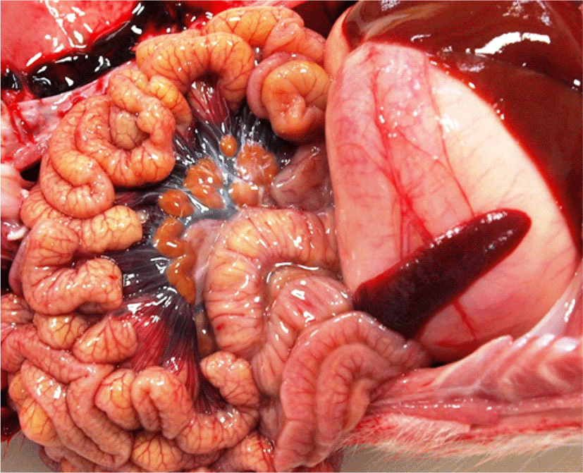
In 2017, four 4–5-day-old piglets with diarrhea from a farm in Korea were sent to the diagnostic laboratory at Optipharm Inc. (Cheongju, Korea). Although the piglets were continuously treated with antibiotics at the farm, their diarrhea did not improve. The piglets were thin and had rough and greasy coats. No disease lesions were observed during the initial post-mortem examination. The stomachs were filled with food material, and the wall thickness of the small intestines appeared normal. Furthermore, the omentum of the intestinal mesentery in each piglet was clearly observed, indicating that the piglets did not experience difficulties with milk intake (Fig. 1). The contents of the small and large intestines were fluid, and kidney stones were scattered across the bilateral sections of the kidneys (unpublished data), indicating that the piglets were severely dehydrated.

jbtr-20-4-115-g1
Fig. 1. No lesions were observed in the intestines during necropsy. The piglet stomachs were filled with large amounts of undigested milk curd, and lacteals were clearly observed in the mesenteries of the small intestines.
Download Original Figure

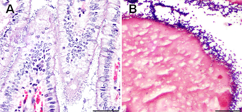
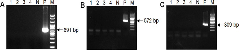
Tissue samples were collected from the jejunum, ileum, and colon of each piglet and fixed in 10% neutral buffered formalin for histopathological examination. The fixed tissues were processed, embedded in paraffin wax, and cut into 3-μm-thick sections. Tissue sections were stained using hematoxylin and eosin and Gram staining. Upon examination, numerous Gram-positive cocci were found to be adhered to the villous epithelial cells throughout the jejunum and ileum of each piglet. However, we did not observe significant changes in villi structure, such as in villi length, in the small intestine, nor any physical damage to the intestinal epithelium (Fig. 2). Polymerase chain reaction (PCR) was performed on small intestine samples to check if diarrhea-causing viral pathogens, such as PEDV, TGEV, and other rotaviruses, were present. Primer sequences and sizes are listed in Table 1. According to the PCR results, these viruses were not present in the piglet small intestines (Fig. 3). PCR results and histopathological examination also revealed that protozoan parasites, such as Isospora suis, were absent from the intestinal contents and intestinal villi (unpublished data).

jbtr-20-4-115-g2
Fig. 2. Microscopic examination of the small intestine of a piglet with diarrhea. (A) Numerous cocci were adhered to the surfaces of villous epithelial cells. Tissues were stained using hematoxylin and eosin. Bar = 50 μm. (B) Gram-positive cocci on the villi surfaces. Tissues were Gram-stained. Bar = 20 μm.
Download Original Figure
Table 1. List of primers used in this study
Target Primer Sequence (5’-3’) Product length (bp) Annealing temperature (°C)
PEDV PED-F GTATCACCACCATCAACAGC 691 63
PED-R ACAAGTCTCGTAACCAGTCC
TGEV S gene-F AGAACTATAGGTAACCATTGG 572 63
S gene-R TTCTAATGTAGTCGCACGCAT
Rotavirus Rota-F AAAGATGCTAGGGACAAAATTG 309 63
Rota-R TTCAGATTGTGGAGCTATTCCA
E. durans (ddlE. durans) DuHiF TTATGTCCCWGTWTTGAAAAATCAA 186 57
DuR TGAATCATATTGGTATGCAGTCCG
E. hirae (ddlE. hirae) DuHiF TTATGTCCCWGTWTTGAAAAATCAA 377 57
HiR TTTTGTTAGACCTCTTCCGGA

E. Enterococcus; PEDV, porcine epidemic diarrhea virus; TGEV, transmissible gastroenteritis virus.

Download Excel Table
jbtr-20-4-115-g3
Fig. 3. The PCR products obtained by amplifying DNA in piglet small intestines. Gel electrophoresis was performed to confirm whether common diarrhea-causing viral pathogens were present. Lanes 1–4 contain PCR products from each piglet, N contains the negative control, and M contains the DNA molecular weight marker. P contains the positive control for (A) the porcine epidemic diarrhea virus (target band size: ~691 bp), (B) the transmissible gastroenteritis virus (target band size: ~572 bp), and (C) rotaviruses (target band size: ~309 bp). These viruses were absent in all four piglet samples. PCR, polymerase chain reaction.
Download Original Figure

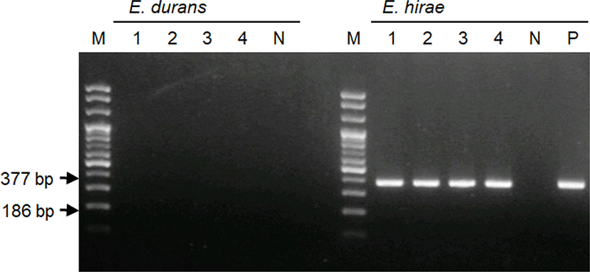
Samples of the intestinal contents of all piglets were inoculated on a sheep blood agar plate and incubated aerobically at 37°C for 24 h. The bacterial colonies produced were small and round, and induced α-hemolysis. The VITEK 2 system (bioMérieux, Marcy-l'Étoile, France) was used to identify the bacteria according to their biochemical properties. The results revealed that 99% of the bacterial colonies were E. hirae. PCR was also performed using DNase/RNase-free distilled water according to the method described by Knijff et al. [16], to identify the cultured colonies more accurately. PCR primers and reaction conditions are summarized in Table 1. Gel electrophoresis was performed using the PCR products, and bands corresponding to E. hirae were present in all four samples, whereas those of E. durans were not present (Fig. 4).

jbtr-20-4-115-g4
Fig. 4. Polymerase chain reaction (PCR) products obtained by analyzing bacteria cultured from piglet small intestines. Gel electrophoresis was performed to confirm the presence of Enterococcus durans (target band size: ~186 bp) and E. hirae (target band size: ~377 bp), respectively. M, DNA molecular weight marker; lanes 1–4, PCR products of bacteria cultured from each piglet; N, negative control; P, positive control (Enterococcus hirae). PCR, polymerase chain reaction.
Download Original Figure

Lastly, we performed an antibiotic susceptibility test using the disk diffusion method. E. hirae was inoculated onto blood agar plates, and discs containing antibiotics (18 types) were placed onto the plates. Plates were then incubated at 37°C for 24 h. E. hirae was susceptible only to amoxicillin and florfenicol, and was resistant to several antibiotics that are used to treat enteric diseases, such as colistin, enrofloxacin, gentamicin, linsmycin, neomycin, tiamulin, trimethoprim/sulfamethoxazole, and tylosin (Table 2).

Table 2. Antibiogram showing the resistance of Enterococcus hirae to various antibiotics (n = 4)
Antibiotics Susceptibility of each sample*
1 2 3 4
Amikacin (AK30) R R R R
Amoxicillin (AML25) S S S S
Ampicillin (AMP10) I R S S
Cefazolin (KZ30) R R R R
Tilmicosin (TIL15) R R R R
Colistin (CT10) R R R R
Enrofloxacin (ENR5) R R R R
Florfenicol (FFC30) S S S S
Ceftiofur (C2.5) R R R R
Gentamicin (CN10) R R R R
Linsmycin (LS109) I R R R
Neomycin (N30) R R R R
Penicillin G (P10) R R I I
Tiamulin (Ti30) R I R R
Streptomycin (S10) R I R R
Tetracycline (T30) R R R R
Trimethoprim/Sulfamethoxazole (STX25) R R R R
Tylosin (Ty30) R R R R

S, susceptible; I, intermediate; R, resistant.

Download Excel Table

Discussion

In general, when diarrhea in piglets is caused by viral enteritis, the villi become shorter and physical damage to the epithelium in the small intestine can be observed by histopathological examination. We did not observe structural changes in the villi or epithelium of the small intestines of any of the piglets. Thus, the diarrhea was not caused by viral infections. Additionally, Gram staining did not reveal the presence of Gram-negative bacteria, such as Escherichia coli, which can cause enteritis without damaging intestinal villi. By contrast, numerous Gram-positive cocci were attached to the intestinal villi. These cocci also exhibited the same morphology as E. hirae. However, E. hirae is difficult to differentiate from E. durans using conventional methods, and their 16S rRNA is 98.8% similar [17]. Specialized diagnostic methods are needed to detect overexpression of specific microorganisms in the small intestine, in the absence of a gold-standard method [18]. In this study, we combined histopathological and microbiological methods to accurately diagnose an E. hirae infection. Thus, we used the VITEK 2 system and PCR to identify E. hirae, and diagnosed the piglets with diarrhea caused by E. hirae infection.

The main determinants of piglet diarrhea include passive immunity to colostrum and milk [19], environmental temperature [20] and humidity [21], management practices, and infections by specific pathogens [22]. Case histories should always be examined, as piglet diarrhea can be caused by a multitude of interacting factors [23]. In pig farms, antibiotics are commonly used to treat piglet diarrhea. The diarrhea in the four piglets in this study did not improve despite continuous antibiotic treatment; this could have been because E. hirae is resistant to most types of antibiotics. Although antibiotics help to prevent and treat clinical diseases, their continuous use in the swine industry has led to reduced microbiota diversity in pigs and the emergence of antibiotic-resistant bacteria [24]. Furthermore, exposure to antibiotics immediately after birth may affect the richness and diversity of intestinal microorganisms. As antibiotic-resistance genes accumulate in the bacterial community, the populations of certain species may start increasing [25]. Bacterial overexpression in the small intestine can inhibit enzymatic action, absorption, and normal metabolism, resulting in digestive or absorptive disorders [26,27]. Therefore, the continuous use of antibiotics may increase the risk of diarrhea in piglets caused by E. hirae, although this species is rarely pathogenic.

In conclusion, the pathoanatomical and histopathological characteristics of individual cases, and bacterial biochemistry, should be evaluated together with case history (e.g., antibiotic treatment) to determine the cause of diarrhea in piglets. Thus, this study presents rare cases of E. hirae infection of the piglet small intestine, which can occur in association with diarrhea possibly by the continuous use of antibiotics. However, further studies are needed to elucidate how E. hirae infection causes diarrhea, and to determine the relationship between antibiotic use and E. hirae overexpression in the small intestine.

References

1.

Katsuda K, Kohmoto M, Kawashima K, Tsunemitsu H. Frequency of enteropathogen detection in suckling and weaned pigs with diarrhea in Japan. J Vet Diagn Invest 2006;18:350-354.

2.

Yaeger M, Funk N, Hoffman L. A survey of agents associated with neonatal diarrhea in Iowa swine including Clostridium difficile and porcine reproductive and respiratory syndrome virus. J Vet Diagn Invest 2002;14:281-287.

3.

Lippke RT, Borowski SM, Marques SMT, Paesi SO, Almeida LL, Moreno AM, Corbellini LG, de Barcellos DESN. Matched case-control study evaluating the frequency of the main agents associated with neonatal diarrhea in piglets. Pesqui Vet Bras 2011;31:505-510.

4.

Sachdev AH, Pimentel M. Gastrointestinal bacterial overgrowth: pathogenesis and clinical significance. Ther Adv Chronic Dis 2013;4:223-231.

5.

Nicklas JL, Moisan P, Stone MR, Gookin JL. In situ molecular diagnosis and histopathological characterization of enteroadherent enterococcus hirae infection in pre-weaning-age kittens. J Clin Microbiol 2010;48:2814-2820.

6.

Farrow JAE, Collins MD. Enterococcus hirae, a new species that includes amino acid assay strain NCDO 1258 and strains causing growth depression in young chickens. Int J Syst Evol Microbiol 1985;35:73-75.

7.

Vela AI, Fernandez A, Moreno B, Casamayor A, Chacon G, Villa A, Comenge J, Fernandez-Garayzabal JF. Isolation of Enterococcus hirae from suckling rabbits with diarrhoea. Vet Rec 2010;167:345-346.

8.

Tzipori S, Hayes J, Sims L, Withers M. Streptococcus durans: An unexpected enteropathogen of foals. J Infect Dis 1984; 150:589-593.

9.

Jones ME, Draghi DC, Thornsberry C, Karlowsky JA, Sahm DF, Wenzel RP. Emerging resistance among bacterial pathogens in the intensive care unit: A European and North American Surveillance study (2000–2002). Ann Clin Microbiol Antimicrob 2004;3:14.

10.

Cheon DS, Chae C. Outbreak of diarrhea associated with Enterococcus durans in piglets. J Vet Diagn Invest 1996;8: 123-124.

11.

Hoover D, Bendele SA, Wightman SR, Thompson CZ, Hoyt JA. Streptococcal enteropathy in infant rats. Lab Anim Sci 1985;35:635-641.

12.

Etheridge ME, Vonderfecht SL. Diarrhea caused by a slow-growing enterococcus-like agent in neonatal rats. Lab Anim Sci 1992;42:548-550.

13.

Collins JE, Bergeland ME, Lindeman CJ, Duimstra JR. Enterococcus (Streptococcus) durans adherence in the small intestine of a diarrheic pup. Vet Pathol 1988;25:396-398.

14.

Vancanneyt M, Snauwaert C, Cleenwerck I, Baele M, Descheemaeker P, Goossens H, Pot B, Vandamme P, Swings J, Haesebrouck F, Devriese LA. Enterococcus villorum sp. nov., an enteroadherent bacterium associated with diarrhoea in piglets. Int J Syst Evol Microbiol 2001;51:393-400.

15.

Larsson J, Lindberg R, Aspan A, Grandon R, Westergren E, Jacobson M. Neonatal piglet diarrhoea associated with enteroadherent Enterococcus hirae. J Comp Pathol 2014;151: 137-147.

16.

Knijff E, Dellaglio F, Lombardi A, Andrighetto C, Torriani S. Rapid identification of Enterococcus durans and Enterococcus hirae by PCR with primers targeted to the ddl genes. J Microbiol Methods 2001;47:35-40.

17.

Devriese LA, Vancanneyt M, Descheemaeker P, Baele M, Van Landuyt HW, Gordts B, Butaye P, Swings J, Haesebrouck F. Differentiation and identification of Enterococcus durans, E. hirae and E. villorum. J Appl Microbiol 2002;92:821-827.

18.

Khoshini R, Dai SC, Lezcano S, Pimentel M. A systematic review of diagnostic tests for small intestinal bacterial overgrowth. Dig Dis Sci 2008;53:1443-1454.

19.

Le Dividich J, Noblet J. Colostrum intake and thermoregulation in the neonatal pig in relation to environmental temperature. Biol Neonate 1981;40:167-174.

20.

Kelley KW, Blecha F, Regnier JA. Cold exposure and absorption of colostral immunoglobulins by neonatal pigs. J Anim Sci 1982;55:363-368.

21.

Pedersen LJ, Malmkvist J, Kammersgaard T, Jorgensen E. Avoiding hypothermia in neonatal pigs: Effect of duration of floor heating at different room temperatures. J Anim Sci 2013;91:425-432.

22.

Fairbrother JM. Neonatal Escherichia coli diarrhea In: Straw BE, D’Allaire S, Mengeling WL, Taylor DJ (eds.). Diseases of swine. 8th ed. Ames, IA: Iowa State University Press; 1999.

23.

Larsson J, Fall N, Lindberg M, Jacobson M. Farm characteristics and management routines related to neonatal porcine diarrhoea: A survey among Swedish piglet producers. Acta Vet Scand 2016;58:77.

24.

Janczyk P, Pieper R, Souffrant WB, Bimczok D, Rothkotter HJ, Smidt H. Parenteral long-acting amoxicillin reduces intestinal bacterial community diversity in piglets even 5 weeks after the administration. ISME J 2007;1:180-183.

25.

Jernberg C, Lofmark S, Edlund C, Jansson JK. Long-term ecological impacts of antibiotic administration on the human intestinal microbiota. ISME J 2007;1:56-66.

26.

Bala L, Ghoshal UC, Ghoshal U, Tripathi P, Misra A, Gowda GA, Khetrapal CL. Malabsorption syndrome with and without small intestinal bacterial overgrowth: A study on upper‐gut aspirate using 1H NMR spectroscopy. Magn Reson Med 2006;56:738-744.

27.

Gasbarrini A, Lauritano EC, Gabrielli M, Scarpellini E, Lupascu A, Ojetti V, Gasbarrini G. Small intestinal bacterial overgrowth: diagnosis and treatment. Dig Dis 2007;25: 237-240.