


Introduction
Impairment of the skin barrier has a critical role in the etiology of a number of diseases of the skin, including atopic dermatitis [1]. As in humans, the canine skin functions as a biophysical barrier between the environment and the body [2]. The epidermis in particular has a crucial protective role in regulating the body’s water content and preventing entry of harmful environmental agents, such as microorganisms and allergens [3]. Ongoing maintenance of the protective function of the epidermis is necessary for prevention of diseases of the skin in both dogs and humans [4].
Recently, a number of non-invasive methods have been developed for measurement and evaluation of the physicochemical properties of the skin barrier and for objective functional assessment of skin conditions [5]. For example, transepidermal water loss (TEWL), skin hydration, and sebum level have been used to evaluate the condition of the skin in dogs and cats as well as in humans [3-9]. TEWL reflects the amount of water lost through the epidermis and is an indicator of the extent of damage to the skin barrier [8, 9]. Skin hydration simply refers to the relative hydration state of the epidermis [9]. Sebum is a waxy material that is secreted by the sebaceous glands and keeps the skin surface soft by forming a lipid layer to retain an appropriate amount of moisture [10].
The skin barrier has been evaluated by non-invasive methods in human medicine [6, 11]. Generally, dogs have a higher incidence of skin diseases than other species, but very little is known about the physiologic indicators of skin condition in dogs [12]. Further, there is little information in the literature on the consistency of repeated skin measure ments at various anatomic sites over time or on the relationship between biophysical parameters indicating skin condition in dogs. The aim of this study was to evaluate the variation in repeated skin measurements and the relationship between TEWL, skin hydration, and sebum levels in normal dogs.
Materials and Methods
Five female Beagle dogs aged 1–3 years and weighing 8–10 kg were evaluated. All dogs were clinically normal without skin or metabolic disease. Before the skin measurements were taken, all dogs were acclimatized for at least 60 min in the same room at a temperature of 20°C–21°C and a relative humidity of 44%–66%. The experiment was approved by the Institution Animal Care of the Laboratory Animal Research Center at Chungbuk National University.
TEWL, skin hydration, and the sebum level were measured at seven anatomic sites, i.e., the left and right pinnae, left and right axillae, left and right groin areas, and ventrum. Each skin parameter was measured five times on two occasions each separated by an interval of two days. TEWL, skin hydration, and the sebum level were measured using a VapoMeter (Delfin Technologies Ltd, Kuopio, Finland), Corneometer (CM825, Courage & Khazaka, Cologne, Germany), and Sebumeter (SM810, Courage & Khazaka, Cologne, Germany), respectively. The sites to be measured were marked with an oil-based pen to ensure measurement at the same anatomic locations. After excluding the maximum and minimum values, the average of the three remaining measurements was recorded.
Variations in TEWL, skin hydration, and sebum levels over time and according to anatomic site were evaluated using the Wilcoxon signed rank test. The coefficient of variation was calculated for each anatomic site measured to estimate the variation in data obtained for each skin parameter. The Spearman rank correlation was used to demonstrate the contribution of skin hydration and sebum level to TEWL. All statistical analyses were performed using SPSS for Windows version 12.0 software (SPSS Inc., Chicago, IL, USA).
Results
The skin measurements were in the range of 2.2–21.3 g/ m2/h for TEWL, 0.5–26.2 units for skin hydration, and 0–240 μg/cm2 for sebum level (Figs 1–3). There was no statistically significant difference between the values obtained on the two different days or between those obtained at the seven anatomic sites (both P>0.05, Figs 1–3).Fig. 2
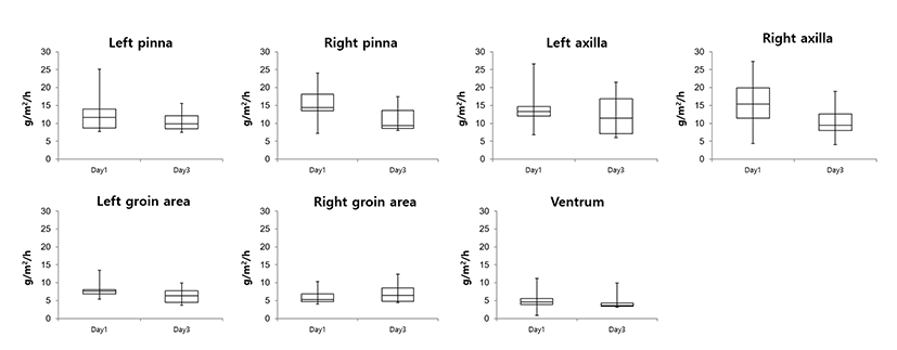
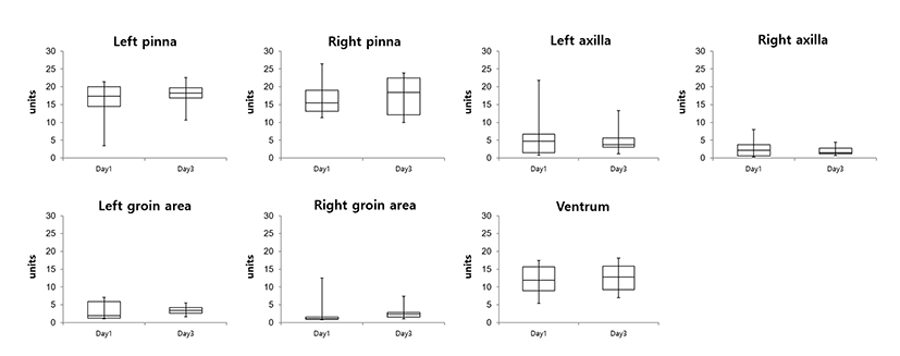
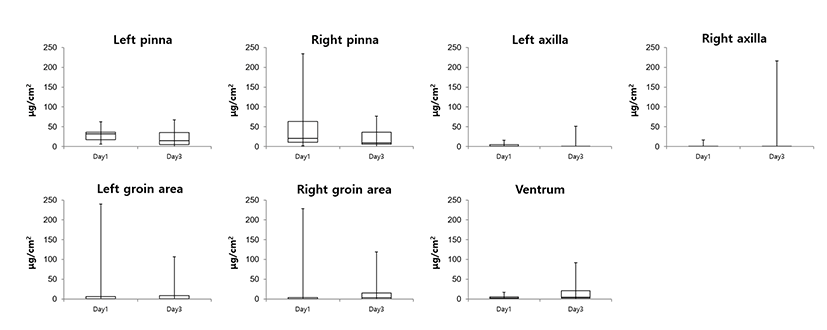
The coefficients of variation for the measurements of the three skin parameters at each anatomic site are shown in Table 1. The mean variation (and standard deviation) was highest for sebum level (209.0 ± 81.8%), followed in descending order by skin hydration (62.7 ± 34.5%) and TEWL (41.1 ± 6.9%). Of the seven anatomic sites, the left and right pinnae showed the least variation in measurements for TEWL (39.2%), skin hydration (29.6%), and sebum level (75.5%). The highest variation in TEWL was observed on the ventrum (52.4%), in the right groin area for skin hydration (110.6%), and the right axilla for sebum level (334.1%).
The correlations between the measurements recorded for the three parameters are shown in Table 2. There was no significant correlation between TEWL and skin hydration or sebum level (P>0.05) or between skin hydration and sebum level (P>0.05).
| Variables | Correlation coefficient | P-value |
|---|---|---|
| TEWL and skin hydration | 0.125 | 0.323 |
| TEWL and sebum level | 0.081 | 0.219 |
| Skin hydration and sebum level | 0.154 | 0.352 |
Discussion
The present data demonstrate no time-related or anatomic variation in repeated measurements of TEWL, skin hydration, or sebum level in normal dogs. Further, no effect of skin hydration or sebum level on TEWL was identified.
Skin barrier function has been studied in humans by measuring TEWL, skin hydration, and sebum level for the purposes of diagnosing skin disease and monitoring the response to treatment [5-7]. Although TEWL has been examined previously in veterinary medicine [1-4, 13], there are few reports on skin hydration and sebum levels in dogs and cats [14, 15]. In view of the similarity in skin structure between humans and dogs, this study was designed to compare the suitability of the above three parameters for assessment of the skin barrier in normal dogs.
In humans, TEWL, skin hydration, and sebum level are determined in relation to anatomic site, sex, and age [6]. In dogs, TEWL is also influenced by the hair covering the skin surface [8]. Therefore, an appropriate methodology is needed to obtain reliable results when using these techniques in dogs [12, 13, 16, 17]. Measurements of indicators of skin barrier function are greatly influenced by environmental factors, particularly ambient temperature and relative humidity [2, 14]. In this study, no time-related variation in TEWL, skin hydration, or sebum level was identified.
Generally, TEWL is a useful indicator of skin barrier function in dogs [3]. Previous research suggests that TEWL is greater in atopic dogs than in normal dogs [1, 18]. In particular, TEWL from the pinna was positively correlated with the severity of atopic dermatitis, and this was attributed to the relatively low density of hairs at this site [1]. In the present study, of the three parameters and seven anatomic sites measured, the coefficient of variation was lowest for TEWL and the pinna, respectively. Given it relatively low variation over time, TEWL might be a more suitable parameter for repeated measurements than skin hydration or sebum level. Although no variation was detected between measurements taken at the seven anatomic sites, measurements taken at the pinna could be useful in terms of convenience and lack of need for clipping.
The variation in repeated measurements of skin hydration was moderate, with lower variation found at the pinna and ventrum in comparison with the values for TEWL and sebum. The low density of hairs at these sites may have contributed to this low variation, given that measurement of skin hydration relies on electric resistance, which is interfered with by hair [19]. Previous studies in humans have reported that skin hydration levels are lower in patients with atopy than in normal subjects [20, 21]. Therefore, to obtain consistent results in dogs with skin diseases, skin hydration might be best measured at anatomic sites with a low density of hairs, such as the pinna and ventrum.
The coefficient of variation was highest for sebum levels. Generally, measurement of sebum depends on the optical reflection of lipids, which are absorbed onto a tape pressed against the skin [7]. Therefore, the data obtained could vary according to the adhesion per unit area and the pressure on the tape applied to the skin. Our finding of the highest variation being in sebum levels may be attributable to the fact that the other two parameters were not fixed because of gravity or other forces.
TEWL has been reported to be inversely proportional to skin hydration in humans and dog with atopic dermatitis [1, 2, 5, 11], but no such correlation has been noted in healthy humans [6, 19, 21]. Similarly, the relevant relationship among three types of measurements was not found in this study evaluating normal dogs.
There are some limitations to this study. The data presented were obtained from healthy dogs only, so further studies are needed to identify the effect of skin hydration and sebum on TEWL in dogs with skin disease. In this study, no time-related variation in TEWL, skin hydration, or sebum level was identified. Whether this finding is related to the time allowed for the dogs to acclimatize to the examination room and/or the measurements being taken under identical temperature and humidity conditions on two occasions is unknown. Further, we evaluated the variation in skin measurements taken on two occasions separated by an interval of only two days, so further studies that include more sets of measurements taken on occasions separated by longer intervals are warranted.
In conclusion, measurements of TEWL, skin hydration, and sebum level did not vary over time or according to the anatomic site of measurement in clinically normal dogs. The coefficients of variation were lowest for TEWL and for skin areas with a low hair density, such as the pinna and ventrum. Because of its relatively low variation on repeated measurements, TEWL might be the most useful indicator of the health of canine skin. The pinna and ventrum are recommended as the best sites for measurement. In healthy skin, the amount of TEWL was not related to degree of skin hydration or the sebum level. Further research is needed to identify the precise relationship between TEWL, skin hydration, and sebum levels in dogs with skin diseases.