Original Article

Effect of P. ginseng on the expression of c-Fos in the brain of Wistar rats with testosterone induced benign prostatic hyperplasia

Hyang Mi Jang*http://orcid.org/0000-0002-7348-622X
Author Information & Copyright
Department of Basic Nursing Science, school of nursing, Kyungdong University, Wonju 26495, Korea
*Corresponding author: Hyang Mi Jang Department of Basic Nursing Science, school of nursing, Kyungdong University, Wonju 26495, Korea +82-33-738-1440, +82-33-738-1449, she_said@kduniv.ac.kr

© Research Institute of Veterinary Medicine, Chungbuk National University. All rights reserved.

Received: Aug 30, 2017; Revised: Sep 14, 2017; Accepted: Sep 14, 2017

Abstract

The lower urinary tract symptoms (LUTS) show in benign prostatic hyperplasia (BPH). The three major micturition centers in brain are pontine micturition center (PMC), ventrolateral periaqueductal gray (vlPAG), and medial preopticnucleus (MPA) regions. Previous study showed that c-Fos expression change was associated with LUTS. In present study, the effect of P. ginseng on c-Fos expression in PMC, vlPAG, and MPA regions in rat brain was tested. P. ginseng is the four year-old Korean ginseng. It was collected at the department of medicinal crop research (Eumsunggun, Chungbuk, Korea) in September 2010. The four groups (n = 6) are control group, BPH-induced group, BPHinduced and P. ginseng-treated group, and BPH-induced and finasteride-treated group. BPH in rats was induced by testosterone. After 4 weeks, all animals were sacrificed to evaluate c-Fos expression in PMC, vlPAG, and MPA regions in rat brain. The c-Fos expression was evaluated in the regions of rat brain by immunohistochemistry (IHC). Present results showed that c-Fos expressions in PMC, vl-PAG, and MPA regions in brain of rats in the BPH-induced group were higher compared to c-fos expression of the control group. The increased c-Fos expression in three regions (PMC, vlPAG, and MPA) were decreased by treatment with P. ginseng (200 mg/kg). These results suggest that P. ginseng has an inhibitory effect on the symptoms of BPH and is associated with regulation of c-Fos expression in the brain in a testosterone induced BPH rat model.

Keywords: Benign prostatic hyperplasia; Panax ginseng; c-Fos; brain; herbal

Introduction

The pathological causes of benign prostatic hyperplasia (BPH) include inflammatory and cellular proliferative factors. Such signals may lead to hyperplasia of the stromal and glandular tissues in the prostate gland [1-3]. The prostate is located around the male urethra and beneath the urinary bladder, therefore enlargement of the prostate may cause obstructive symptoms in urinations, termed lower urinary tract symptoms (LUTS), which are including frequency, urgency, nocturia, voiding difficulty, and dribbling [4]. However, the prostate is a male specific organ, and its growth is affected by androgenic hormones, such as testosterone and dihydrotestosterone [5-7], and therefore, BPH and LUTS are common diseases that occur after middle-age [4].

Androgen-induced increased signal may include the c- Fos [7-9]. Androgen may not directly induce c-Fos [7], however, androgen receptor may regulate epidermal growth factor (EGF) or insulin like growth factor 1 (IGFI) [10-12], which is closely related to c-Fos signal [13, 14]. The c-Fos may act as a regulator of cell proliferation, differentiation, and transformation [15-17].

Moreover, c-Fos play a role in controlling neuronal activity and survival. Interestingly, it has been suggested that neurological factors cause LUTS [18]. And micturition is under the influence of diverse neurological circuits, because brain, spinal cord, and peripheral nervous system and their neurotransmitters are involved in development of urinatary control [19]. There are evidences suggesting that c-fos is related to the control of micturition [20, 21]. A previous study reported that c-Fos expression is related to bladder reflex micturition [22].

Urinary center of central nervous system includes pontine micturition center (PMC), periaqueductal gray (PAG), and medial preopticnucleus (MPA) [19, 23, 24]. The c-Fos expression has been used as a marker of neuronal activity [25], and c-Fos expression changes in stress urinary incontinence has been reported [26]. These previ ous studies suggest that LUTS may affect the expression of c-Fos [26].

A previous study showed that the Panax ginseng C.A. Mayer (P. ginseng) has a protective effect on enlargement of the prostate in testosterone induced BPH rat model [27]. However, PMC, vlPAG, and MPA regions in brain were not investigated. Therefore, this study investigated the effects of P. ginseng on c-Fos expression in the brain in testosterone induced BPH rat model.

Materials and Methods

Preparation of the P. ginseng

P. ginseng is a four year-old Korean ginseng. It was collected from the Department of Medicinal Crop Research (Eumsung-gun, Chungbuk, Korea) in September 2010. To obtain the water extract of ginseng, 100 g of ginseng root was added to 600 mL of distilled water, and the mixture was extracted by heating at 95°C. It was then filtered through a muslin cloth and lyophilized. The resulting powder (yield 32 g) was dissolved in distilled water and sterilized by passing through a 0.22 μM filter sequentially [27, 28].

Animals

In the present study, seven-week-old Wistar rats (Central Lab Animal Inc, Korea) were used with an average weight of 250 ± 10 g. The animal room was maintained at 22 ± 2°C and relative humidity of 40~70%. Indoor lighting consists of 12 periods of light and dark cycles. All experiments were carried out in accordance with procedures approved by the Animal Care Committee of the Animal Center at Kyung Hee University and in accordance with guidelines of the Korean National Health Institute of Health Animal Facility.

Induction of BPH and treatments

BPH was induced by subcutaneous injection of testosterone (20 mg/kg) for 4 weeks. Following sentinel resection, rats were divided into four groups (n = 6): (A) control group; (B) BPH derived group subcutaneously injected with testosterone; (C) P. ginseng group treated with 200 mg/kg. (D) 1 mg/kg of the finasteride was orally administered (Sigma-Aldrich, St Louis, MO, USA) as a positive anti-BPH drug. All materials were administered to the animals once a day for 4 weeks, and body weights were measured weekly. After 4 weeks, all animals were fasted overnight. Animals were sacrificed, and fresh prostate was stored in formaldehyde solution for optical microscopy. The remaining prostate was stored at -70°C for later analysis.

Immunohistochemistry

Immunostaining was performed with 35 μM sections of brain tissues. Peroxidase activity quenching was performed with 3% H2O2 in PBS for 10 min. The sections were then washed with water and pre-blocked with normal goat or rabbit serum for at least 1 hour. In the primary antibody reaction step, slides were diluted 1: 200 overnight at 4°C and incubated with anti-c-Fos (Santa Cruz Biotechnology Inc., Santa Cruz, CA, USA). The sections were then incubated with biotinylated secondary antibody (1: 1000) for 1 hour. After the wash step with PBS, streptavidin-HRP was applied. Finally, the sections were rinsed with PBS and developed with a diaminobenzide dehydrochloride (DAB) substrate for 10 minutes. At least three random fields per section were checked at × 100.

Statistical Analyses

All values were presented as mean ± S.E. Significant differences among groups were statistically analyzed using one-way analysis of variance (ANOVA) and non-parametric post Tukey test. All p values were double-tailed, and significance was set at P<0.05. All statistical analysis was performed using SPSS for Windows.

Results

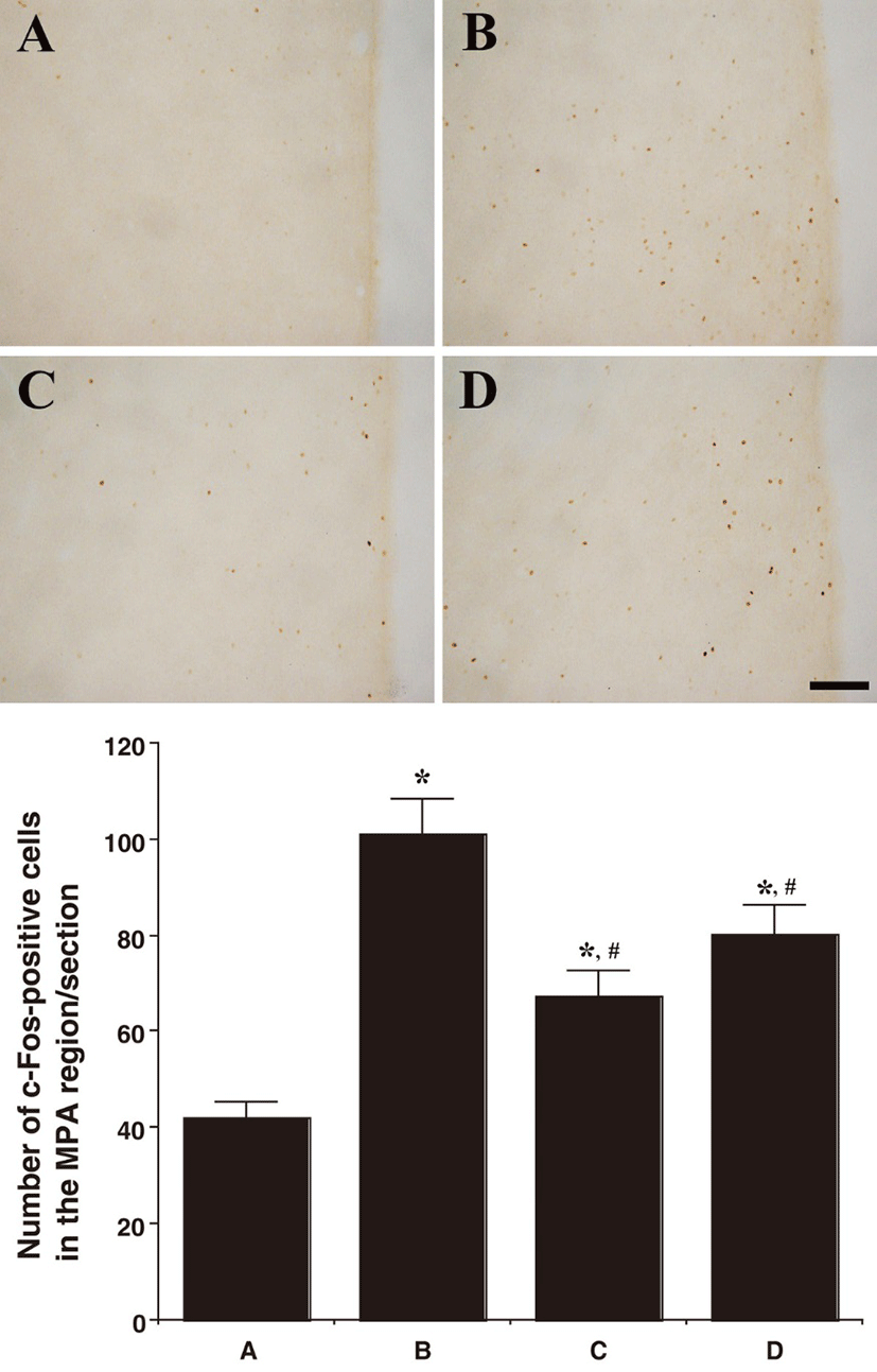
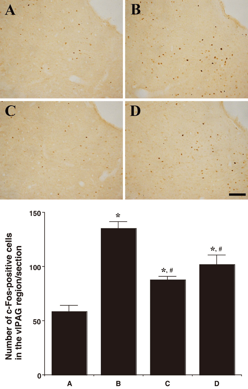
Fig 1, 2, and 3 showed photomicrographs of c-Fos-positive cells in the neuronal voiding centers of the brain (MPA, vlPAG, and PMC). In the MPA region, the number of c-Fos-positive cells was 42.00 ± 3.38/section in the control group, 101.16 ± 7.29/section in the BPH-induced group, 67.58 ± 5.15/section in the BPH-induced and P. ginseng-treated group, and 80.25 ± 6.18/section in the BPH-induced and finasteride-treated group. In the vlPAG region, the number of c-Fos-positive cells was 58.50 ± 5.57/section in the control group, 135.16 ± 6.22/section in the BPH-induced group,87.66 ± 3.47/section in the BPH-induced and P. ginseng-treated group, and 102.00 ± 8.78/section in the BPH-induced and finasteride-treated group.
jbtr-18-3-108-g1
Fig. 1. Effect of P. ginseng on c-Fos expressions in MPA region after the induction of BPH. Upper: Photomicrographs of c-Fospositive cells in the neuronal voiding centers. The sections were stained for c-Fos immunoreactivity (brown). The scale bar represents 150 μM. (A) Control group, (B) BPH-induced group, (C) BPH-induced and P. ginseng-treated group, (D) BPH-induced and finasteride-treated group. Lower: Number of c-Fos-positive cells in each group. *represents P<0.05 compared to the control group. #represents P<0.05 compared to the BPH-induced group.
Download Original Figure
jbtr-18-3-108-g2
Fig. 2. Effect of P. ginseng on c-Fos expressions in vlPAG region after the induction of BPH. Upper: Photomicrographs of c-Fos-positive cells in the neuronal voiding centers. The sections were stained for c-Fos immunoreactivity (brown). The scale bar represents 150 μM. (A) Control group, (B) BPH-induced group, (C) BPH-induced and P. ginseng-treated group, (D) BPH-induced and finasteride-treated group. Lower: Number of c-Fos-positive cells in each group. *represents P<0.05 compared to the control group. #represents P<0.05 compared to the BPH-induced group.
Download Original Figure
jbtr-18-3-108-g3
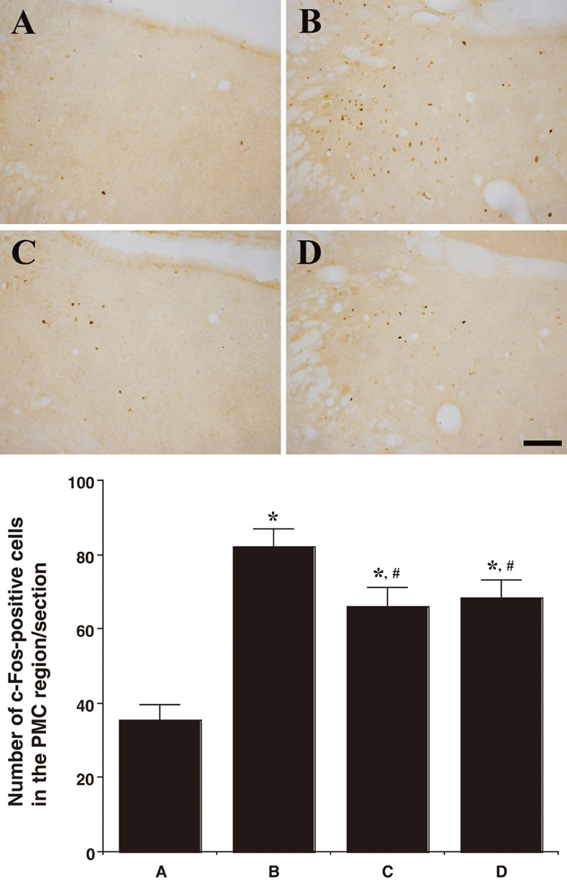
Fig. 3. Effect of P. ginseng on c-Fos expressions in PMC region after the induction of BPH. Upper: Photomicrographs of c-Fospositive cells in the neuronal voiding centers. The sections were stained for c-Fos immunoreactivity (brown). The scale bar represents 150 μM. (A) Control group, (B) BPH-induced group, (C) BPH-induced and P. ginseng-treated group, (D) BPH-induced and finasteride-treated group. Lower: Number of c-Fos-positive cells in each group. *represents P<0.05 compared to the control group. #represents P<0.05 compared to the BPH-induced group.
Download Original Figure

In the PMC region, the number of c-Fos-positive cells was 35.66 ± 3.82/section in the control group, 82.33 ± 4.69/section in the BPH-induced group, 66.00 ± 5.11/ section in the BPH-induced and P. ginseng-treated group, and 68.50 ± 4.78/section in the BPH-induced and finasteride- treated group.

In summary, the c-Fos expression in the neuronal voiding centers (MPA, vlPAG, and PMC) was increased by the induction of BPH (P<0.05) and P. ginseng treatment significantly decreased the BPH-induced c-Fos expression in the neuronal voiding centers (MPA, vlPAG, and PMC) (P<0.05).

Discussion

The results of this study showed that c-Fos expression in PMC, MPA, and vlPAG regions of brain was increased in the BPH group compared to those of the normal group, whereas the increased c-Fos expression in PMC, MPA, and vlPAG was significantly attenuated in finasteridetreated. In P. ginseng-treated group, the c-Fos expression was statistically significantly decreased similarly to the group treated with the positive drug.

In recent study [27] of P. ginseng, they showed that administration of P. ginseng in the testosterone induced BPH rat model significantly prevent prostate enlargement. Their observation in the study revealed that decreased expression of alpha-1D adrenergic receptor (Adra1d), EGFR, and BCL2 correlate with the protective effect of P. ginseng [27]. In addition to previous study, results of this study suggest biologic evidences that P. ginseng may have a protective effect on development of LUTS, therefore supporting the claim that P. ginseng helps ameliorate BPH symptoms.

The correlation of c-Fos expression and neuronal activation of central micturition centers in LUTS rat model was reported by Cho et al [29]. They reported that altered c-Fos expression is associated with neurogenic lower urinary tract dysfunction caused by intracerebral hemorrhage. Additionally, another study by Chung et al. [20] showed that c-Fos expressions in PMC, vlPAG, and MPA were correlated with stress urinary incontinence.

These evidences are related to bladder activity and c-Fos in brain [19, 22-25], however, they may partially support our result, because LUTS in BPH are caused by bladder outlet obstruction [30], and bladder obstruction is related to spinal cord c-Fos expression [31]. It may suggest that c-Fos is related to stress in bladder, which also occurs in LUTS due to BPH.

Moreover, overactive bladder is associated with BPH in approximately 40–75% of cases [32]. It is a very common symptom, and these two conditions share common bladder storage problems [33, 34]. Thus, these previous reports also support our observation that attenuated expression of c-Fos in P. ginseng treated BPH group may represent the decreased severity of LUTS in rats with BPH.

In summary, P. ginseng, which has a protective effect against BPH, may also have protective effect against LUTS, because c-Fos expression was increased in micturition centers of rats with BPH, and P. ginseng significantly decreased the c-Fos expression. Therefore, the present study suggests that P. ginseng could be used as an effective treatment for BPH.

References

1.

Wang HH, Wang L, Jerde TJ, Chan BD, Savran CA, Burcham GN, Crist S, Ratliff TL. Characterization of autoimmune inflammation induced prostate stem cell expansion. The Prostate. 2015; 75:1620-1631.

2.

Porcaro AB, Rubilotta E, Petrozziello A, Ghimenton C, Migliorini F, Zecchini Antoniolli S, Lacola V, Monaco C, Curti P, Cavalleri S, Pianon R, Artibani W. Chronic inflammation of the prostate type IV with respect to risk of prostate cancer. Archivio italiano di urologia, andrologia : organo ufficiale [di] Societa italiana di ecografia urologica e nefrologica / Associazione ricerche in urologia. 2014; 86:208-211.

3.

Rodrigues MM, Rema A, Gartner MF, Laufer-Amorim R. Role of adhesion molecules and proliferation hyperplasic, pre neoplastic and neoplastic lesions in canine prostate. Pakistan journal of biological sciences (PJBS). 2013; 16:1324-1329.

4.

Nickel JC. The overlapping lower urinary tract symptoms of benign prostatic hyperplasia and prostatitis. Current opinion in urology. 2006; 16:5-10.

5.

Enatsu N, Miyake H, Haraguchi T, Chiba K, Fujisawa M. Effects of dutasteride on serum free-testosterone and clinical significance of testosterone changes. Andrologia. 2016; 48:1195-1201.

6.

Bauman TM, Sehgal PD, Johnson KA, Pier T, Bruskewitz RC, Ricke WA, Huang W. Finasteride treatment alters tissue specific androgen receptor expression in prostate tissues. The Prostate. 2014; 74:923-932.

7.

Levine AC, Ren M, Huber GK, Kirschenbaum A. The effect of androgen, estrogen, and growth factors on the proliferation of cultured fibroblasts derived from human fetal and adult prostates. Endocrinology. 1992; 130:2413-2419.

8.

Brum IS, Morsch DM, Pozzobon A, Boeri VA, Geib G, Spritzer PM. Androgen-dependent expression of c-jun and c-fos in human non-transformed epithelial prostatic cells association with cell proliferation. Hormone research. 2003; 60:209-214.

9.

Chipuk JE, Cornelius SC, Pultz NJ, Jorgensen JS, Bonham MJ, Kim SJ, Danielpour D. The androgen receptor represses transforming growth factor-beta signaling through interaction with Smad3. The Journal of biological chemistry. 2002; 277:1240-1248.

10.

Oliver VL, Poulios K, Ventura S, Haynes JM. A novel androgen signalling pathway uses dihydrotestosterone, but not testosterone, to activate the EGF receptor signaling cascade in prostate stromal cells. British journal of pharmacology. 2013; 170:592-601.

11.

Robaire B, Hamzeh M. Androgen action in the epididymis. Journal of andrology. 2011; 32:592-599.

12.

Davies P, Eaton CL, France TD, Phillips ME. Growth factor receptors and oncogene expression in prostate cells. American journal of clinical oncology. 1988; 11Suppl 2:S1-7.

13.

Peng C, Zeng W, Su J, Kuang Y, He Y, Zhao S, Zhang J, Ma W, Bode AM, Dong Z, Chen X. Cyclin-dependent kinase 2 (CDK2):is a key mediator for EGF-induced cell transformation mediated through the ELK4/c-Fos signaling pathway. Oncogene. 2016; 35:1170-1179.

14.

Izumi K, Zheng Y, Li Y, Zaengle J, Miyamoto H. Epidermal growth factor induces bladder cancer cell proliferation through activation of the androgen receptor. International journal of oncology. 2012; 41:1587-1592.

15.

Grigoriadis AE, Wang ZQ, Wagner EF. Fos and bone cell development lessons from a nuclear oncogene. Trends in genetics : TIG. 1995; 11:436-441.

16.

Candeliere GA, Glorieux FH, Prud'homme J, St-Arnaud R. Increased expression of the c-fos proto-oncogene in bone from patients with fibrous dysplasia. The New England journal of medicine. 1995; 332:1546-1551.

17.

Ruther U, Garber C, Komitowski D, Muller R, Wagner EF. Deregulated c-fos expression interferes with normal bone development in transgenic mice. Nature. 1987; 325:412-416.

18.

Skolarikos A, Thorpe AC, Neal DE. Lower urinary tract symptoms and benign prostatic hyperplasia. Minerva urologica e nefrologica = The Italian journal of urologyand nephrology. 2004; 56:109-122.

19.

Fowler CJ, Griffiths D, de Groat WC. The neural control of micturition. Nature reviews. Neuroscience. 2008; 9:453-466.

20.

Chung IM, Kim YS, Sung YH, Kim SE, Ko IG, Shin MS, Park HJ, Ham DH, Lee HJ, Kim KJ, Lee SW, Jee YS, Kim KH, Kim CJ. Effects of acupuncture on abdominalleak point pressure and c-Fos expression in the brain of rats with stress urinary incontinence. Neuroscience letters. 2008; 439:18-23.

21.

Cho YS, Ko IG, Kim SE, Hwan L, Shin MS, Kim CJ, Kim SH, Jin JJ, Chung JY, Kim KH. Caffeine enhances micturition through neuronal activation in micturitioncenters. Molecular medicine reports. 2014; 10:2931-2936.

22.

Dinis P, Charrua A, Avelino A, Cruz F. Intravesical resiniferatoxin decreases spinal c-fos expression and increases bladder volume to reflex micturition in rats with chronic inflamed urinary bladders. BJU international. 2004; 94:153-157.

23.

Yum KS, Na SJ, Lee KY, Kim J, Oh SH, Kim YD, Yoon B, Heo JH, Lee KO. Pattern of voiding dysfunctionafter acute brainstem infarction. European neurology. 2013; 70:291-296.

24.

Kavia RB, Dasgupta R, Fowler CJ. Functional imaging and the central control of the bladder. The Journal ofcomparative neurology. 2005; 493:27-32.

25.

Lee TH, Jang MH, Shin MC, Lim BV, Kim YP, Kim H, Choi HH, Lee KS, Kim EH, Kim CJ. Dependence of rat hippocampal c-Fos expression on intensity and duration of exercise. Life sciences. 2003; 72:1421-1436.

26.

Ko IG, Kim SE, Kim CJ, Jung JH, Lee SJ, Kim DH, Lee KY, Kim KH. Effect of Treadmill Exercise on Leak-point pressure and Neuronal Activation in Brain of Rats with Stress Urinary Incontinence. International neurourology journal. 2010; 14:141-148.

27.

Kim SK, Chung JH, Lee BC, Lee SW, Lee KH, Kim YO. Influence of Panax ginseng on Alpha-Adrenergic Receptor of Benign Prostatic Hyperplasia. International neurourology journal. 2014; 18:179-186.

28.

Kim SK, Seok H, Park HJ, Jeon HS, Kang SW, Lee BC, Yi J, Song SY, Lee SH, Kim YO, Chung JH. Inhibitoryeffect of curcumin on testosterone induced benign prostatic hyperplasia rat model. BMC complementary and alternative medicine. 2015; 15:380.

29.

Cho YS, Ko IG, Kim CJ, Kim KH. A novel intracerebral hemorrhage-induced rat model of neurogenic voiding dysfunction Analysis of lower urinary tract function. Molecular medicine reports. 2015; 12:2563-2569.

30.

Lin-Tsai O, Clark PE, Miller NL, Fowke JH, Hameed O, Hayward SW, Strand DW. Surgical intervention for symptomatic benign prostatic hyperplasia is correlated with expression of the AP-1 transcription factor network. The Prostate. 2014; 74:669-679.

31.

Yazaki J, Aikawa K, Shishido K, Yanagida T, Nomiya M, Ishibashi K, Haga N, Yamaguchi O. Alpha1-adrenoceptorantagonists improve bladder storage function through reduction of afferent activity in rats with bladder outlet obstruction. Neurourology and urodynamics. 2011; 30:461-467.

32.

Blaivas JG, Marks BK, Weiss JP, Panagopoulos G, Somaroo C. Differential diagnosis of overactive bladder inmen. The Journal of urology. 2009; 182:2814-2817.

33.

Singh I, Agarwal V, Garg G. 'Tamsulosin and Darifenacin' Versus 'Tamsulosin Monotherapy' for 'BPH withAccompanying Overactive Bladder'. Journal of clinical and diagnostic research : JCDR. 2015; 9:PC08-11.

34.

Caremel R, Cornu JN, Kerdraon J, Castel-Lacanal E, Bastide C, Bruyere F, Guy L, Karsenty G. Drug therapy of bladder dysfunction. Progres en urologie. journal de l'Association francaise d'urologie et de la Societe francaise d'urologie. 2013; 23:1271-1286.