Introduction
Shiga toxin-producing Escherichia coli (STEC) is characterized by the release of Shiga toxin 2e variant (Stx2e), causing edema disease (ED) and resulting in significant death and economic losses in the pig industry [1, 2, 3]. To diagnose ED, it is necessary to identify Stx2e in E. coli isolates and gut lumen. Structurally, Shiga toxin (Stx) is composed of an enzymatically active A subunit and the pentamer B receptor-binding subunit [4]. The B-pentamer binds to a glycolipid receptor (GB4) to gain access to the host cytoplasm. The A subunit serves as an RNAglycohydrolase and removes an adenine residue from rRNA, resulting in an impairment of protein synthesis. The A and B subunits are synthesized and then transferred to the periplasm, where the toxin is assembled. Although the toxin is enough for assembly in periplasm, there is a special process of phage-mediated lysis encoded in a prophage of E. coli, which is induced by certain antibiotics. The toxin is released into the intestines, and it enters the bloodstream, damaging the endothelial cells of small arterioles [5, 6].
Various enrichment broths have been used for the detection of STEC. Several protocols include the use of selective agents (antibiotics and bile salts) in the broths [7, 8], but others use nonselective broths [9]. Some use long incubation periods, while others prefer short incubation periods [10, 11]. In addition, several efforts have been made to prevent ED in experimental trials using subunit vaccines containing inactivated or attenuated Stx2e [12, 13]. However, the mass production of Stx2e is difficult because only a small amount of toxins is released into the bacterial culture supernatant. Some antibiotics have been used to induce the release of the toxin packed in the periplasm of E. coli [14]. However, few stud ies have compared the efficiency of enrichment media, and the results of enrichment vary between reports. Accordingly, in the current study, comparisons between commercial media were used to determine the performance of different enrichment media in order to find suitable and reliable conditions for Stx2e production.
Materials and Methods
E. coli KEFS1302 (F18ab:Stx2e:AIDA) was isolated from grower pigs showing signs of ED, including ataxia, edema of the eyelid, and mesenteric edema of the spiral colon. Stx2e production was determined using a Vero cell assay with filtered culture supernatant in LB broth (Beckton Dickinson, USA). A total of four media namely ISOSensitest broth (ISB; Oxoid, UK), E. coli broth (ECB; Oxoid, UK), trypticase soy broth (TSB; Beckton Dickinson, USA), and Mueller Hinton broth (MHB; Beckton Dickinson, USA) were selected for monitoring Stx2e production. Two to three colonies of KEFS1302 were inoculated into 100 mL of LB broth and incubated at 37°C for 18 h. After centrifugation at 5,000 × g for 10 min, the pellet was suspended in sterile phosphate-buffered saline (PBS; pH 7.2) and adjusted to McFarland No. 1.
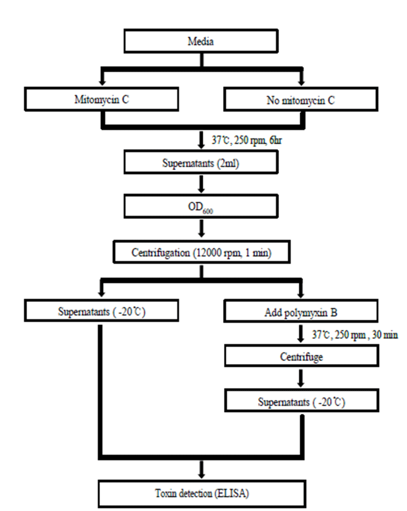
The scheme for mass production of Stx2e is represented in Fig. 1. The each media containing 100 mL was divided into groups with and without mitomycin C (10 ng/mL) treatment. After inoculation of 1% of the bacterial suspension, the cultures were incubated with agitation at 250 rpm at 37°C for 6 h. First, 2-mL samples were collected every hour, and cell growth was determined in terms of optical density (OD) at 600 nm (OD600) using a spectrophotometer (Infinite M200 PRO; Tecan, Australia). After centrifugation at 8,000 × g for 1 min, supernatants were stored at -20°C. The pellets were then mixed with 1 mL of polymyxin B (0.2 mg/mL) and incubated for 30 min. After centrifugation, this supernatant was stored at -20°C until use in the toxin assay.

An ELISA was used to detect the toxin level [17]. The culture supernatants were incubated in a 96-well plate (Corning, USA) with calcium carbonate buffer (pH 9.0) at 4°C overnight. The plates were washed three times with PBS with 1% Tween 20 (PBST). After blocking using 10% bovine serum albumin (BSA; Sigma, USA) for 1 h, the plate was incubated with anti-Stx2 rabbit polyclonal antibody (GeneTex, USA) diluted 1:1000, followed by incubation with goat anti-rabbit IgG conjugated to peroxidase (Sigma, USA) diluted 1:5000. O-phenylenediamine (OPD, Sigma) plus hydrogen peroxide was used to develop the color, and the reaction was terminat- ed by adding 1 N H2SO4. Absorbance was determined by an ELISA reader (Infinite M200 PRO; Tecan, Australia).
To purify the edema toxin, the supernatant of ECB culture was fractioned by FPLC (Waters, USA) using a Hi- TrapTM SP55 column (GE, USA), and the edema toxin was eluted by a 5 M NaCl linear gradient with 10 mM Tris-HCl (pH 7.6) buffer. The protein concentration of the purified Stx2e fraction was determined using a BCA kit (Thermo, USA). The eluted fractions were applied to a 12% sodium dodecyl sulfate-polyacrylamide gel electrophoresis (SDS-PAGE) gel (Mini-Cell kit, Invitrogen), and the gels were stained with Coomassie Blue G-250 to visualize the toxin. After PAGE, the proteins were electroblotted from a duplicate gel to a nitrocellulose membrane (GE Healthcare, USA). After blocking with 2% BSA, primary antibody (1:1000, anti-Stx2 rabbit polyclonal antibody, Genetex) was added, and the membrane was incubated for 1 h. This was followed by a secondary antibody using peroxidase conjugated goat anti-rabbit IgG (1:1,000, KPL) for 1 h. Protein bands were detected using an ECL detection kit (Amersham, Germany).
Results
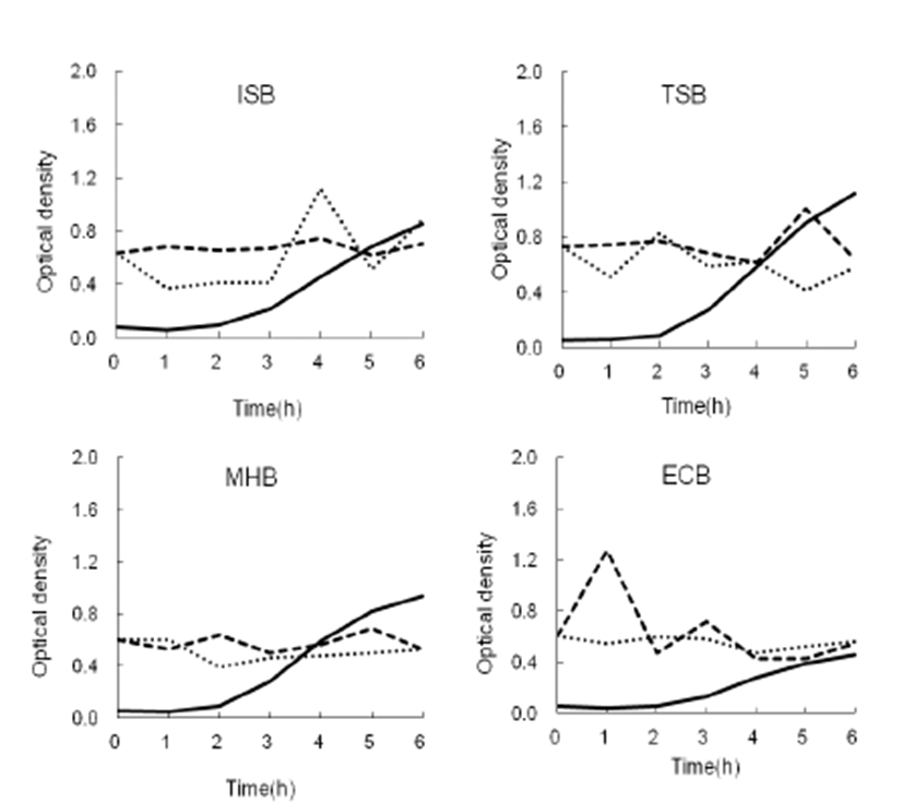
The growth and toxin production of KEFS1302 in various media with mitomycin C which is used for toxin induction were shown in Fig. 2. The growth peak was observed when the OD600 reached 1.1 in TSB, followed by that in MHB (0.9), MHB (0.8), and ECB (0.4) at 6 h. Stx2e was detected at OD490 in the supernatant of KEFS1302 between 1 and 3 h, reaching a production peak at 1 h, when the bacterial strain was grown in ECB. In ISB, Stx2e was observed in the supernatant between 4 and 6 h. For the polymyxin B-treated pellet, the toxin was detected between 3 and 5 h, reaching a peak at 4 h from cultures incubated in ISB (polymixin B was used for induction of the toxin located in periplasm of the bacterial cell where the toxin will be stored [14].).

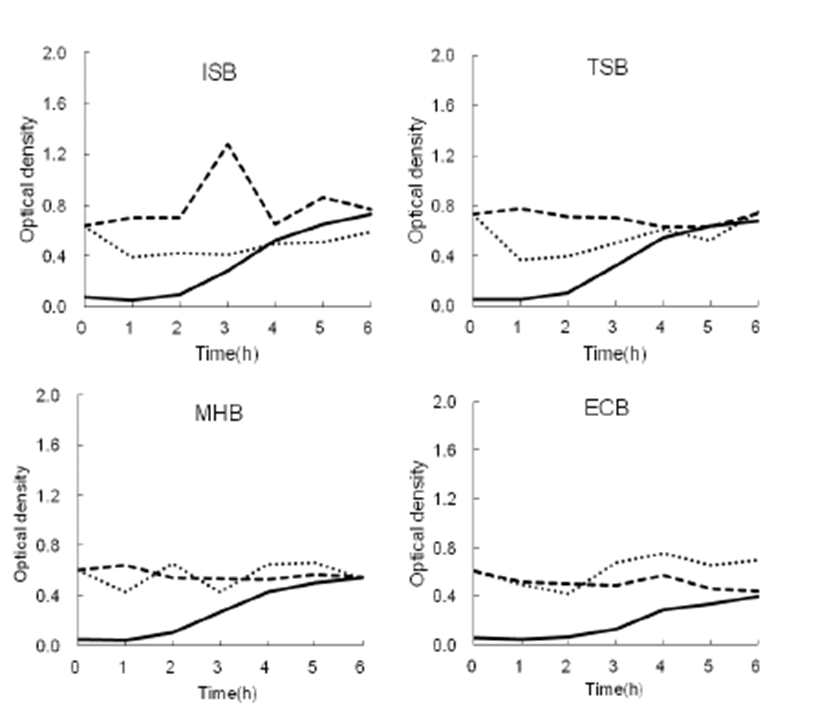
The growth and toxin production of KEFS1302 in various media without mitomycin C were shown in Fig. 3. The growth peaks at OD600 of ISB, TSB, and MHB cultures ranged from 0.4 to 0.7 after incubation at 4 to 6 h. However, ECB cultures reached 0.2 to 0.4 during the same periods of time. Stx2e was detected at OD490 in the supernatant of KEFS1302 between 2 and 5 h, reaching a production peak at 3 h when the bacterial strain was grown in ISB. There was no the production of toxin in the supernatant of TSB, ISO, or ECB. In polymyxin Btreated pellet, toxin was only detected after 3 h incubation in ECB.

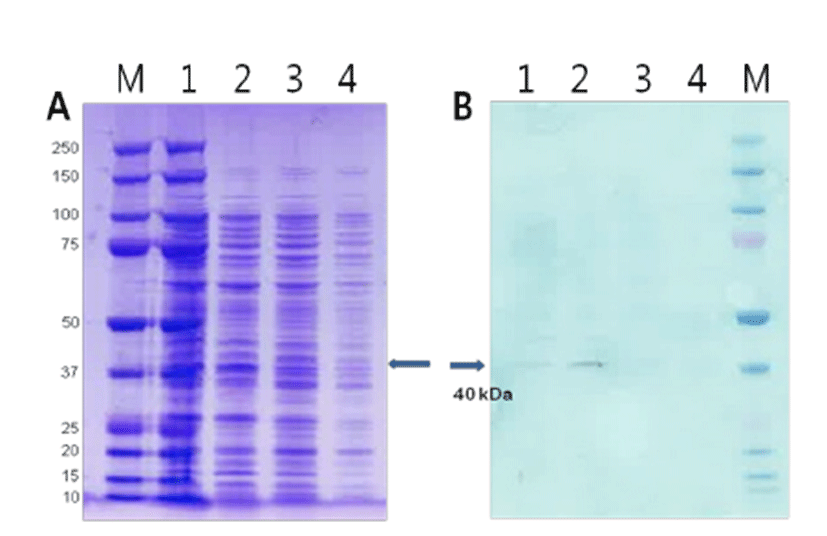
Stx2e was purified by fast protein liquid chromatography (FPLC) using anion-exchange chromatography. The eluted protein was electrophoresed in a 12% SDS-PAGE gel, and Stx2e was detected by western blotting using a polyclonal STX2 antibody (Fig. 4). A 43-kDa band was observed at in the fraction no. 2 after purification using FPLC. The Stx2e was detected to 151 μg/mL of ECB culture.

Discussion
Stx2e is the key virulence factor of STEC, causing ED, ataxia, and sudden death in weaned pigs. To diagnose ED, it is important that Stx2e be detected in the bacterial culture and affected pigs. However, it is difficult to detect Stx2e toxin in infected pigs because the lethal dose is as low as 3 ng/kg [18]. In regard to bacterial growth, the growth curves were found to be similar in three tested broths (ISB, TSB, and MHB), with the exception for ECB with an OD600 approximately 50% lower than the other conditions, regardless of the addition of mitomycin C.
Concerning Stx2e production, the toxin was secreted into the supernatant of ISB after 3 h of incubation without mitomycin C, but was also picked in the supernatant of ECB within 1 h containing mitomycin C These results are consistent with those of Racha and Piazza [17], although instead of mitomycin C, ciprofloxacin and novobiocin were used. In addition, Stx2e expression could be dependent on the media in which the STEC are grown [17, 19]. In general, the expression of Stx2e in ECB with mitomycin C was higher when compared with that in the other broths tested, but the time and level of Stx production were variable. Additionally, ECB contains bile salts, which are used for selecting the Enterobacteriaceae strains, like pathogenic bacteria including E. coli and Salmonella. It is thought that bacterial stress conditions such as bile salts, oxidative stress, and antibiotics induce the expression of virulence factors including Stx [20, 4, 14, 15, 16].
Stx2e was detected either in the supernatant or in the polymyxin-treated pellets; however, the amount of toxin was variable and even lower than the level in the supernatant of KEFS1302 grown in the presence of mitomycin C. These results indicate that Stx2e production is unlikely to be associated with bacterial growth. Stx2e expression in the supernatant of ECB was confirmed by anion-exchange chromatography and western blotting (Fig. 4). The 43-kDa band was observed in the western blot, which is consistent with other reports.
To our knowledge, this is the first report of the determination of Stx2e production in different media using an antibiotic to act as a stimulator of Stx2e production. Accordingly, the results described here could contribute to ED diagnosis and mass production of the toxin for use as a subunit vaccine candidate, since the production of Stx2e in STEC isolates could be determined in the laboratory.


