


Introduction
Speech and language are human-specific traits medi Reviewated by brain function. Speech is regarded as a physical vocalization in the form of voice, articulation, and fluency generated by cooperative movement of the tongue and regulation of breath. Disruptions in normal speech function result in speech disorders, including stuttering, verbal dyspraxia, spasmodic dysphonia, articulation disorder and aphasia [1-3]. In contrast, human language is defined as a non-physical system of signs governed by grammatical rule, and individuals with abnormal language functions exhibit language disorders such as specific language impairment (SLI) and dyslexia [4, 5]. Although speech and language disorders are distinct, they are not easily differentiated due to overlap of symptoms across different disorders, which has hindered researchers investigating the etiologies of these disorders. In addition, since highly-organized speech and language functions are unique to humans and deficits originate in the human brain, researchers face challenges in their efforts to reveal the molecular mechanisms underlying these disorders.
About 2~5% of children worldwide are reported to exhibit functional abnormalities in normal speech and language, despite having population average intelligence and sufficient opportunities for education [6]. However, genetic causes of speech and language disorders are not well understood. Speech and language disorders often cluster in families, suggesting that genetic factors are involved in occurrence of these disorders. This has encouraged geneticists to perform linkage studies, followed by sequencing of candidate genes. Several genetic causes of speech and language disorders were recently discovered [4, 7, 8]. The functional roles of causative genes that are discovered should be investigated in an animal disease model. Although speech and language are human-specific traits, several studies have revealed that mice can communicate with each other by emitting ultrasonic vocalization (USV), which has led to the generation of knock-in (KI), knock-out(KO), and humanized mouse models [9-11]. These mouse models are now used to elucidate the molecular network involved in speech and language function.
Here, I describe recent discoveries related to the genetic causes of speech and language disorders as well as altered vocalization patterns in mice resulting from genetic mutations associated with these disorders.
Genetic studies on verbal dyspraxia and FOXP2
A single large family, named KE, affected by severe developmental verbal dyspraxia and characterized by severe impairment of orofacial movements has enabled researchers to investigate the genetic causes of speech and language disorders (OMIM #602081) [12]. In this three-generation family, 15 of 37 members exhibited deficits in selecting words, coordinating sequences of words, and properly articulating, which are characteristics of verbal dyspraxia. However, the non-verbal intelligence quotients (IQs) of the affected members were close to the population average [13], which implies that the abnormal speech and language functions were not due to other systematic brain disorders such as mental retardation, autism, or stroke. In an effort to identify the genetic causes of verbal dyspraxia, Fisher et al. [14] hypothesized that this disorder was transmitted in the KE family in a dominant manner and thus performed genome-wide linkage analysis by genotyping microsatellite markers evenly distributed throughout the genome of 27 members of this family. Significant linkage (maximum logarithm of odds ratio (LOD) score=6.62 at θ=0.0) of verbal dyspraxia was detected for markers residing within the 5.6-cM locus (designated SPCH1) and flanked by D7S2459 and D7S643 on chromosome 7 [14]. However, identification of the causative mutation remains challenging since there are about 50 genes in this locus, and further efforts are required to reveal which of these candidate genes are the true genetic cause of verbal dyspraxia.
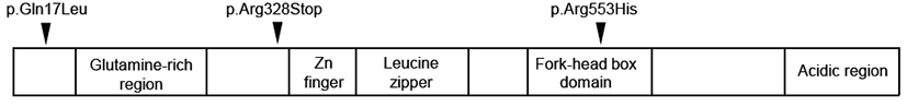
Independent of the genome-wide linkage analysis of the KE family described above, there was another molecular cytogenetic report of an unrelated individual, named CS, affected by a similar speech and language disorder and carrying a de novo reciprocal translocation t(5;7)(q22;q31.2) [15]. This cytogenetic study suggested that the chromosomal breakpoint resides within the gene responsible for verbal dyspraxia. Thus, combining the results from the genome-wide linkage analysis of the KE family and the cytogenetic study on the CS individual revealed that the translocation breakpoint at chromosome 7q31.2 was mapped to the intronic region between exon 3b and exon 4 of the forkhead box p2 (FOXP2) gene. FOXP2 encodes a transcription factor harboring a polyglutamine repeat track and forkhead DNA-binding domain. Further, a missense mutation substituting histidine for arginine (p.Arg553His) was detected in the forkhead domain of the protein product (Fig. 1) [4]. All affected individuals were found to carry one copy of this mutation; however, this mutation was not detected in any of the unaffected individuals in the KE family [4]Table 1.
| References | Study methods | Major findings | Genes or chromosomal loci |
|---|---|---|---|
| Verbal dyspraxia | |||
|
|
|||
| [15] | Fluorescence in situ hybridization assay | Located chromosomal break point t(5;7)(q22;q31.2) from an individual affected by verbal dyspraxia | chromosome 7q31 |
| [4, 14] | Genome-wide linkage analysis and sanger sequencing of KE family | Found significant linkage on chromosome 7q31, and found mis-sense mutation in FOXP2 | FOXP2 on chromosome 7q31 |
| [11, 27] | Generation and characterization of Foxp2 knock-out mouse | Found altered USV pattern in KO | – |
| [10] | Generation and characterization of humanized Foxp2hum/hum mouse | Found altered USV and behavioral patterns of in humanized Foxp2hum/hum mouse | – |
|
|
|||
| Specific language impairment | |||
|
|
|||
| [31] | Transcriptional targets of FOXP2 using chromatin immunoprecipitation | Found that FOXP2 binds and down-regulates CNTNAP2. This These genes was were associated with nonsense-word repetition | CNTNAP2 |
| [8] | Targeted association study of SLI1 region | Found significant association of the SNPs with phonological short-term memory | CMIP, ATP2C2 |
|
|
|||
| Stuttering | |||
|
|
|||
| [39] | Genome-wide linkage analysis | Found significant linkage on chromosome 12q | 12q22.2 |
| [7] | Sequencing of genes in linkage interval | Found genetic mutations in the genes involved in lysosomal enzyme-targeting pathway | GNPTAB, GNPTG, NAGPA |
Further sequencing of 49 individuals affected by verbal dyspraxia in the coding region of the FOXP2 gene revealed three other structural mutations: p.Gln17Lys, p.Arg328Stop, and small expansion of polyglutamine tract (Gln40 to Gln44). This denotes that the genetic load of dysfunctional FOXP2 in verbal dyspraxia is about 6% in the unrelated affected individuals with this disorder [16]. Thus, FOXP2 was suggested as the first known gene associated with human speech function in humans.
Functional effect of FOXP2 at the cellular level
The protein encoded by the FOXP2 gene is known as a transcription factor containing multiple domains and motifs, such as the N-terminal glutamine-rich region, zinc and leucine zipper motif, fork-head box domain, and C-terminal acidic region. Among them, the forkhead domain, also known as the FOX domain, is an 80~110 amino acid DNA-binding motif [17]. As a transcription factor, FOXP2 regulates gene expression in downstream pathways involved in developmental processes [18], thereby influencing neuronal circuits involved in sensory-motor processing and motor-skill learning in vertebrates [19, 20].
In an effort to explore the functional effects of the three mutations, p.Arg553His, p.Arg328Stop, and p.Gln17Leu, found in verbal dyspraxia patients, Vernes et al. investigated the intracellular localization of both wild-type and mutant FOXP2 using immunofluorescence. Data revealed that the majority of FOXP2 proteins carrying p.Arg553His or p.Arg328Stop mutation were localized to the cytoplasm rather than the nucleus, where wild-type transcription factors such as FOXP2 normally reside [21]. In addition, it was reported that these two mutations abolished the DNA-binding capacity of FOXP2, resulting in loss of function as a transcription factor as revealed by electromobility shift assay. However, FOXP2 carrying p.Gln17Leu mutation maintained normal intracellular localization and DNA binding, which implies that this mutation is a rare polymorphism without any effect on normal FOXP2 function [21].
Transcriptional targets of FOXP2 in the human brain
Since FOXP2 is involved in verbal dyspraxia, its transcriptional targets might also influence the normal development of human speech and language in the human brain. In a study performed by Spiteri et al., chromatin immunoprecipitation followed by microarray analysis was used to identify FOXP2 targets in the human basal ganglia (BG) and inferior frontal cortex (IFG), and 285 transcriptional target genes of FOXP2 were initially identified in the fetal human brain [22]. Further detailed analysis to identify core transcriptional targets revealed that 34 genes, including ANK1 and TAGLN, were statistically significant overlapping targets in the BG and IFG. Many of these target genes are involved in patterning of the central nervous system, neurite growth, or brain plasticity, which may provide insights into the functional network of genes regulated by FOXP2 in the human brain [22].
Animal model of speech and language disorders
Once genetic mutations responsible for verbal dyspraxia were identified, subsequent follow-up studies to generate an animal model harboring the mutations identified in human patients were performed to help reveal the molecular mechanism underlying this disorder. Generation and characterization of a genetically manipulated FOXP2 mouse were highly challenging, and researchers did not expect to recapitulate the phenotype in a mouse model since speech and language are human-specific traits. However, mice produce USV with frequencies ranging between 30-110 kHz, which is not audible to the human hearing system [9, 23]. Typically, mouse vocalization is emitted when pups are isolated from their mother, when adult males try to attract females, or when mice detect their urinary pheromones [24-26]. Based on previous ideas that mice communicate with each other by emitting USV, Shu et al. knocked out the FOXP2 gene in mice and characterized the resulting altered vocalization pattern and brain structure [27]. In their study, complete disruption of both copies of the mouse FOXP2 gene resulted in severe motor impairment, premature death, and absence of isolation calls when pups were removed from the nest [27]. Interestingly, premature death is also expected to occur in human subjects based on the finding that the KE family had no one carrying two copies of the mutation [4]. In contrast, heterozygous FOXP2 knock-out mice experienced moderate developmental delay and an altered vocalization pattern, including reduced frequency of whistle type vocalization. However, the frequencies of click-type vocalization and behavioral patterns, including response to shock and Morris water maze performance, were within normal ranges [27]. Another animal study on KI mice generated by introducing FOXP2 p.Arg552His, which corresponds to the human FOXP2 p.Arg553His mutation, showed that both the homozygous and heterozygous mouse with this mutation displayed altered USV patterns, reduced weight, and disrupted normal development of the cerebellum with incompletely folded folia in the brain [11].
Thus, animal studies with KO and KI mice demonstrated that disruptions in the normal function of FOXP2 result in abnormal USV and suggested that emission of USV and human speech may share a molecular network that is regulated by FOXP2 in the brain [11, 27]. It is still unclear which brain sub-regions are regulated by FOXP2, thereby enabling the mouse to generate USV. However, evidence that the FOXP2 (p.Arg552His) mutation hinders normal development of the cerebellum and maturation of dendrites of Purkinje cells suggests that the cerebellum, which is known to be involved in coordinating motor neurons, might be the brain region involved in speech and language function in mice. However, further studies are needed to support these findings.
Mouse model with humanized version of FOXP2
Human communication skills, mediated by speech and language, are the most complex and well organized among all species, and thus it is interesting to reveal specific genes or alleles that enable humans to have complex communication skills. From an evolutionary aspect, genes or alleles involved in speech and language functions, such as human FOXP2, may play roles in positive selection, thereby driving the human ability to speak. However, there are questions remaining as to why other mammals, including chimpanzees and mice, cannot speak even though they have orthologous versions of FOXP2. To answer this question, several studies were performed to compare the protein sequences of human FOXP2 and its orthologues in chimpanzees and mice, and it was found that there were two amino acids substitutions (p.Thr303Asn, p.Asn325Ser) in human FOXP2 [10, 28]. Thus, it was proposed that these two human-specific amino acid substitutions may affect the downstream protein network involved in speech function.
To investigate the functional roles of these two human-specific substitutions, Enard et al. generated a humanized KI mouse (FOXP2hum/hum) by introducing human alleles into the two copies of endogenous FOXP2 [10]. The phenotypes were characterized, in terms of behavior, USV, neurotransmitter production, and brain anatomy, and this humanized FOXP2hum/hum mouse was generally healthy. However, these mice displayed reduced exploratory behavior, reduced dopamine production, qualitatively altered USV patterns, and increased synaptic plasticity [10]. In addition, these mice had longer dendritic trees of striatal neurons. In contrast, mice carrying one copy of non-functional FOXP2 (FOXP2hum/wt) showed the opposite phenotype. Comparative study of humanized FOXP2hum/hum and FOXP2hum/wt mice demonstrated that alteration of the cortico-basal ganglia network might be crucial to the evolution of speech and language in humans [10].
SLI and CNTNAP2, CMIP, and ATP2C2 genes
SLI is characterized by the presence of delayed language development without any other deficits preventing proper language learning, such as hearing deficits, autism spectrum disorder, brain damage, and other neurological disorders [29]. Prevalence of SLI was reported to be 5~8% in preschoolers. Investigation of the concordance rate in both monozygotic (100%) and dizygotic (50%) twins suggested a genetic basis for this disorder [6]. Familial clustering of this disorder also supports the genetic basis hypothesis [30].
The first gene associated with SLI was discovered by studying downstream genes regulated by human FOXP2 protein [31]. In this study, Vernes et al. performed a chromatin immunoprecipitation assay and found that FOXP2 binds to the intronic region of CNTNAP2 encoding contactin-associated protein-like 2 (CASPR2). Subsequent genotyping of 38 SNPs in this gene from 184 families affected by SLI revealed a statistically significant association of these SNPs with incidence of repeating nonsense words [31]. However, the functional effects of these SNPs on the CNPNAP2 gene remain poorly understood, and further efforts to reveal the mechanisms of action of these variants in SLI are still necessary.
There was another genome-wide linkage analysis of large-scale independent samples consisting of 490 affected cases and 211 families, and significant linkage of the markers on chromosome 16q to SLI was detected [8]. Two genes, CMIP (c-maf-inducing protein) and ATP2C2 (calcium-transporting ATPase, type2C, member 2), were shown to be associated with repetition of non-words. Therefore, discovery of CMIP and ATP2C2 provided molecular evidence for the involvement of phonological short-term memory in SLI [8].
Genetic evidence of stuttering
Stuttering is the most common speech disorder and is characterized by the presence of uncontrollable prolongations and repetitions of words or syllables as well as interruptions in the smooth flow of speech, known as blocks. This disorder arises in young children, where it is common with a typical onset age ranging 3~4 years, thereby affecting 5% of the population at this age. Male- to-female ratio of this disorder at preschool age is 2:1 and changes to 4:1 at age 9, which implies that stuttering is more common in males than in females. Alteration of the sex ratio results from a different spontaneous recovery ratio, as females resolve more frequently than males. Overall prevalence of stuttering in the adult population is known to be 1% [32, 33]. Stuttering may occur in all populations and language groups, and its severity can be affected by anxiety, fatigue, and other factors.
Several associations of low birth weight with increased susceptibility to stuttering have been reported. However, it took tremendous efforts to reveal the genetic causes of developmental stuttering due to the puzzling clinical features of this disorder. For example, singing or speaking in unison mitigates symptoms, and this disorder presents only in humans and thereby hinders ex vivo study. Further, its etiology might originate in the human brain.
There have been several suggestions that genes are involved in occurrence of stuttering, as revealed by twin and family studies. Howie et al. observed that the concordance rate of stuttering in monozygotic twins (63%) was higher than that in dizygotic twins (9%), and half of all stutters had a family history, with risk to first degree relatives estimated to be 15% [34]. In addition, an adoption study showed that there was no evidence that stuttering is a learned behavior [35].
Genome-wide linkage studies of stuttering
Several genome-wide linkage analyses were performed to identify genetic causes of stuttering [36-42]. In studies on stuttering families with Caucasian ancestry, multiple chromosomal loci were suggested to harbor genetic mutations. However, linkage of microsatellite markers to stuttering was only nominally significant, and linkage loci were not replicated [40-42].
The most notable research on stuttering was performed by a group led by Dr. Drayna at National Institutes on Deafness and Other Communication Disorders (NIDCD), National Institutes of Health (NIH), USA [36-39]. In their studies, multiple families affected by persistent developmental stuttering were recruited from Pakistan with the expectation that the pedigree structure has greatly elevated incidence of a recessive disorder since 70% of all marriages in this country are between either 1st or 2nd cousins (inbred marriage). The first significant genome-wide linkage (LOD score=4.61) was identified between microsatellite markers on chromosome 12q and stuttering in the analysis of 44 Pakistani inbred families [39]. Kang et al. investigated 44 genes localized in this 10 Mb linkage region by combining Sanger sequencing and comparative genomic hybridization assay and discovered a causative mutation (p.Glu1200Lys) in GNPTAB encoding GlcNAc-1-phosphotransferase α/β [7]. Further sequencing of 123 unrelated Pakistani patients in the GNPTG and NAGPA genes, which are functionally related with GNPTAB, revealed six other mutations [7]. GNPTAB and GNPTG together encode an enzyme named GlcNAc-1-phosphotransferase, which catalyzes the first step in the synthesis of the mannose 6-phosphage (Man-6-P) tag on lysosomal enzymes destined to reside in lysosomes [43]. In contrast, GlcNAc-1-phosphodiester α-N-acetylglucosaminidase (encoded by NAGPA) mediates the second step of this lysosomal enzyme-targeting pathway [43]. Thus, disruptions in the normal trafficking of lysosomal enzymes from the Golgi complex to the lysosome may be relevant to the underlying etiology of stuttering, which is due to lysosomal storage disorders. In the sequencing analysis of worldwide samples of 1013 unrelated stutterers, 164 individuals carried 81 different mutations in either the GNPTAB, GNPTG, or NAGPA gene, which implies that 16% of the cases are attributed to genetic deficits in this lysosomal enzyme-targeting pathway. However, functional effects of those variants remain to be investigated.
Conclusion
Speech and language are human-specific and complex traits. Disruptions in normal speech and language functions result in diverse disorders such as verbal dyspraxia, SLI, stuttering, and dyslexia. Genetic linkage and association studies have suggested multiple genes associated with these disorders. Specifically, discovery of the FOXP2, GNPTAB, GNPTG, and NAGPA genes as well as their respective associations with verbal dyspraxia and stuttering have opened the gate to investigation of the molecular mechanisms underlying speech and language disorders. In addition, development of tools analyzing USV patterns and neural circuits of genetically manipulated mouse models has accelerated the discovery of the network associated with human speech and language function. However, identification of specific brain regions or neurons affected by these mutations remains to be carried out.