ARTICLE

Scanning electron microscopic observation of lingual papillae in a Bengal tiger (Panthera tigris tigris)

Munki Kim, Chong-Sup Kim, Gon-Sup Kim, Chung-Kil Won
Author Information & Copyright
Institute of Animal Medicine & Department of Veterinary Medicine, Gyeongsang National University, Jinju 660-701, Korea
Corresponding author: Chung-Kil Won, Institute of Animal Medicine & Department of Veterinary Medicine, Gyeongsang National University, Jinju 660-701, Korea, Tel: +82-55-772-2351, E-mail: wonck@gnu.ac.kr

© Research Institute of Veterinary Medicine Chungbuk National University. This is an Open Access article distributed under the terms of the Creative Commons Attribution Non-Commercial License (http://creativecommons.org/licenses/by-nc/3.0/) which permits unrestricted non-commercial use, distribution, and reproduction in any medium, provided the original work is properly cited.

Received: Aug 21, 2014; Revised: Sep 16, 2014; Accepted: Sep 22, 2014

Abstract

The morphology of the lingual papillae in a female Bengal tiger (Panthera tigris tigris) was examined by scanning electron microscopy (SEM). The tongue was 22.3 cm in length and 7.1 cm in width. Numerous filiform papillae were distributed over the entire dorsal surface of the tongue. SEM examination of the tongue revealed two types of mechanical papillae, i.e. filiform and conical papilla, and two types of gustatory papillae, i.e. fungiform and vallate papilla, on the dorsal surface of the tongue. Each filiform papilla consisted of one primary papilla and several secondary papillae. The filiform papillae on the anterior part of the tongue were divided into one primary and 6~14 secondary papillae. Unlike other mammalians, however, secondary papillae in the mid-part of the tongue showed pineal-like papillae. In the posterior part of the tongue, secondary papillae were rare or absent. Fungiform papillae were surrounded by filiform papillae and densely distributed on the lingual surface. There were two vallate papillae on the borderline between the lingual body and root of the tongue. A vallate papilla contained two secondary papillae inside the grooves. Conical papillae were located in the area of the vallate papillae and covered the posterior part of the tongue root. No foliate papillae were seen on both margins of the posterior part of the tongue. Our results indicate that the structure on the lingual papillae of the Bengal tiger is somewhat different from that of other mammals.

Keywords: Bengal tiger; lingual papillae; morphology; scanning electron microscopy; tongue

Introduction

Morphological studies on the distribution and structure on the lingual papillae of the tongue in mammals revealed a variability of morphological features related to the type of food i.e. herbivorous versus carnivorous animals, and the adaptation of animals to environmental conditions [1, 2]. Scanning electron microscopy (SEM) observations of the morphology on the tongue conducted so far have mainly focused on laboratory and domestic animals. In carnivores, observations of the tongue structure on domestic animals have been conducted on dogs [3-5] and cats [6-8]. Morphological investigations of the tongue surface on wild animals in carnivores have been conducted on the Asian black bear [9], adult bush dog [10], and silver fox [11].

The structures and distributions of the papillae on the dorsal lingual surface of the tongue are varied and highly specific for individual systematic groups as well as the animal species [2]. However, there is no morphological study on the distribution and structure of the lingual papillae in the Bengal tiger. The purpose of this study was to examine the location and structure of dorsal and ventral lingual surface of the Bengal tiger using SEM, in order to compare the results with those of previous studies on other mammals.

Materials and Methods

The tongue of one female Bengal tiger (Panthera tigris tigris; 12 years old; 143 kg of body weight) donated from Jinyangho zoo in Jinju was used in this study. The tongue was removed and fixed in 10% neutral formalin. For SEM study, small blocks containing papillae were cut with razor blade and the samples were washed in 0.1 M phosphate buffer (pH 7.4) and fixed in 2.5% glutaraldehyde in 0.1 M phosphate buffer for 24 hours at 4°C. The fixed tissues were washed three times at an interval of 10 minutes in 0.1 M phosphate buffer. Thereafter, the specimens were dehydrated through a graded series of ethanol, critical point dried, and sputter coated with gold palladium. The tissues were placed on metallic stubs and observed under SEM (XL 30S FEG, Philips, Netherlands) operated at an accelerating voltage of 10 kV. The animal experiment was performed according to a protocol set out in the guidelines of the Animal Experiments Ethics Committee at Gyeongsang National University (Approval No. GNU-LA-12).

Results

Macroscopically, the anteroposterior length of the tongue in the Bengal tiger was 22.3 cm and the maximum width of the middle of the tongue was 7.1 cm.

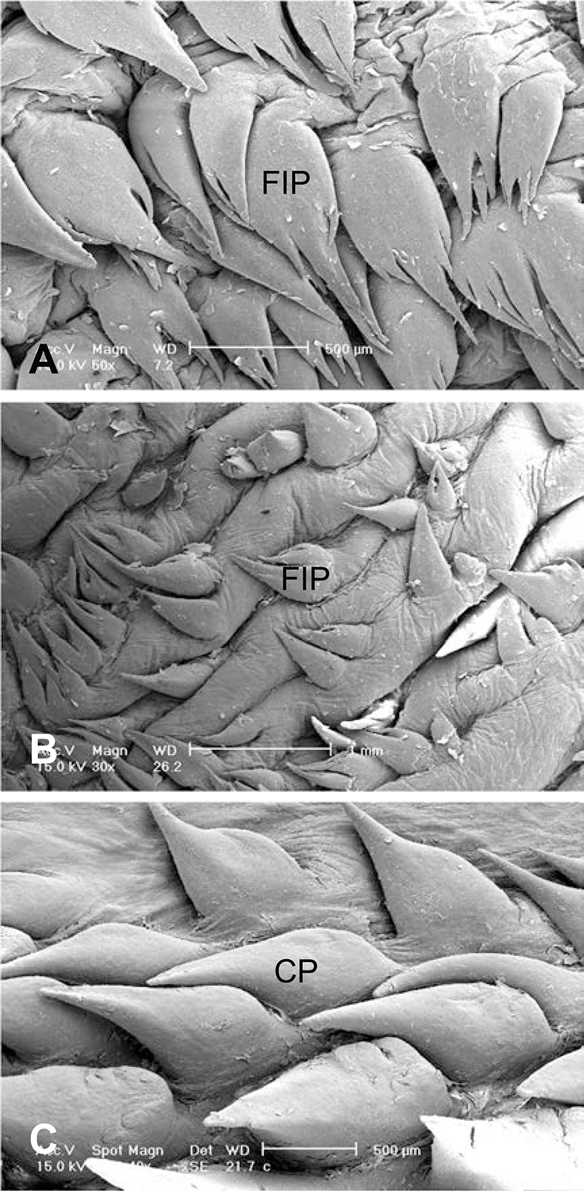
Under SEM, the tongue of the Bengal tiger showed numerous lingual papillae such as filiform, conical and fungiform and vallate papillae distributed on its dorsal and dorsolateral aspect. The apex and mid part of the dorsal lingual surface in Bengal tiger were covered by two types of lingual papillae, i.e. filiform and fungiform papillae. Filiform papillae were distributed over the entire dorsal surface of the lingual body and the processes of filiform papillae were tilted toward the root of the tongue. (Fig. 1). The filiform papillae on the anterior part of the tongue had a pen-like shape and they were consisted of 1 primary papillae and 6~14 smaller secondary papillae (Figs. 1B~D). Whereas, the papillae on the mid part of the dorsal lingual surface were also consisted of rod-like primary papillae and pineal-like smaller secondary papillae (Figs. 2A~D). In the posterior part of the dorsal surface of the tongue, secondary papillae were rare or absent (Figs. 2E and 2F). Two types of filiform papillae presented sharp or blunt free apical tips. The densely packed large numbers of sharp tip papillae were distributed throughout the tongue (Fig. 1), except in the middle portion raised area of tongue, which was mainly covered with blunt tip papillae (Fig. 2). The blunt tip papillae had a smooth surface with occasional shallow grooves.

jbtr-15-3-135-g1
Fig. 1. Scanning electron micrographs on anterior part of dorsal lingual surface. The filiform papillae on apical part of tongue have a pen-like shape and consist of one primary papillae and 6~14 secondary papillae. (A) The filiform papillae on the anterolateral part of the tongue. (B, C, D) Fungiform papillae are surrounded by filiform papillae. FIP: filiform papilla, FUP: fungiform papilla, PP: primary papilla, SP: secondary papilla.
Download Original Figure
jbtr-15-3-135-g2
Fig. 2. Scanning electron micrographs on mid part of dorsal lingual surface. (A-F) The filiform papillae on the mid-part of the lingual dorsal surface consist of rod-like primary papillae and pineal-like secondary papillae. (A, E, F) Fungiform papillae are scattered among the filiform papillae, round in shape, and more densely distributed on the lingual body. FIP: filiform papilla, FUP: fungiform papilla, PP: primary papilla, SP: secondary papilla.
Download Original Figure

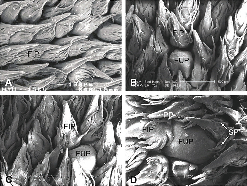
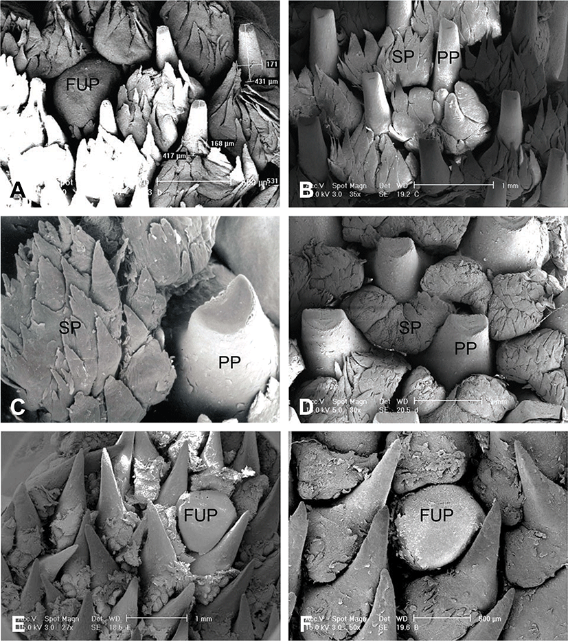
Fungiform papillae were scattered among the filiform papillae, round in shape (Fig. 1). The papillae were also found on the dorsolateral aspect of middle of the tongue (Fig. 2A). They were larger than those located on the apex.

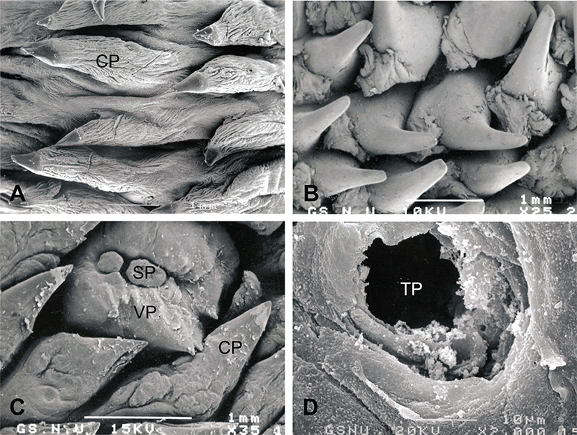
Conical papillae are seen in the root of the tongue (Figs. 3A and 3B). Two vallate papillae in total were located on both sides of the posterior end of the lingual body. The vallate papillae were composed of a primary papilla divided into two secondary papillae by inside grooves, and the papilla was surrounded by the conical papillae (Fig. 3C). The lingual papillae were also found on the ventral surface of the tongue (Fig. 4). Filiform papillae were scattered on the anterolateral and midiolateral part of the ventral lingual surface, respectively (Figs. 4A and 4B). Conical papillae were located on the posterolateral part of the ventral lingual surface (Fig. 4C). There were no foliate papillae on both margins of posterior part of the tongue.

jbtr-15-3-135-g3
Fig. 3. Scanning electron micrographs on posterior part of dorsal lingual surface. (A) Conical papillae with cap in the posterior part are seen on the root of the tongue. (B) Conical papillae in posterolateral part are seen. (C) Vallate papillae are surrounded by conical papillae. (D) Magnified taste pore on the vallate papilla. CP: conical papilla, SP: secondary papilla, TP: taste pore, VP: vallate papilla.
Download Original Figure
jbtr-15-3-135-g4
Fig. 4. Scanning electron micrographs on ventral part of tongue. (A, B) Filiform papillae are scattered on the anterior and mid-part of the ventrolateral surface of the tongue, respectively. (C) Conical papillae are located on the posterior part of the ventrolateral surface of the tongue. CP: conical papilla, FIP: filiform papilla.
Download Original Figure

Discussion

Regarding the three-dimensional morphological structure of the lingual papillae, there are many differences in mammalian species among those with a carnivorous, omnivorous or herbivorous diet [12-14]. Filiform papillae consist of primary papillae and smaller secondary papillae in the mammals. Secondary papillae show relatively restricted distribution, being present in the anterior and middle of the tongue, but rare or absent in the posterior region [15]. Filiform papillae in mammals have secondary papillae in dog, cat, Formosan serow, goat and giant panda [3, 6, 16-18]. In a cat, a marked transition occurred between the apical and mid part of the dorsal lingual surface [6]. We demonstrated that several secondary papillae were also located in the filiform papillae of the Bengal tiger. Especially, the filiform papillae on the middle of the tongue were blunt and thicker than those of the papillae in the anterior region, suggesting that filiform papillae have marked regional variation in size and structure which is contrast with herbivorous animals.

The fungiform papillae located on the lingual torus were larger than those located on the apex and body [16]. In the lesser mouse deer, the fungiform papillae were larger and more abundant than those located on the lingual body of the tongue [15]. In bovines, the fungiform papillae had a clear groove surrounding their base and separating them from the rest of the lingual surface [19]. In the present study, the fungiform papillae in the middle of the lingual surface were more densely distributed than those of the lingual apex. The papillae on the lingual apex were smaller and thinner than those of the mediolateral sides of the lingual body. Therefore, fungiform papillae may be similar to those found in other mammals, rounded and encircled by filiform papillae.

In most of mammalians, vallate papillae are observed between the body and root of both sides of the tongue [3, 7, 9-12, 20-22]. Equine vallate papillae are composed of a primary papilla divided into several secondary papillae by intermediate grooves [20]. In bovine vallate papillae, twin papillae are sometimes surrounded only by a primary papillary groove [20], whereas the vallate papillae of dogs and cats are only encircled by the conical papillae in the posterior region of the tongue [3, 7]. In this study, vallate papilla contained two secondary papillae located on the posterolateral region of the tongue. Distribution pattern of the papilla was observed to be similar to that of the other animals; however morphological structure was somewhat different. The number of the vallate papillae has been reported in many vertebrates. There were about 24 in total in the Formosan serows [16], 9~12 in the one-humped camel [22], 10 in the giant panther [18] and 7~8 in the Asian black bear [9], 5 in the bush dog [10]. In the present study, the Bengal tiger showed two vallate papillae in total, indicating that the number of vallate papillae tends to be smaller than that of other mammals. The results of this study show the SAM structure of lingual papillae on the tongue of Bengal tiger, at the same time indicating differences in their structure in comparison to the lingual papillae of the other mammals.

Acknowledgements

This Work (RPP-2011-012) was Supported by the fund of Research Promotion Program, Gyeongsang National University, 2011.

REFERENCES

1.

Doran GA. Review of the evolution and physiology of the mammalian tongue. Acta Anat. 1975; 91 p. 118-129.

2.

Iwasaki S. Evolution of the structure and function of the vertebrate tongue. J Anat. 2002; 20 p. 1-13.

3.

Iwasaki S, Sakata K. Scanning electron microscopy of the lingual dorsal surface of the beagle dog. Okajimas Folia Anat Jpn. 1985; 62 p. 1-13.

4.

Kobayashi K, Miyato K, Iino T. Three-dimensional structures of the connective tissue papillae of the tongue in newborn dogs. Arch Histol. 1987; 50 p. 347-357.

5.

Iwasaki S, Miyata K. Fine structure of the filiform papillar of beagle dogs. J Morphol. 1989; 201 p. 235-242.

6.

Boshell JL, Wilborn WH, Singh BB. Filiform papillae of cat tongue. Acta Anat (Basel). 1982; 114 p. 97-105.

7.

Chamorro CA, Sandoval J, Fernández JG, Fernández M, de Paz P. Comparative study of the lingual papillae of cats (Felis catus) and rabbits (Oryctolagus cuniculus) using the scanning electronic microscope. Anta Histol Embryol. 1987; 16 p. 37-47.

8.

Iwasaki S. Fine structure of the dorsal lingual epithelium of the domestic, newborn kitten, Felis catus. Ann Anat. 1992; 174 p. 293-300.

9.

Emura S, Hayakawa D, Chen H, Shoumura S. Morphology of the dorsal lingual papillae in the newborn panther and Asian black bear. Okajimas Folia Anat Jpn. 2001; 78 p. 173-177.

10.

Emura S, Tamada A, Hayakawa D, Chen H, Shoumura S. Morphology of the dorsal lingual papillae in the bush dog (Speothos venaticus). Okajimas Folia Anat Jpn. 2000; 77 p. 137-141.

11.

Jackowiak H, Godynicki S. The scanning electron microscopic study of lingual papillae in the silver fox (Vulpes vulpes fulva, Desmarest, 1820). Ann Anat. 2004; 186 p. 179-183.

12.

Iwasaki S, Miyata K, Kobayashi K. Scanning-electron-microscopic study of the dorsal lingual surface of the squirrel monkey. Acta Anat (Basal). 1988; 132 p. 225-229.

13.

Kobayashi K, Iwasaki S. Comparative studies on the stereo architecture of the connective tissue papillae in some mammalian tongues. Prog Clin Biol Res. 1989; 295 p. 303-308.

14.

Kobayashi K, Wanichanon C. Stereo architecture of the connective tissue cores of the lingual papillae in the treeshrew (Tupaia glis). Anat Embryol. 1992; 186 p. 511-518.

15.

Agungpriyono S, Yamada J, Kitamura N, Nisa C, Sigit K, Yamamoto Y. Morphology of the dorsal lingual papillae in the lesser mouse deer, Tragulus javanicus. J Anat. 1995; 187 p. 635-640.

16.

Atoji Y, Yamamoto Y, Suzuki Y. Morphology of the tongue of a male formosan serow (Capricornis crispus swinhoei). Anat Histol Embryol. 1998; 27 p. 17-19.

17.

Lee HS, Lee IS, Kang TC. Morphological study on the tongue of Korean native goat. Korean J Vet Res. 1996; 36 p. 225-264.

18.

Pastor JF, Barbosa M, De Paz FJ. Morphological study of the lingual papillae of the giant panda (Ailuropoda melanoleuca) by scanning electron microscopy. J Anat. 2008; 212 p. 99-105.

19.

de Paz Cabello P, Chamorro CA, Sandoval J, Fernandez M. Comparative scanning electron-microscopic study of the lingual papillae in two species of domestic mammals (Equus caballus and Bos taurus). II. Mechanical papillae. Acta Anat. 1988; 132 p. 120-123.

20.

Chamorro CA, de Paz P, Snadval J, Fernandez JG. Comparative scanning electron-microscopic study of the lingual papillae in two species of domestic mammals (Equus caballus and Bos taurus). I. Gustatory papillae. Acta Anat. 1986; 125 p. 83-87.

21.

Emura S, Tamada A, Hayakawa D, Chen H, Jamali M, Taguchi H, Shoumura S. SEM study on the dorsal lingual surface of the flying squirrel, Petaurista leucogenys. Ann Anat. 1999; 181 p. 495-498.

22.

Qayyum MA, Fatani JA, Mohajir AM. Scanning electron microscopic study of the lingual papillae of the one humped camel, Camelus dromedaries. J Anat. 1988; 160 p. 21-26.