ARTICLE
Association between carotid intima-media thickness and Ankle-Brachial Index in patients with ischemic stroke
Young Mi Yoon, Hyung-Suk Lee, Shin-Hye Baek, Hye Lim Lee, Minju Yeo, Seo Young Choi, Ji Seon Kim, Sung-Hyun Lee, Sang-Soo Lee, Dong-Ick Shin*
Author Information & Copyright ▼
Department of Neurology, Chungbuk National University Hospital, Chungbuk National University, College of Medicine, Cheongju 361-711, Korea
*Corresponding Author :Dong-Ick Shin, Department of Neurology, Chungbuk National University Hospital, Chungbuk National University, College of Medicine, 776 1Sunhwan-ro, Heungdeok-gu, Cheongju 361-711, Korea, Tel: +82-43-269-6372, Fax: +82-43-273-7591; E-mail:
sdi007@hanmail.net
© Research Institute of Veterinary Medicine, Chungbuk National University. All rights reserved. This is an Open-Access article distributed under the terms of the Creative Commons Attribution Non-Commercial License (http://creativecommons.org/licenses/by-nc/3.0/) which permits unrestricted non-commercial use, distribution, and reproduction in any medium, provided the original work is properly cited.
Received: May 15, 2014; Revised: Jun 15, 2014; Accepted: Jun 18, 2014
Abstract
Background and Purpose: The Ankle-Brachial Index (ABI) is the ratio of blood pressure in the lower legs to that in the arms. The intima-media thickness (IMT) of extracranial carotid arteries determined by B-mode ultrasound is a measurable index of the presence of atherosclerosis. A low ABI and a high carotid IMT are independently related to increased risk of cardiovascular events. This study examined the association between carotid IMT and ABI in patients with ischemic stroke. Materials and Methods: Retrospectively, 116 hospitalized patients with ischemic stroke were recruited. Using a pulse wave velocity ABI device along with carotid duplex sonography, we measured carotid IMT and ABI and investigated the correlation between average values. Results: There was a significant difference in carotid IMT between the normal and abnormal ABI groups (P=0.0262). The group with an abnormal ABI was more than five times as likely to have increased carotid IMT as the group with a normal ABI (age, sex-adjusted OR 5.67 (95% CI 1.85~17.38)). The ABI and carotid IMT showed a weak inverse linear correlation in patients with ischemic stroke (correlation coefficient –0.378 after adjusting for age and sex). Conclusion: Our study suggests that an abnormal ABI is associated with a high carotid IMT in patients with ischemic stroke.
Keywords: ischemic stroke; carotid; intima media thickness; ankle brachial index; atherosclerosis
Introduction
최근 한국 통계에 의하면, 매년 약 105,000명의 환자들이 뇌졸중을 경험하고 이중 26,000명 이상이 사망한다[1]. 뇌졸중의 대표적인 위험인자로는 고혈압, 당뇨병, 고지질혈증, 동맥 경화증, 흡연, 비만 등이 있으며, 이 중 동맥경화증은 뇌졸중의 주요 위험 인자로 알려져 있다.
발목상완지수(ankle-brachial index, ABI)는 동맥경화증을 조기에 선별할 수 있는 빠르고 쉬우며 비침습적인 방법이다[2]. 일반적으로 ABI가 0.9 이하인 경우 동맥경화증이 있는 것으로 판단하며, 이런 경우 말초동맥질환의 위험도가 증가하고 심혈관계 이환율과 사망률의 위험이 2.5배 증가하는 것으로 알려져 있다(95% 신뢰구간: 1.5~4.3) [3-5]. 44,590명을 분석한 한 연구에 의하면 낮은 ABI가 관상동맥질환, 뇌졸중을 포함한 심혈관 질환의 이환율 및 사망률 증가와 관련이 있으며, 이 중 뇌졸중의 상대위험도는 1.35로 나타났다[6]. 그리고 뇌출혈 군과 뇌경색 군을 비교한 연구에서는 뇌경색 군의 ABI가 뇌출혈 군에 비하여 유의하게 낮으면서 허혈성 뇌혈관 질환의 진행과 상관이 있었다[7].
ABI와 함께 내막-중막 두께(intima-media thickness, IMT) 역시 동맥경화증의 조기 지표로 사용되고 있다[8]. 증가된 IMT는 관상동맥 질환을 포함한 진행된 동맥경화증, 혈관 질환의 위험요소와 연관이 있다[9-11]. 6년 이상 추적 조사한 Atherosclerosis Risk in Communities (ARIC) 연구에서는 IMT가 1 mm 이상일 때 1 mm 미만인 경우에 비해서 뇌졸중이 남자에서 3.6배, 여자에서 5.5배 더 발생한다고 보고하였다[12]. 또 다른 연구에서는 경동맥 IMT가 0.1 mm 증가할 때마다 심근경색과 뇌졸중의 상대 위험도가 각각 1.15, 1.18배 증가한다고 보고하였고, 55세 이상의 연령인 7983명을 조사한 Rotterdam 연구에서는 IMT가 0.16 mm 증가할 때 뇌졸중이 1.4배 증가하였다{교차비: 1.42 (95% 신뢰구간: 1.25~1.82)} [13]. 이러한 결과들은 IMT의 증가가 뇌졸중과 의미 있는 상관 관계가 있음을 보여준다.
심혈관 질환 위험 인자를 가진 일반 인구 집단을 대상으로 조사한 연구에서는 ABI가 1.09 이하인 군의 경동맥 IMT는 ABI가 1.10 이상인 군의 경동맥 IMT에 비해 높게 측정되었으며, ABI와 경동맥 IMT가 역상관 관계를 보이는 것으로 나타났다[14]. 그리고 전 인구 집단을 대상으로 조사한 ARIC 연구 또한 ABI가 감소함에 따라 경동맥 IMT가 증가함을 보 여 ABI와 경동맥 IMT가 역상관 관계에 있을 것으로 추정된다[15].
지금까지 많은 연구들을 통해 낮은 ABI와 높은 IMT는 뇌졸중을 포함한 심혈관계 질환의 높은 위험도와 연관이 있다는 사실이 보고되었다[8, 16]. 그러나 실제 뇌졸중 환자에서의 ABI와 IMT의 관계를 분석한 연구는 보고된 바가 없다. 그러므로 본 연구에서는 허혈성 뇌졸중 환자에서 ABI와 경동맥 IMT를 측정하고 그 상관 관계를 알아보고자 한다.
Materials and Methods
연구대상
2007년 1월부터 2008년 10월까지 허혈성 뇌졸중(일과성 허혈 발작과 뇌허혈 포함)으로 본원 신경과에 입원한 환자 116명을 연구대상으로 하였다. 모든 환자들은 신경과 전문의에 의해 병력 등의 문진, 신경학적 검사가 시행되었으며, 자기공명영상(magnetic resonance imaging, MRI) 등의 영상검사를 통해 허혈성 뇌졸중으로 확진하였다. 연구 대상은 남자 72명(62.1%), 여자 44명(37.9%)으로 평균 나이는 65.31 ± 10.36세였다.
발목상완지수(ankle-brachial index, ABI)의 측정
ABI 측정을 위해 PWV/ABI device (VP-1000, Colin Medical Technology, Komaki, Japan)를 이용하였다. 누운 자세에서 5분 휴식 후에 양측 발등 동맥(dorsalis pedis artery)과 위팔 동맥(brachial artery)의 수축기 혈압을 도플러를 이용하여 측정하였다. ABI는 발등 동맥의 수축기 혈압을 위팔 동맥의 수축기 혈압으로 나눈 값이며 이 때 양측 ABI의 평균값을 통계에 이용하였다. ABI가 0.9 초과 시 정상 ABI로 정의하였다.
경동맥 내막-중막 두께(intima-media thickness, IMT)의 측정
경동맥의 IMT는 10-MHz 선형 탐촉자가 장치된 B-방식 초음파(T3000, Terason, USA)를 이용하여 측정하였다. 경동맥 내막-중막 두께는 처음의 반사면의 고음영선과 두 번째 반사면의 고음영선 사이의 거리를 측정하였다. 환자를 반듯이 눕히고 머리는 검사하는 쪽의 반대쪽으로 45도 정도 돌린 상태에서 탐촉자를 지면에서 45도 각도를 이루게 하여 lateral angle에서 세로 영상(longitudinal image)를 얻었다. 영상은 전벽과 후벽이 모두 관찰되는 지점에서 얻은 것을 원칙으로 하였으며 최대 IMT를 구하기 위하여 경동맥 분지 부위의 근위부에 위치한 총경동맥의 후벽에서 내막-중막 두께가 최고최고인 지점을 정하고 이를 중심으로 근위 10 mm, 원위 10 mm 위치에서 IMT를 측정하여 세 군데의 산술평균치를 구해 한쪽 경동맥의 IMT를 구하였으며 같은 조작을 반대편에서도 하여 양측의 IMT를 구한 뒤 산술평균의 각 환자의 좌우평균 IMT를 구하였으며 이를 분석에 사용하였다. 만약 경동맥 IMT의 측정이 예정된 부위가 석회화되었거나 내강 내로 돌출된 병변이면서 초음파상 이질성을 보이는 죽전(plaque)이 있는 경우에는 죽전이 포함되지 않은 근위부에서 측정을 시행하였다.
이전 대규모 연구를 통해 심근경색과 뇌졸중의 위험이 증가하는 IMT의 cut-point가 0.9로 나타나, 본 연구 역시 경동맥 IMT가 0.9 미만 시 정상 IMT로 정의하였다[17, 18].
통계학적 방법
모든 통계는 SPSS version 12.0 for Windows on a PC을 통해서 시행하였다. 모든 자료는 평균 ± 표준편차의 값으로 표현하였다. ABI의 정상유무에 따른 평균 경동맥 IMT는 t-test에 의해 분석하였으며, ABI의 정상유무에 따른 경동맥 IMT 정상유무간의 교차비(odds ratio)는 나이, 성 보정 logistic regression을 통해 분석하였다. ABI와 경동맥 IMT의 상관관계는 나이, 성 보정 partial correlation으로 통계 처리하였다. 통계적 유의수준은 P값이 0.05 이하인 경우로 하였다.
Results
평균 ABI는 1.11 ± 0.13, 평균 경동맥 IMT는 0.94 ± 0.46 mm로 측정되었다. ABI는 정상 ABI (>0.9)인 사람이 94%(109명), 비정상 ABI (≤0.9)인 사람이 6.0%(7명)이었으며, 경동맥 IMT는 정상 경동맥 IMT (<0.9)인 사람이 61.2%(71명), 비정상 경동맥 IMT (≥0.9)인 사람이 38.8%(45명)이었다(Table 1).
Table 1.
Relationship between carotid Intima-media thickness and ankle-brachial index
|
|
ankle-brachial index
|
sum |
| ≤ 0.9 |
>0.9 |
|
intima-media thickness (mm)
|
<0.9
|
1.7% |
59.5% |
61.2% |
| (N=2) |
(N=69) |
(N=71) |
|
≥0.9
|
4.3% |
34.5% |
38.8% |
| (N=5) |
(N=40) |
(N=45) |
| sum |
6.0% |
94% |
100% |
| (N=7) |
(N=109) |
(N=116) |
Download Excel Table
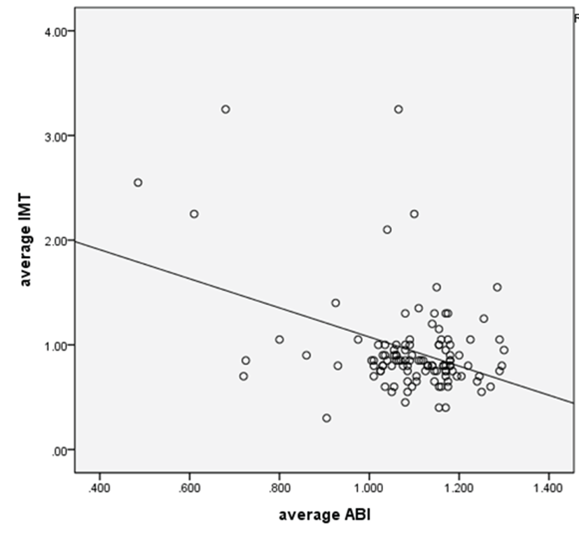
ABI가 비정상인 경우(≤0.9)에는 정상인 경우에 비해 평균 경동맥 IMT가 비정상적으로 두꺼웠다(P=0.0262). 또한 나이, 성별 보정 시 비정상 ABI (≤0.9)인 경우 정상 ABI일 때보다 경동맥 IMT가 5.67배 더 증가하였다{교차비: 5.67(95% 신뢰구간: 1.85~17.38)}. 나이, 성별에 따른 변화를 보정하고 ABI와 IMT간의 상관 관계를 연속적으로 분석한 결과, ABI와 경동맥 IMT는 상관계수 −0.378로 역상관 관계를 나타내었다(P=0.0001) (Fig. 1).
Discussion
이전 연구들을 통해 발목상완지수(Ankle-brachial index, ABI)와 내막-중막 두께(Intima-media thickness: IMT)는 잠재적인 동맥경화증의 지표로서 증가된 심혈관 질환의 위험도와 연관이 있다고 알려졌다[9]. 그리고 뇌경색 환자의 ABI값이 정상인에 비해 유의하게 낮았으며 심혈관계 질환자의 IMT가 정상인에 비해 유의하게 높았다고 보고하였다[6, 19].
본 연구에서 허혈성 뇌졸중 환자의 ABI와 경동맥 IMT를 조사한 결과 ABI가 정상인 경우가 비정상인 경우보다 현저하게 많았다(94%(109명) vs. 6.0%(7명)). 경동맥 IMT에서는 정상인 경우가 비정상인 경우보다 많았으나{71명(61.2%) vs. 45명(38.8%)} ABI에서의 비정상 비율보다는 상대적으로 높은 비정상 비율을 나타내었다.
일반 인구집단을 대상으로 한 연구에서 비정상 ABI의 비율은 연구에 따라 3.8~18.2%로 다양하게 보고되었고, 비정상 IMT는 28.2%였다[16, 20-23]. 본 연구에서는 허혈성 뇌졸중환자의 비정상 ABI는 6.0%로 일반 인구집단에서와 비슷하였으나 비정상 IMT는 38.8%로 일반 인구집단보다 더 많은 비율로 나타났다.
두 가지 검사 결과 모두가 동맥경화를 반영하는 지표로 사용이 되고 있지만 본 연구와 같이 이미 뇌혈관 질환이 발생한 경우에는 ABI에 비해서 IMT가 뇌혈관 질환을 조금 더 민감하게 반영하는 것 같다. 그러나 해석에 있어서 허혈성 뇌졸중 초기에는 혈압이 상승되는 경우가 있는데 이에 따라 ABI가 전반적으로 높아져 비정상군이 줄어드는 효과일 가능성을 주의해야 한다. 이를 확인하기 위해서 뇌졸중 후 혈압이 정상화되는 시기의 ABI에 대해 재조사할 필요가 있어 보인다.
ABI가 비정상일 때 평균 경동맥 IMT는 유의미하게 증가하였으며 나이, 성별 보정할 때 비정상 ABI (≤0.9)인 경우 경동맥 IMT가 5.67배 증가하였다. 또한 허혈성 뇌졸중 환자 군에서의 나이와 성별 보정 시 평균 ABI와 평균 경동맥 IMT는 역상관 관계를 나타냈다. 이는 전체 인구를 대상으로 시행한 기존 연구에서와 일치하는 결과이나 상관도(r=−0.378)가 약해 그 의미 해석에는 한계가 있다[16]. 이는 연구대상 군의 수가 적고, 나이, 성별 이외에 뇌졸중의 발생에 영향을 미칠 수 있는 다른 인자인 흡연, 고혈압, 당뇨병, 고지혈증 등 여러 요인을 고려하지 않은 결과로 생각된다.
본 연구에서는 뇌졸중 아형 분류를 시행하지 않았다. 기존 연구에서는 낮은 ABI가 허혈성 뇌졸중의 위험요인으로 알려져 있으나 ABI는 두개 내 내경동맥과 중뇌혈관의 협착 정도와 강한 관련성을 보이며 작은 혈관 질환에 의한 허혈성 뇌졸중보다 큰 뇌혈관동맥의 동맥경화증에 의한 허혈성 뇌졸중과 연관이 깊다고 보고된 바 있다[7, 24]. 그리고 경동맥 IMT 역시 작은 혈관 질환보다 큰 뇌혈관동맥의 허혈성 뇌졸중에서 더 증가하였다[25, 26]. 이러한 결과들은 ABI와 경동맥 IMT 모두 뇌졸중의 아형 중 큰 뇌혈관 질환과 관련 있음을 보여준다. 이에 추후 뇌졸중 아형에 따른 ABI와 IMT에 대한 보다 광범위한 환자를 대상으로 한 연구가 필요할 것이다.
Acknowledgements
본 연구는 2013년도 정부(교육과학기술부)의 재원으로 한국연구재단의 기초연구사업(NRF-2010-0021940)을 지원받아 수행되었으며, 이에 감사 드립니다.
REFERENCES
Hong KS, Bang OY, Kang DW, Yu KH, Bae HJ, Lee JS, Heo JH, Kwon SU, Oh CW, Lee BC, Kim JS, Yoon BW. Stroke statistics in korea: Part I. Epidemiology and risk factors: A report from the Korean stroke society and clinical research center for stroke. J Stroke. 2013; 15 p. 2-20.

Espinola-Klein C, Rupprecht HJ, Bickel C, Lackner K, Savvidis S, Messow CM, Munzel T, Blankenberg S. Different calculations of ankle-brachial index and their impact on cardiovascular risk prediction. Circulation. 2008; 118 p. 961-967.
Sander D. Stroke risk prediction beyond classical risk factors: The role of the ankle-brachial index. Cerebrovasc Dis. 2010; 29 p. 555-556.
Stoffers HE, Kester AD, Kaiser V, Rinkens PE, Kitslaar PJ, Knottnerus JA. The diagnostic value of the measurement of the ankle-brachial systolic pressure index in primary health care. J Clin Epidemiol. 1996; 49 p. 1401-1405.
Newman AB, Sutton-Tyrrell K, Vogt MT, Kuller LH. Morbidity and mortality in hypertensive adults with a low ankle/arm blood pressure index. JAMA. 1993; 270 p. 487-489.
Heald CL, Fowkes FG, Murray GD, Price JF. Risk of mortality and cardiovascular disease associated with the ankle-brachial index: Systematic review. Atherosclerosis. 2006; 189 p. 61-69.
Nakano T, Ohkuma H, Suzuki S. Measurement of ankle brachial index for assessment of atherosclerosis in patients with stroke. Cerebrovasc Dis. 2004; 17 p. 212-217.
Bots ML, Hofman A, Grobbee DE. Increased common carotid intima-media thickness. Adaptive response or a reflection of atherosclerosis? Findings from the rotterdam study. Stroke. 1997; 28 p. 2442-2447.
Bots ML, Hoes AW, Hofman A, Witteman JC, Grobbee DE. Cross-sectionally assessed carotid intima-media thickness relates to long-term risk of stroke, coronary heart disease and death as estimated by available risk functions. J Intern Med. 1999; 245 p. 269-276.
Simons PC, Algra A, Bots ML, Grobbee DE, van der Graaf Y. Common carotid intima-media thickness and arterial stiffness: Indicators of cardiovascular risk in high-risk patients The smart study (second manifestations of arterial disease). Circulation. 1999; 100 p. 951-957.
Baldassarre D, Amato M, Bondioli A, Sirtori CR, Tremoli E. Carotid artery intima-media thickness measured by ultrasonography in normal clinical practice correlates well with atherosclerosis risk factors. Stroke. 2000; 31 p. 2426-2430.
Chambless LE, Heiss G, Folsom AR, Rosamond W, Szklo M, Sharrett AR, Clegg LX. Association of coronary heart disease incidence with carotid arterial wall thickness and major risk factors: The atherosclerosis risk in communities (ARIC) study, 1987-1993. Am J Epidemiol. 1997; 146 p. 483-494.
Lorenz MW, Markus HS, Bots ML, Rosvall M, Sitzer M. Prediction of clinical cardiovascular events with carotid intima-media thickness: A systematic review and meta-analysis. Circulation. 2007; 115 p. 459-467.
Allison MA, Laughlin GA, Barrett-Connor E. Association between the ankle-brachial index and carotid intimal medial thickness in the rancho bernardo study. Am J Cardiol. 2006; 98 p. 1105-1109.
Zheng ZJ, Sharrett AR, Chambless LE, Rosamond WD, Nieto FJ, Sheps DS, Dobs A, Evans GW, Heiss G. Associations of ankle-brachial index with clinical coronary heart disease, stroke and preclinical carotid and popliteal atherosclerosis: The atherosclerosis risk in communities (ARIC) study. Atherosclerosis. 1997; 131 p. 115-125.
Price JF, Tzoulaki I, Lee AJ, Fowkes FG. Ankle brachial index and intima media thickness predict cardiovascular events similarly and increased prediction when combined. J Clin Epidemiol. 2007; 60 p. 1067-1075.
Bots ML, Hoes AW, Koudstaal PJ, Hofman A, Grobbee DE. Common carotid intima-media thickness and risk of stroke and myocardial infarction: The rotterdam study. Circulation. 1997; 96 p. 1432-1437.
van der Meer IM, Bots ML, Hofman A, del Sol AI, van der Kuip DA, Witteman JC. Predictive value of noninvasive measures of atherosclerosis for incident myocardial infarction: The rotterdam study. Circulation. 2004; 109 p. 1089-1094.
Du LG, Qiu J, Ruan YJ, Dong FY, Hong CJ, Ma J, Xu L. association between multi-noninvasive indexes and mild coronary stenosis. Zhonghua Xin Xue Guan Bing Za Zhi. 2010; 38 p. 31-34.
Kornitzer M, Dramaix M, Sobolski J, Degre S, De Backer G. Ankle/arm pressure index in asymptom-atic middle-aged males: An independent predictor of ten-year coronary heart disease mortality. Angiology. 1995; 46 p. 211-219.
Leng GC, Fowkes FG, Lee AJ, Dunbar J, Housley E, Ruckley CV. Use of ankle brachial pressure index to predict cardiovascular events and death: A cohort study. BMJ. 1996; 313 p. 1440-1444.
Newman AB, Siscovick DS, Manolio TA, Polak J, Fried LP, Borhani NO, Wolfson SK. Ankle-arm index as a marker of atherosclerosis in the cardiovascular health study. Cardiovascular heart study (chs) collaborative research group. Circulation. 1993; 88 p. 837-845.
Price JF, Stewart MC, Douglas AF, Murray GD, Fowkes GF. Frequency of a low ankle brachial index in the general population by age, sex and deprivation: Cross-sectional survey of 28,980 men and women. Eur J Cardiovasc Prev Rehabil. 2008; 15 p. 370-375.
Ratanakorn D, Keandoungchun J, Tegeler CH. Prevalence and association between risk factors, stroke subtypes, and abnormal ankle brachial index in acute ischemic stroke. J Stroke Cerebrovasc Dis. 2012; 21 p. 498-503.
Yoon HJ, Jeong MH, Kim KH, Ahn Y, Cho JG, Park JC, Kang JC, Bae JH. Carotid intima-media thickness, not carotid plaque, is associated with large territory cerebral infarction in patients with ischemic stroke. Korean Circ J. 2010; 40 p. 272-276.
Pruissen DM, Gerritsen SA, Prinsen TJ, Dijk JM, Kappelle LJ, Algra A. Carotid intima-media thickness is different in large- and small-vessel ischemic stroke: The smart study. Stroke. 2007; 38 p. 1371-1373.