


Introduction
Alterations in the normal differentiation patterns of gonads, genital duct and/or external genitalia often results in intersexuality. Intersex animals are usually considered hermaphrodites with genital organs showing features of both sexes. Intersex animals are also called pseudohermaphrodites depending on their gonads [1]. Pseudoherkrmaphroditism is a condition of ambiguous genitalia with a part or all of the genital organs of both sexes present [2]. Depending on testosterone levels in the developing fetus, the gonads become testes or ovaries. Problems associated with intersex animals are not unique but can be found in animals with normal chromosomal karyotypes [1, 3]. These developmental disorders are caused by abnormalities of genetic or chromosomal origin, or inappropriate hormonal or chemical exposure [2]. The most common breeds affected with pseudohermaphroditism are miniature schnauzers, poodles and Pekingese. Pseudohermaphrodites are divided into two categories, male and female. Male pseudohermaphrodites most often occur in miniature schnauzers. Male pseudohermaphrodites have XY chromosomes and testicles but the genitals appear feminine. In cases with a normal appearing penis and descended testicles, these dogs may not be diagnosed unless abdominal surgery is performed and the vestigial female organs are found. Female pseudohermaphrodites have XX chromosomes and ovaries but the internal and external genitals appear masculine due to excess amounts of testosterone. Usually, female pseudohermaphrodites occur when testosterone-type medications or progesterone are given to the mother during pregnancy.
This report describes a case of male pseudohermaphrodite in a Maltese/poodle mixed dog diagnosed using physical, radiological, ultrasonic, surgical, and anatomopathological examinations.
Case report
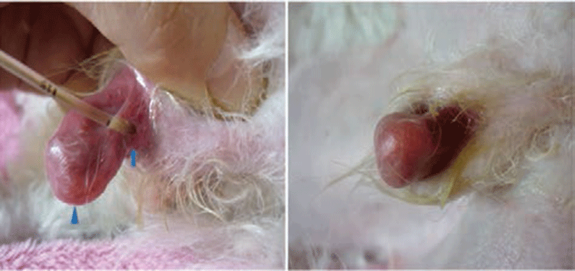
A 3-year-old Maltese/poodle mixed dog presented with malformed external genitalia and episodic hematuria. Inspection of the external genitalia showed them to be exclusively testis-like in character. The dog had a malformed penis and cryptorchidism. There was no urethral meatus at the tip of the penis. The urethral opening was situated between the prepuce and the penis (Fig. 1A). The anterior half of the prepuce was absent, and the penis was free and exposed to trauma and licking (Fig. 1B).
A peripheral blood sample drawn into an EDTA bottle was analyzed with a complete blood count machine. Complete cell count and blood chemistry profiles were within the normal reference ranges. The pH of urine was 6.0 and the specific gravity was 1.031. Erythrocytes and leukocytes were found in urine samples.
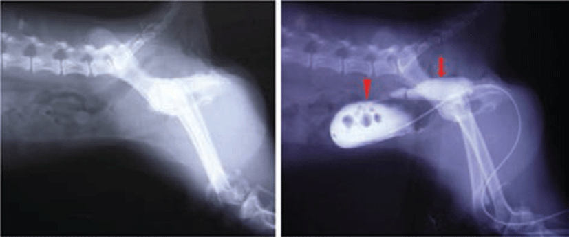
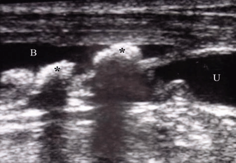
Plain radiographic examination showed absence of an os penis in the penis (Fig. 2A). The dog was administrated air and iodixanol (Visipaque®, GE healthcare, USA) into the urethra using a urethral catheter. A double-contrast cystograph showed the suspected uterus and the cystic calculi (Fig. 2B). A hypoechoic space was seen at dorsal portion of the urinary bladder. The space was suspected to be the uterus. A sagital ultrasonograph showed cystic calculi in the urinary bladder (Fig. 3).
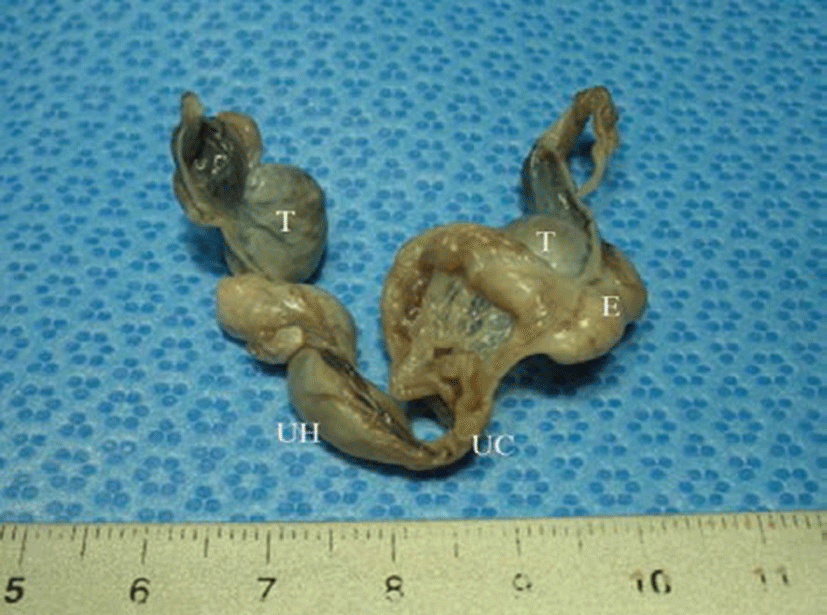
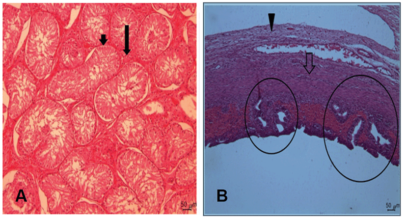
During surgery to remove cystic calculi, the internal genitals were found to include two uterine horns and two gonads (Fig. 4). The surgically removed gonads and uterus were fixed in 10% neutral buffered formalin and embedded in paraffin. Sections were cut at 3 μm and stained with hematoxylin and eosin for histological examination. Histological sections of the gonads showed structures similar to those of immature testis (only Sertoli cells were present in the epithelium of the seminiferous tubules). Germ cells were totally absent. The glandular interstitial tissue was reduced. The uterus has striated squamous epithelial cells and the smooth muscle cells. There were the glands formed by the columnar cells (Fig. 5).
Discussion
Hermaphrodite means animals and humans in which male and female sex organs are present simultaneously, or in which the sex organs contain both ovarian and testicular tissue [4]. Hermaphrodites are diagnosed when chromosomal abnormalities occur and the normal XX or XY does not occur. Hermaphroditism is very rare in dogs and humans. Normal genital development does not occur in the hermaphrodite. Pseudohermaphrodites have matching chromosomes and gonads but mismatching external genitals. In true hermaphroditism, both testicular and ovarian tissues are present in various combinations. A testis may be found on one side in combination with an ovary on the contralateral side, an ovotestis only may be present, or an ovotestis may be paired with a testis or ovary. Of all intersex cases in dogs, 25 percent are true hermaphrodites. These dogs may appear to have a large clitoris but otherwise normal female genitals. Others may have what appears to be a small but otherwise normal penis. Often, the testicles or ovotestis remain within the abdomen and do not descend into the scrotal sacs. Sometimes, the dog is never diagnosed as a hermaphrodite and lives life with few problems. Typically, female dogs do not exhibit heat cycles and do not reproduce. In addition to true hermaphrodites, male and female pseudohermaphrodites and unclassified cases can be diagnosed.
Pseudohermaphrodites are cases when the chromosomes and the gonads match but the external genitals do not. For example, a dog has an XX chromosome and ovaries but also has a penis. Male pseudohermaphrodites have XY chromosomes and testicles but the genitals appear feminine. Male pseudohermaphroditism is a sex differentiation disorder in which the gonads are testes and the genital ducts are incompletely masculinized. In the present case the dog possessed vestigial oviducts and uterus with male appearing external genitals. This dog has a normal appearing penis, but the testicles remain in the abdomen. This dog is not diagnosed unless abdominal surgery is performed. These were in full agreement with our case. The simultaneous presence of testicular tissue, vulva, male karyotype were compatible with a male pseudohermaphrodite condition [5].
Abnormalities in sexual development can arise from a defect at any step in sexual development, the chromosomal, gonadal or phenotypic level of differentiation [6]. Abnormalities of gonadal sex occur in an individual if the chromosomal and gonadal sex does not agree [7]. The pig is one species in which hermaphroditism has been well described. In pigs, this condition is common, affecting 0.1~0.5% of XX females [8]. Testis formation is followed by the development of Sertoli cells, Leydig cells and male germ cells. The absence of germ cells results in complete sterility in the intersexual animal. Sertoli cells secrete the anti-Mullerian hormone (AMH) responsible for the regression of Mullerian ducts and perhaps for the inhibition of follicular development [9]. The presence of developed derivatives of the Miillerian ducts in female pseudohermaphrodites suggests the absence of production of the AMH. In contrast, the presence of testicular tissue in male pseudohermaphrodites is not necessarily associated with an inhibition of Mullerian ducts by AMH. Female pseudohermaphrodites have ovaries but phenotypically they have a masculine appearance [1, 10]. These animals are often sterile, but should not be bred even if fertile. Ovariohysterectomy is recommended to eliminate the risk of uterine or ovarian disease. A female pseudohermaphrodite has ovaries but male external genitalia, and for a male pseudohermaphrodite the reverse is true.
This present case was classified as male pseudohermaphroditism because of the presence of testis and ambiguity of internal genitalia.