
INTRODUCTION
The gut microbiome is considered an endocrine organ comprising bacteria, fungi, viruses, and protozoa that have a symbiotic relationship with the host [1]. Gut microbiota plays a vital role in host functions such as digestion, hormone regulation, metabolism, detoxification, and immune cell maturation, while also protecting the female genital tract from pathogens [2, 3]. The health of the female reproductive system is influenced directly and/or indirectly by the gut microbiome, which regulates immune function, metabolic activities, stress levels, hormones, inflammation, and emotional states [4, 5].
Dysbiosis, defined as an imbalance in the composition and diversity of gut microbiota, is typically associated with many diseases including cancers, inflammation, metabolic disorders, immune system dysfunction, cardiovascular diseases, and neurological issues [6, 7]. Additionally, dysbiosis contributes to female reproductive disorders such as endometritis, endometrial hyperplasia, polycystic ovarian syndrome (PCOS), insulin resistance, sub/infertility, and obesity [8, 9]. Several factors, such as poor nutrition, physical and psychological stress, radiation, genetics, and drugs, can lead to dysbiosis [1]. Furthermore, overdose or frequent administration of antibiotics can lead to dysbiosis, which is negatively associated with fertility, hormone regulation, the oestrus cycle, and the quality of oocytes in the female reproductive structure and functions in mammals [10]. A research study described dysbiosis induced by amoxicillin that disrupts the oestrus cycle and reduces cell counts across all phases by altering gut homeostasis in antibiotic-treated female mice compared with controls [10].
The mammalian (or mechanistic) target of rapamycin (mTOR) is a central regulatory pathway in mammals. It is a conserved serine/threonine kinase protein (290 kDa) that creates two structurally and functionally different complexes: mTOR complexes 1 (mTORC1) and mTOR complexes 2 (mTORC2) [11, 12]. mTOR can sense various environmental conditions, such as nutrient and energy availability. It controls key cellular functions, including cell growth, differentiation, protein synthesis, cytoskeletal organization, and autophagy [13, 14]. Research highlights its importance in regulating folliculogenesis, oocyte maturation, ovarian somatic cell maturation, steroidogenesis, puberty, and embryonic development [15, 16]. Overactivation of the mTORC1 stimulates the activation of primordial follicle granulosa cells, which in turn influence the early maturation of dormant oocytes through the activation of the phosphatase and tensin homolog/phosphoinositide 3-kinase/protein kinase B/forkhead box O3 (PTEN/PI3K/AKT/FOXO3) pathway. This dysregulation can negatively impact female reproductive health [17].
This study aimed to investigate the relationship between antibiotic-induced intestinal microbiome imbalance and mTOR pathway regulation in female reproductive dysfunction. By analyzing changes in serum luteinizing hormone (LH), follicle-stimulating hormone (FSH), and glucose levels, as well as mTOR pathway gene expression in the ovaries and uterus, this study sought to elucidate how microbiome-derived metabolic and inflammatory signals might affect female reproductive health in animals.
MATERIALS AND METHODS
Ten- to eleven–week–old C57BL/6 female mice were obtained from Samtaco BIO (Osan, Korea) and allowed to acclimatize for 1 week before use in the experiment. Mice had access to ad libitum deionized water and a standard diet (Samtaco BIO) under laboratory conditions (temperature: 2 0°C –25°C, relative humidity: 50% (± 5), 12:12 h light-dark cycle). To induce dysbiosis, antibiotics were administered orally to the mice via their drinking water [18]. The antibiotic mixture was prepared by reconstituting ampicillin (1 g/L), neomycin sulfate (1 g/L), vancomycin (1 g/L), and metronidazole (0.5 g/L) in deionized water [18, 19]. Previous studies have reported that administration of this antibiotic cocktail promotes gut dysbiosis in mice through broad alterations in the composition of Gram-positive and Gram-negative, as well as anaerobic and aerobic, bacterial communities [19, 20].
After the acclimatization period, mice were administered this antibiotic solution for 4 weeks to induce dysbiosis (WD), while the control group (W/O) received water without antibiotics for the same duration. Five mice were housed per cage (n = 20 per group), with the bottle of antibiotics solution (WD) or water (W/O) provided in each cage. Tissues were collected at different stages of the estrous cycle. This approach is consistent with previous studies investigating gut microbiota-reproductive interactions in female mice, which similarly did not report cycle staging [21, 22]. Body weight and water consumption of the mice were recorded weekly.
All animal experiments were conducted in accordance with the Institutional Animal Care and Use Committee (IACUC) guidelines and approved by the IACUC of Sunchon National University (SCNU IACUC-2024-06). Unless specified, chemicals were acquired from Thermo Fisher Scientific (Waltham, MA, USA) and Sigma-Aldrich (St. Louis, MO, USA).
Mice were euthanized after 4 weeks, using CO2 inhalation, followed by the prompt isolation of blood and tissues [23]. Following collection, blood samples were centrifuged at 1,800 × g for 10 min at room temperature (1730R, Labogene, Seoul, Korea). The separated serum was harvested and kept at –20°C for further experiments. The ovary, oviduct, uterus, intestine, liver, kidney, and spleen were weighed on an electronic balance and reported as absolute weights. Ovary and uterus samples were preserved in RNAiso (Takara Bio, Shiga, Japan) and stored at –80°C.
Serum glucose, total cholesterol, and total ammonia levels were determined using commercial kits (AM201K, AM202K & AM505K; Asan Pharmaceutical, Seoul, Korea), respectively, as three separate assays performed according to the manufacturer’s instructions. For each assay, respective enzyme solutions were added into tubes containing serum sample, standard solution and blank and the reactions were incubated as specified in the kit protocol. Subsequently, a fixed aliquot of each reaction mixture was added into a 96-well microplate for absorbance measurement. Absorbance at 500 nm was measured for glucose and cholesterol, and total ammonia was measured at 630 nm with a microplate reader (Absorbance 96, Byonoy, Hamburg, Germany). The manufacture confirmed the detection range, sensitivity, and reproducibility of each assay.
The levels of LH and FSH in serum samples were determined using mouse enzyme-linked immunosorbent assay (ELISA) kits, according to the manufacturer’s guidelines (MBS2514287 & MBS2700327; MyBioSource, San Diego, CA, USA). Serum samples and blanks were loaded into microplate wells for each hormone and incubated with specific detection reagents and enzyme conjugates. A substrate solution was then added to generate a colour reaction, which was subsequently hated by the addition of stop solution. Hormone concentrations were quantified by optical density readings using a microplate reader (Byonoy) at 450 nm.
RNA was isolated from frozen ovary and uterus samples using RNAiso Plus reagent (Takara Bio) according to the manufacturer’s guidelines. Ovary and uterus samples were homogenized using a bead beater. After homogenization of the tissue samples, the homogenate was extracted with chloroform, and RNA was precipitated with isopropanol. RNA yield was quantified using a nanodrop spectrophotometer (DS-11FX, DeNovix, Wilmington, DE, USA). Complementary DNA was produced from RNA using TOYOBO ReverTra Ace qPCR RT Master Mix with the manufacturer’s protocol (TOYOBO, Osaka, Japan). Subsequently, quantitative real-time PCR (qPCR) was performed using SYBR™ Premix Ex Taq™ II (Bioneer, Daejeon, Korea) and analysis was carried out on a MyGo Pro cycler (Diagnostic Technology, Belrose, Australia). Relative mRNA expression levels of target genes were normalized to the endogenous control glyceraldehyde-3-phosphate dehydrogenase (GAPDH). Specific primers targeting mTOR-related genes were used to evaluate the relative mRNA expression levels in ovary and uterus samples (Table 1).
| Gene name | Primer sequence (5’–3’) | References |
|---|---|---|
| p53 |
F: GGGACAGCCAAGTCTGTTATG R: GGAGTCTTCCAGTGTGATGAT |
[46] |
| PI3K |
F: AGGAGCGGTACAGCAAAGAA R: GCCGAACACCTTTTTGAGTC |
[47] |
| Akt1 |
F: TGAAAACCTTCTGTGGGACC R: TGGTCCTGGTTGTAGAAGGG |
[47] |
| Raptor |
F: TTTGTCTACGACTGTTCCAATGC R: GCTACCTCTAGTTCCTGCTCC |
[48] |
| Rictor |
F: ACAGTTGGAAAAGTGGCACAA R: GCGACGAACGTAGTTATCACCA |
[49] |
| Mtor |
F: AGGCCGCATTGTCTCTATCAA R: GCAGTAAATGCAGGTAGTCATCCA |
[50] |
| Rheb |
F: TTGATTCCTACGATCCAACCATAG R: TGCTGTGTCTACAAGCTGAAGATG |
[51] |
| p70s6k |
F: GGTGGAGTCTGGGAGCATTA R: GCTTGGACTTCTCCAGCATC |
[51] |
| GAPDH |
F: ACTCCACTCACGGCAAATTC R: TCTCCATGGTGGTGAAGACA |
[47] |
p53, tumor protein p53; PI3K, phosphoinositide 3-kinase; Akt1, serine/threonine protein kinase 1; Raptor, regulatory-associated protein of mTOR; Rictor, rapamycin-insensitive companion of mTOR; Mtor, mechanistic target of rapamycin kinase; Rheb, Ras homolog enriched in brain; p70s6k, ribosomal protein S6 kinase B1; GAPDH, glyceraldehyde-3-phosphate dehydrogenase.
Data were expressed as mean ± S.E.M., and analyzed using analysis of variance (ANOVA) in GraphPad PRISM® (GraphPad Software, San Diego, CA, USA). A completely randomized design was used. When significant differences were detected by ANOVA, Tukey’s multiple-comparison test was performed to compare the values of individual groups. Statistical significance was considered at p-values: *p<0.05, and **p<0.01.
RESULTS
Body weight and water consumption were monitored weekly throughout the 4-week treatment period (Fig. 1). No significant differences in body weight were observed between the W/O and WD groups at any time point (Fig. 1A). Similarly, weekly water consumption, recorded at the cage level and averaged per mouse, did not differ significantly between groups (Fig. 1B). These findings indicate that antibiotic supplementation did not affect food or fluid palatability, and that the subsequent differences in serum hormones, metabolic parameters, and gene expression were unlikely to reflect differences in nutritional or fluid intake.
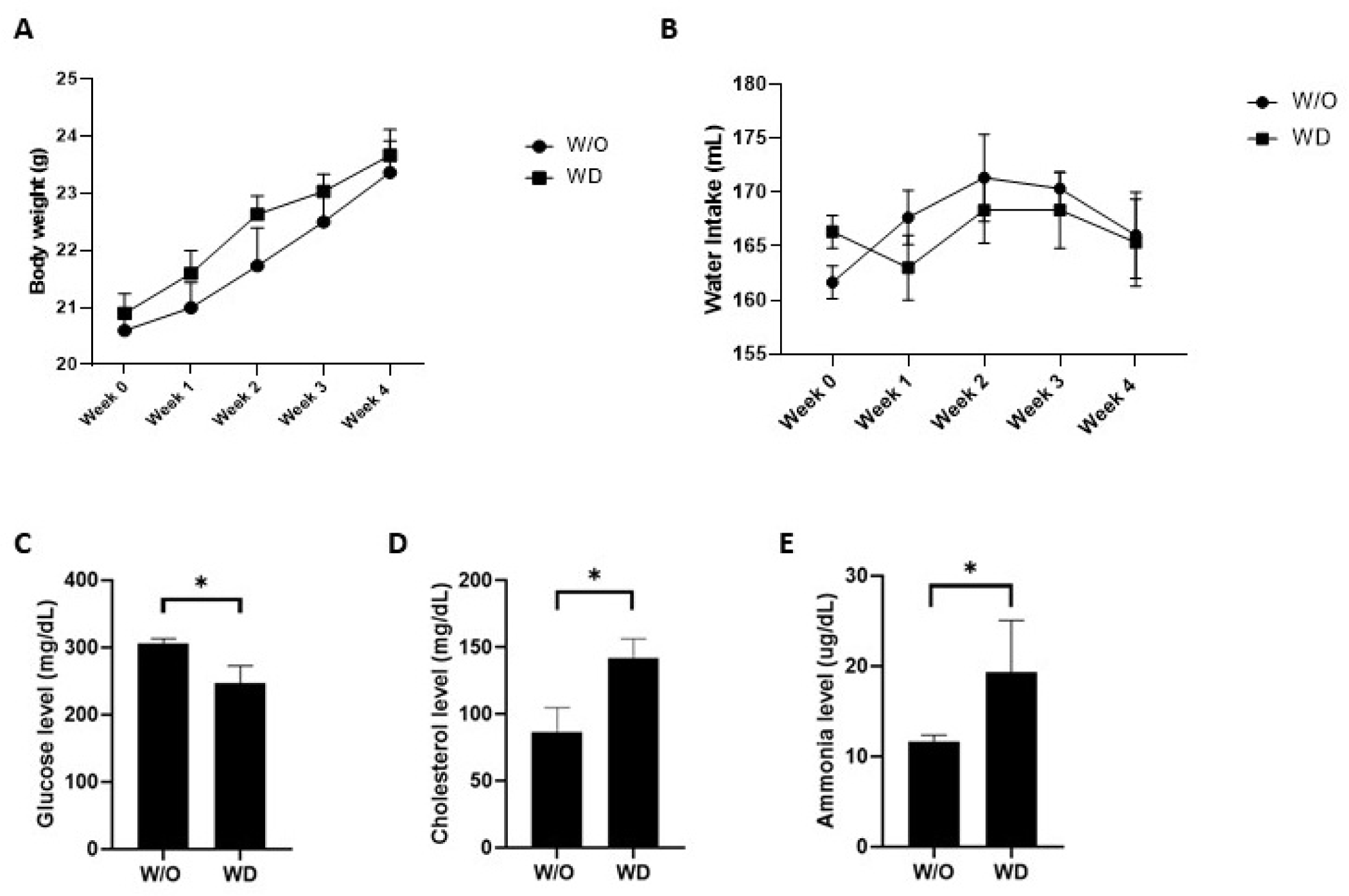
The glucose levels were markedly lower in the dysbiosis-induced mice (WD group) compared to the mice in the control (W/O group; *p<0.05; Fig. 1C). In contrast, the mice in the WD group had statistically higher cholesterol levels than the W/O group (*p<0.05; Fig. 1D). Similarly, the mice in the WD group showed significantly elevated ammonia levels as compared with the mice in the W/O group (Fig. 1E). These significant changes suggest that gut microbiome imbalance may contribute to systemic metabolic alterations.
Serum FSH levels exhibited a significant difference between the experimental and W/O group, with the levels being lower in the WD group (*p<0.05; Fig. 2A). Similarly, serum LH levels were significantly decreased in the WD group compared to the W/O group (*p<0.05; Fig. 2B).
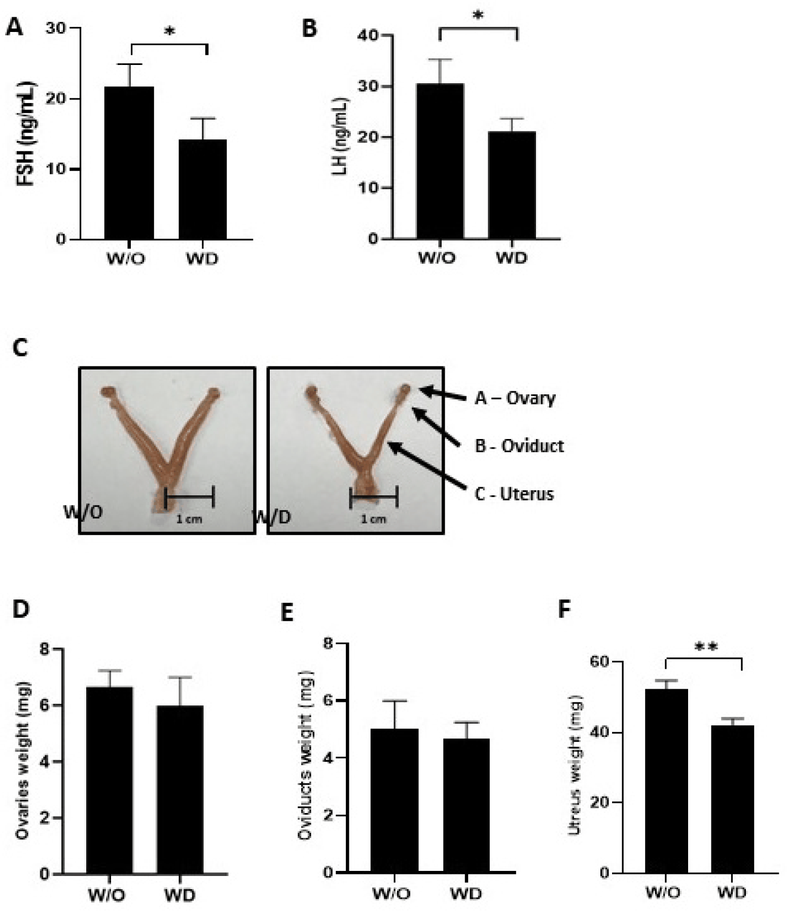
The morphology and weight of the reproductive organs were compared between the W/O and WD groups. The ovary, oviduct and uterus appeared smaller in the WD group. Ovary and oviduct weights were numerically lower in the WD group; however, only uterine weight was significantly reduced in WD group compared to the W/O group (**p<0.01; Fig. 2C, D, E, and F).
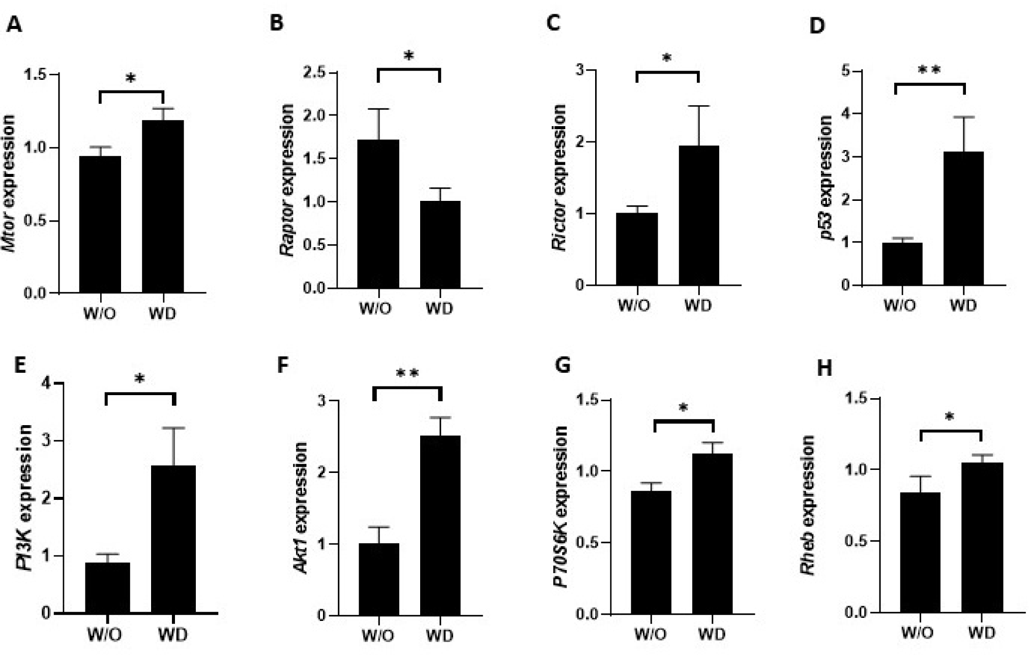
The mRNA expression levels of the mTOR-related genes, mechanistic target of rapamycin kinase (Mtor), regulatory-associated protein of mTOR [Raptor], rapamycin-insensitive companion of mTOR [Rictor], tumor protein p53 [p53], phosphoinositide 3-kinase [PI3K], serine/threonine protein kinase 1 [Akt1], ribosomal protein S6 kinase B1 [p70s6k], and Ras homolog enriched in brain [Rheb] were measured in ovary samples to evaluate the impact of dysbiosis on mTOR signaling-pathway related gene expression. The relative expression levels of Mtor, Rictor, p53, PI3K, Akt1, P70S6K, and Rheb were significantly upregulated in the WD group compared to the W/O group (*p<0.05; Fig. 3A, C, D, E, F, G, and H). Specifically, Raptor expression, a component of the mTORC1 complex, was statistically higher in the W/O group than in the ovary samples of the WD group (*p<0.05; Fig. 3B).
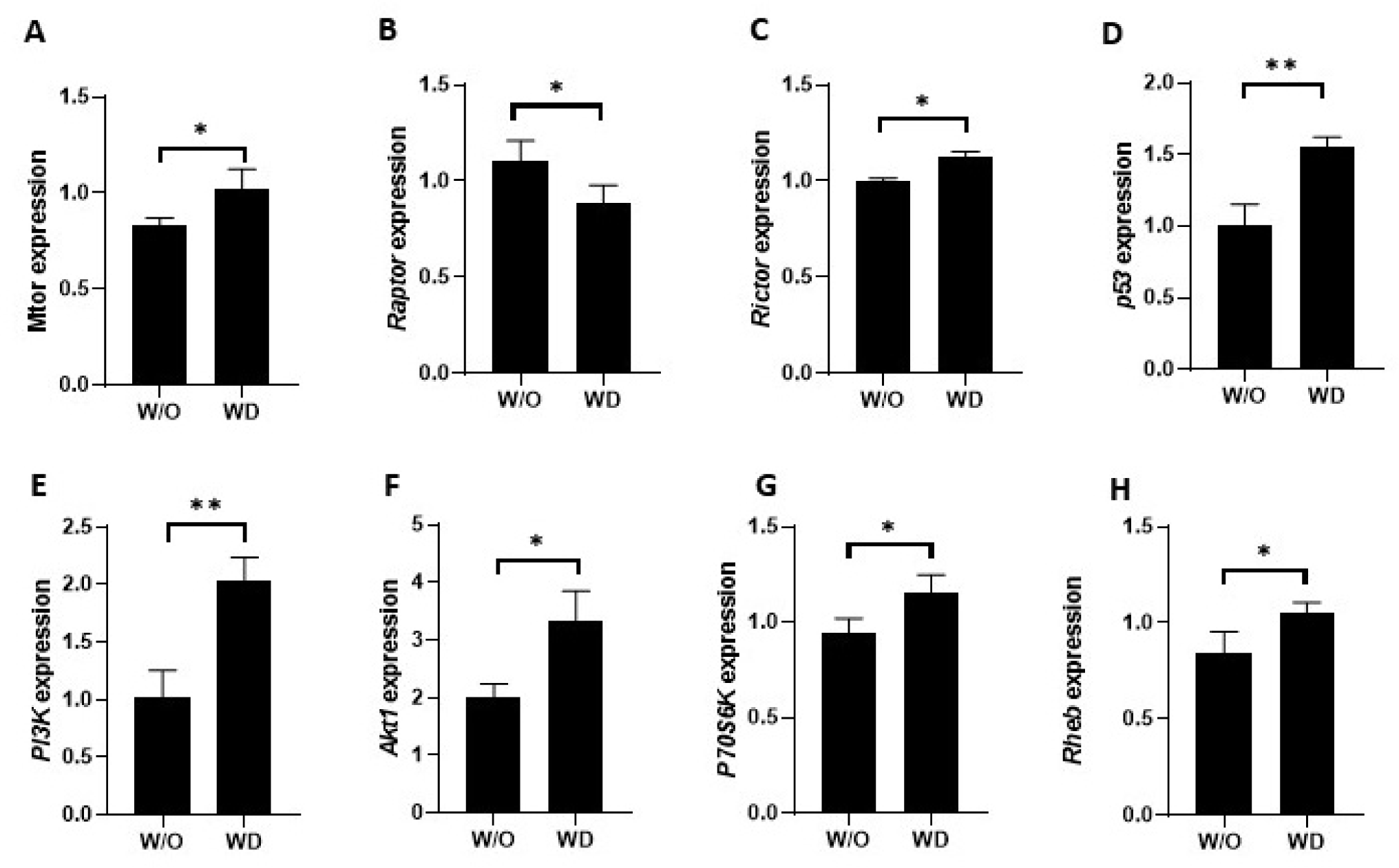
The mRNA expression levels of the mTOR-related genes (Mtor, Raptor, Rictor, p53, PI3K, Akt1, p70s6k, and Rheb) were measured in the uterus samples to evaluate the impact of dysbiosis on the mTOR signaling-pathway related gene expression. In the WD group, the relative mRNA expression levels of Mtor, Rictor, p53, PI3K, Akt1, p70s6k, and Rheb were significantly higher compared to the W/O group (*p<0.05; Fig. 4A, C, D, E, F, G, and H). Importantly, Raptor expression, a component of the mTORC1 complex, was statistically higher in the W/O group than in the uterus samples of the WD group (*p<0.05; Fig. 4B).
DISCUSSION
Numerous signaling pathways are associated with alterations in the gut microbiota and its metabolites, which can disrupt normal host functions [2]. The gut microbiome plays a vital role in digestion, metabolism, hormone regulation, detoxification, immune development, and protection of the female reproductive tract. It influences female reproductive health by regulating immunity, metabolism, hormones, inflammation, stress, and emotional well-being [24]. Antibiotics are commonly used to treat infectious diseases. However, they also affect beneficial gut microbes in addition to targeting pathogens [25]. Frequent or excessive antibiotic use reduces the taxonomic richness and diversity of the gut microbiota, leading to dysbiosis, which is associated with inflammation, altered lipid metabolism, disturbed intestinal barrier, and a negative impact on reproductive functions [26, 27]. Previous studies found that microbial imbalance in the vagina is associated with an immune response, leading to increased pro-inflammatory cytokine expression, triggering inflammation-related pathways [28]. However, researchers have highlighted that antibiotic treatments alter the estrous cycle and ovarian functions and are associated with infertility, as evidenced in animal models [29].
The mTOR signaling pathway involves many processes, is sensitive to environmental changes, and controls cellular activities, such as gene expression, cell growth, cell structure, and autophagy [30]. Previous studies have shown that mTOR is present in the mouse ovary and is predominantly expressed in the cytoplasmic/perinuclear region of granulosa cells. It plays a key role in regulating reproductive functions [31]. Previous research has described dysbiosis of vaginal microflora and bacterial vaginosis as being associated with elevated pro-inflammatory cytokines and mTOR expression, and dysfunction of the epithelial barrier in females [32]. In our study, we found that the relative expression levels of Mtor, Akt1, PI3K, p70s6k, and Rheb were significantly elevated in the ovaries and uterus of mice in the WD group compared with those in the W/O group (Figs. 3 and 4). Similarly, another study found that compared with the control group, endometritis-induced female mice showed a significant increase in gene expression levels of tumor necrosis factor (TNF)-α, interleukin (IL)-1β, and mTOR, as examined by western blotting of the uterine tissue [33]. Sleep deprivation (SD) alters the gut microbiome and negatively affects the reproductive system [34, 35]. SD-induced female mice showed significantly increased protein levels of mTOR, AKT, PI3K, IL-6, and TNF-α in ovarian tissue, as determined by western blotting, compared with the control group [36]. A study demonstrated that administering sulfamonomethoxine (SMM) to pregnant mice and their female pups resulted in behavioral dysfunctions. They found significantly elevated gene expression levels of Mtor and Akt1 in the brains of female pups from the SMM treatment group. Additionally, female pups in the SMM treatment group exhibited lower blood glucose levels than those in the vehicle group [37]. Researchers highlighted that overactivation of PI3K leads to mTOR activation, which, in turn, activates p70s6k, a downstream regulator of mTOR [38, 39].
p53 is an important gene that suppresses tumor development [40] and is activated by hypoxia, metabolic stress, and DNA damage, and protects body cells from harmful stress conditions [41]. Fluoride-induced mature female mice showed significantly increased p53 expression levels in the ovary compared to the control group [42]. Similarly, we observed higher p53 expression in the ovaries and uterus of the WD group compared to the W/O group.
Researchers have highlighted that PCOS, ovarian neoplasms, and ovarian aging lead to alteration of the mTOR signaling pathway. PCOS is associated with low-grade chronic inflammation. The expression of AKT, PI3K, raptor, and p70S6K has been found to be significantly elevated, and Rictor expression decreased in PCOS and ovarian neoplasm [43]. In contrast, we found that Rictor expression was significantly increased, whereas raptor expression was downregulated in the WD group compared with the control. OoRptor-/- mice, in whom raptor was deleted in all oocytes, were shown to block mTORC1 signaling but express follicular development and fertility similar to the wild-type mice [44].
Reproductive hormones are essential for maintaining reproductive function and hormone signaling. However, researchers have highlighted that these antibiotic treatments alter the estrous cycle and ovarian functions and are associated with infertility, as evidenced in animal models [29].
In our study, we found that antibiotic-induced gut dysbiosis is associated with significant alterations in mTOR-related gene expression in the ovaries and uterus. Mtor, Rictor, p53, PI3K, Akt1, P70S6K, and Rheb were upregulated, while hormonal and metabolic disturbances were observed, including reduced LH, FSH, glucose levels and increased cholesterol levels in the WD group. These changes suggest that disruption of the gut microbiota may contribute to metabolic imbalance and impaired reproductive function through modulation of the mTOR signaling pathway-related gene expression.
A limitation of this study is that we did not directly assess inflammatory parameters such as cytokine levels, intestinal barrier integrity, or immune cell filtration. Nevertheless, numerous studies have shown that antibiotic-induced gut dysbiosis can promote low-grade inflammation and barrier dysfunction, which may contribute to the metabolic and reproductive changes we observed [19]. Future studies will include direct inflammatory and histopathological analyses to further clarify these mechanisms. Although mice of the same age were randomly assigned to groups and housed under identical conditions, estrous-cycle stage was not monitored and tissues were collected at random stages, as in several previous studies of gut microbiota and female reproduction in mice. Because FSH, LH, uterine weight, ovarian morphology, and gene expression vary across the cycle, we cannot exclude that unequal stage distribution contributed to some observed differences.
A further limitation is that mTOR pathway involvement was assessed only at the transcript level; mRNA changes alone do not establish pathway activation, which is primarily regulated by post-translational phosphorylation. Protein and phosphorylation analyses of key mTORC1/2 targets would be required to confirm pathway activity. Nevertheless, our transcript-level findings are consistent with prior reports in analogous models where mRNA changes in mTOR components coincided with protein or phosphorylation changes, supporting the biological plausibility of our result [45].


