Journal of Biomedical Translational Research
Research Institute of Veterinary Medicine, Chungbuk National University
Original Article

Subchronic inhalation toxicity study of diethylbenzene in Wistar rats

Eun-Sang Cho1,*https://orcid.org/0000-0001-7297-0063, Dae-Sik Rha1https://orcid.org/0009-0001-2662-1249
1Inhalation Toxicity Research Center, Occupational Safety and Health Research Institute, KOSHA, Daejeon 34122, Korea
*Corresponding author: Eun-Sang Cho, Inhalation Toxicity Research Center, Occupational Safety and Health Research Institute, KOSHA, Daejeon 34122, Korea. Tel: +82-42-869-8536, E-mail: escho@kosha.or.kr

© Research Institute of Veterinary Medicine, Chungbuk National University.

Received: Aug 01, 2024; Revised: Sep 06, 2024; Accepted: Sep 07, 2024

Abstract

Diethylbenzene (DEB) is a colorless flammable liquid composed of a benzene ring and two ethyl substituents. DEBs mostly exist as a mixture of isomers and are mainly used as intermediates and solvents occupationally. Workers may be exposed to DEB inhalation during their occupational activities including manufacturing or processing of materials; however, limited data are available on the risk assessment of DEB mixtures. In this study, male and female Wistar rats were exposed to vapors of a DEB mixture for 13-weeks (6 hr/day, 5 days/week) at concentrations of 0, 40, 80, and 160 ppm in a whole-body inhalation chamber. Clinical signs, mean body weight, food consumption, bronchoalveolar lavage fluid (BALF), hematology, blood biochemistry, gross findings, organ weights, and microscopic findings were examined to determine the toxicity of DEB mixture. The exposure concentrations in chambers were 39.48 ± 1.13 ppm, 80.43 ± 2.06 ppm, and 160.20 ± 4.42 ppm for the low, medium, and high dose groups, respectively. No changes related to the test substance were observed, including changes in clinical observation, body weight, food consumption, BALF and blood analysis, necropsy findings, absolute and relative organ weights or histopathological analysis. Based on these results, the NOAEC (no-observed-adverse-effect-concentration) of DEB was defined as 160 ppm under the study conditions.

Keywords: inhalation; toxicity; diethylbenzene; no-observed-adverse-effect-concentration; occupational exposure

INTRODUCTION

Diethylbenzene (DEB, CAS No. 25340-17-4) is an almost colorless and flammable liquid with an aromatic odor. DEB is a byproduct of the synthesis of ethylbenzene from ethylene and benzene and contains a benzene ring and two ethyl substituents. It is primarily manufactured as a mixture of isomers (ortho: 1,2-; meta: 1,3-; and para: 1,4-) [1]. It is widely used in the production of synthetic rubber and resins as an intermediate or solvent in powderless etching [2], and is manufactured and/or imported in the European Economic area about 10 tonnes per year [3].

There are no data on DEB toxicity in humans, including skin or respiratory irritation, sensitization, or reported cases of occupational exposure. There is a high risk of exposure to workers through inhalation or skin absorption, with an estimated 60% of DEB is being absorbed through the respiratory tract, particularly in cases involving structurally similar chemicals [1]. There are few studies on the DEB mixtures because of the ratio difference between the three isomers; however, several studies on the toxic effects of each isomer in animals are available.

1,2-DEB is known to be the most toxic among isomers and causes central and peripheral neurotoxicity such as limb weakness associated with nerve fiber changes [4, 5]. Decreased leukocyte and lymphocyte counts were observed in rats exposed to the isomer mixture, and even though neurotoxic effect was not observed at the highest concentration (252 ppm), the researchers reported 10% of the highest concentration as no-observed-adverse-effect-concentration (NOAEC) of clinical neurotoxicity [1, 6]. Neither in vitro nor in vivo genotoxicity has been observed for the DEB isomer mixture [3]. Depending on the use of the substance to be manufactured, DEB is mainly exposed through the route of inhalation; however, there are limited data on repeated inhalation exposure, which was performed recently, compared to the oral exposure of animals. In addition, limited animal toxicity studies based on the OECD guidelines have been conducted. Therefore, we performed a 13-week repeated toxicity study to determine the toxic effects of DEB by inhalation exposure, according to the OECD Test Guideline No. 413, in accordance with Good Laboratory Practice (GLP) [7].

MATERIALS AND METHODS

Experimental animals and ethics

Five-week-old specific-pathogen-free Wistar rats (40 males and 40 females) were obtained from SPF Biotechnology (Beijing, China) and acclimatized for two week in polysulfone cages (up to three animals of the same sex). The animal room was maintained at 22 ± 3°C, 50 ± 20% relative humidity, and a 12-hr light/dark cycle. Male and female rats weighed 235–275 g and 167–197 g, respectively, before exposure. The whole-body inhalation chambers (WITC-14M, HCT, Icheon, Korea) that were maintained at 22 ± 3°C and 50 ± 20% relative humidity with individual multi-compartment stainless steel wire mesh cages (W240 × L1200 × H200) were used for exposure. The chambers were vented 10–15 times per hour, and the oxygen concentration was maintained at least 19% during the exposure period. Animals were housed in polysulfone cages after the initial exposure and fed an animal diet (Teklad certified irradiated global 18% protein Rodent Diet 2918C, Envigo RMS, Indianapolis, IN, USA) and filtered water ad libitum. We also provided wooden chewing sticks to each rat as an environmental enrichment item. All animal care and inhalation studies were performed under GLP conditions, and the procedures were in accordance with the National Institutes Guide for the Care and Use of Laboratory Animals [8]. This study was approved by the Institutional Animal Care and Use Committee of the Inhalation Research Center, Occupational Safety and Health Research Institute, and all experiments were conducted according to the established institutional animal care and use protocol (Approval No. IACUC-2203).

Test material and exposure conditions

DEB (mixture of isomers; 99.2% purity) was purchased from Alfa Aesar (Thermo Fisher Scientific, Waltham, MA, USA), and the ratio of isomer was unknown. The vaporized material was generated using a liquid-vapor generator (LVG-04-A, HCT), and the concentrations of the materials were analyzed using a gas chromatograph (Model No. TRACE1310, Thermo Fisher Scientific). The environmental conditions in the chambers were monitored every 30 min (Model No. ITC Manager, HCT).

Subchronic toxicity assessment design

The OECD guidelines (TG413) and the previously published method of Cho were used for the 13-week repeated inhalation study of DEB with modifications [9, 10]. A preliminary 28-day inhalation toxicity study was performed to determine the exposure concentration and showed no toxicological effect at a maximum concentration of 160 ppm, and a 90-day test was planned with reference to this results (unpublished). Male and female rats were randomly divided into four groups according to sex, with each group consisting of ten animals. Each group was exposed to 0, 40, 80, or 160 ppm DEB for 6 hr/day, 5 days/week, for 13 weeks. Concentrations of low (40 ppm), medium (80 ppm), and high (160 ppm) exposure groups were selected. All animals were observed once or twice daily (before and after exposure) to confirm detailed clinical signs, and body weight data were collected once or twice a week. Food consumption was measured once a week. At the end of the study, all the rats were anesthetized with isoflurane (I-Fran liquid, Hana Pharm, Hwaseong, Korea) and euthanized via exsanguination of the abdominal aorta and vein.

Blood analyses

Blood samples were collected from the abdominal aorta before euthanasia. The samples were collected in tubes containing an anticoagulant (ethylenediaminetetraacetic acid or sodium citrate) for hematological parameters, or in serum-separating tubes for biochemical examination. The anti-coagulant tubes containing sodium citrate and serum separating tubes were centrifuged at 3,000 × g at 4°C for 10 min to separate the serum. Hematological or biochemical parameters were examined using blood cell analyzer (ADIVA 2120i, Siemens Diagnostics, Tarrytown, NY, USA), coagulation analyzer (ACL Elite Systems, Instrumentation Laboratory, Bedford, MA, USA), or blood chemistry analyzer (TBA-120FR, Toshiba, Tokyo, Japan): white blood cell (WBC) count, red blood cell (RBC) count, hemoglobin (HGB) concentration, hematocrit (HCT), mean corpuscular volume (MCV), mean corpuscular hemoglobin (MCH), mean corpuscular hemoglobin concentration (MCHC), platelet (PLT) count, reticulocyte (RET) count, differential WBC count (neutrophils, lymphocytes, monocytes, eosinophils, and basophils), activated partial thromboplastin time (APTT), prothrombin time (PT), sodium (Na), potassium (K), chloride (Cl), total protein (TP), albumin (ALB), creatinine (CREA), blood urea nitrogen (BUN), glucose (GLU), calcium (Ca), inorganic phosphorus (IP), total bilirubin (TBIL), total cholesterol (TCHO), triglyceride (TG), aspartate aminotransferase (AST), alanine aminotransferase (ALT), alkaline phosphatase (ALP), and albumin/globulin (A/G) ratio.

Bronchoalveolar lavage fluid examination

Bronchoalveolar lavage fluid (BALF) samples were collected immediately after euthanasia. The samples were collected from the right-sided lung by lavage with phosphate buffered saline and centrifuged at 3,000 × g at 4°C for 10 min to separate the cells and supernatant. Biochemical parameters including TP, ALB and lactate dehydrogenase (LDH), were examined using a blood chemistry analyzer, and total cells were counted using an automatic cell analyzer (NC-200, ChemoMetec, Allerod, Denmark). At least 400 cells, including alveolar macrophages, monocytes, lymphocytes, neutrophils, and eosinophils, were counted under a light microscope (Axio Scope, Carl Zeiss, Oberkochen, Germany).

Organ weights, gross, and histopathological parameters

During necropsy, the adrenal glands, brain, epididymis, heart, kidneys, liver, lungs, ovaries, spleen, testes, thymus, and uterus were examined and weighed. And the following tissues were also examined and collected in 10% neutral buffered formalin or Davison’s fixative (for the testes and eyes/optic nerves) for the histopathologic examination: aorta, bone marrow, cecum, colon, duodenum, esophagus, eyes/optic nerves, femur, gallbladder, harderian glands, ileum, jejunum, larynx, lung (left lobe only), lymph nodes (tracheobronchial and mesenteric), mammary gland, nasopharyngeal tissue, pancreas, parathyroids, pituitary, prostate, rectum, salivary glands (submandibular, sublingual, and parotid), sciatic nerve, seminal vesicle, skeletal muscle, skin, spinal cord, sternum, stifle joint, stomach, teeth, thyroid, tongue, urinary bladder, and vagina. According to the OECD TG413, all of the fixed tissues from control and high exposure groups of male and female rats were paraffin-embedded, 4 μm thickness-sectioned, stained with hematoxylin and eosin (Automatic Stainer, DAKO, Glostrup, Denmark), and examined under a light microscope (DM3000, Leica, Wetzlar, Germany). However, histopathologic examinations from low and medium concentrations were not performed in consideration of the effect of the test substance.

Statistical analysis

Data are expressed as mean ± S.D. Analysis of parameters, including body weight, food consumption, organ weights, and hematological or blood biochemical data was performed using the Pristima® system (Version 7.1.0, Xybion, Princeton, NJ, USA). Data were checked for homogeneity using Levene’s test and analyzed using with one-way analysis of variance (Dunnett’s multiple test) or Kruskal–Wallis (Dunn rank sum test). Differences were considered significant at p<0.05 or p<0.01.

RESULTS

Concentration in exposure chambers

The analytical concentrations of DEB in chambers during the exposure period for the low, medium, and high exposure groups were 39.48 ± 1.13 ppm, 80.43 ± 2.06 ppm, and 160.20 ± 4.42 ppm, respectively.

Clinical signs, body weight, and food consumption changes

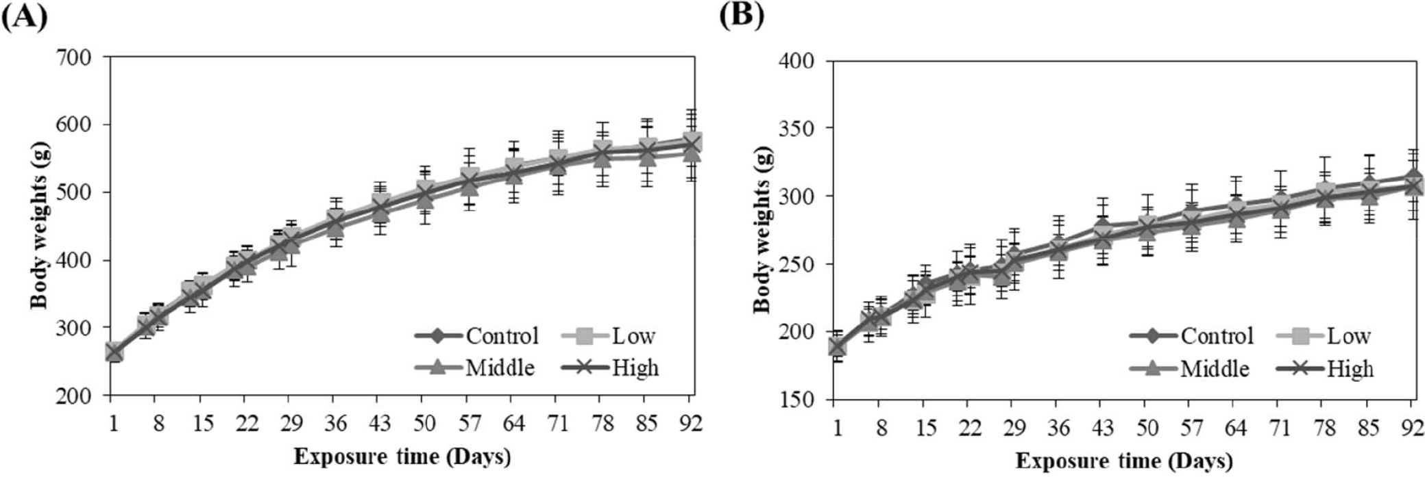
No animals died or unusual behavioral or clinical signs were observed during the study period (data not shown). In addition, there were no significant changes in the body weight (Fig. 1) or food consumption (data not shown) of male and female rats during the study.

jbtr-25-3-119-g1
Fig. 1. Body weight changes in male (A) and female (B) Wistar rats. The animals were exposured to diethylbenzene (0–160 ppm) by inhalation during 90 days. The body weights present as the mean ± S.D. (n = 10/group).
Download Original Figure
Hematology, blood biochemistry, and bronchoalveolar lavage fluid examination

The PT level was significantly increased in the medium- and high-exposure groups in male rats compared to that in control males (medium: p<0.01; high: p<0.05). Additionally, ALP levels in the male high-exposure group were significantly high (p<0.05). No significant differences were observed in other hematological or biochemical parameters between the control and experimental groups (Tables 1, 2, 3, and 4). In addition, there were no significant differences in BALF data between the control and experimental groups (Table 5).

Table 1. Summary of hematological data in male rats inhaling diethylbenzene for 90 days (n = 10)
Parameters Groups
Control Low (40 ppm) Middle (80 ppm) High (160 ppm)
WBC (× 103/μL) 5.232 ± 1.4371 5.23 ± 1.0705 5.818 ± 1.1856 6.079 ± 1.506
RBC (× 106/μL) 8.817 ± 0.3198 8.504 ± 0.2251 8.719 ± 0.273 8.631 ± 0.2518
HGB (g/dL) 14.61 ± 0.534 14.43 ± 0.38 14.59 ± 0.463 14.48 ± 0.319
HCT (%) 43.71 ± 1.859 42.83 ± 1.12 43.8 ± 1.359 43.47 ± 1.106
MCV (fL) 49.55 ± 1.396 50.41 ± 1.603 50.22 ± 1.025 50.43 ± 1.578
MCH (pg) 16.57 ± 0.517 16.97 ± 0.648 16.74 ± 0.532 16.79 ± 0.599
MCHC (g/dL) 33.43 ± 0.579 33.67 ± 0.419 33.33 ± 0.485 33.29 ± 0.504
PLT (× 103/μL) 879.9 ± 346.5 1,014.5 ± 120.31 910 ± 262.3 1,026.7 ± 84.71
NEU% (%) 24.38 ± 7.3 20.82 ± 3.707 22.51 ± 9.948 20.16 ± 3.311
LYM% (%) 70.53 ± 7.694 74.07 ± 3.811 71.86 ± 9.558 74.68 ± 3.633
MON% (%) 2.52 ± 0.745 2.43 ± 0.748 2.5 ± 0.897 2.19 ± 0.53
EOS% (%) 1.67 ± 0.706 1.49 ± 0.407 1.83 ± 0.726 1.93 ± 0.517
BAS% (%) 0.21 ± 0.099 0.17 ± 0.082 0.22 ± 0.063 0.21 ± 0.074
NEUA (× 103/μL) 1.23 ± 0.419 1.096 ± 0.3614 1.314 ± 0.6451 1.206 ± 0.2847
LYMA (× 103/μL) 3.74 ± 1.2199 3.868 ± 0.7686 4.176 ± 1.0253 4.561 ± 1.2814
MONA (× 103/μL) 0.129 ± 0.0479 0.122 ± 0.0305 0.142 ± 0.0492 0.132 ± 0.0382
EOSA (× 103/μL) 0.085 ± 0.035 0.079 ± 0.0351 0.108 ± 0.0457 0.114 ± 0.036
BASA (× 103/μL) 0.012 ± 0.0079 0.01 ± 0.0047 0.013 ± 0.0048 0.014 ± 0.007
RET% (%) 2.018 ± 0.2126 2.187 ± 0.3717 2.251 ± 0.2593 2.369 ± 0.5667
RETA (× 109/μL) 177.52 ± 14.933 185.84 ± 31.11 196.4 ± 24.496 204.23 ± 48.818
APTT (sec) 13.26 ± 1.643 14.74 ± 3.133 15.65 ± 3.267 14.23 ± 2.365
PT (sec) 12 ± 0.76 12.1 ± 0.69 12.9 ± 0.58+ 12.7 ± 0.47*

Data are expressed as the mean ± S.D.

Dunnett’s LSD test significant at the 0.05 level;

Dunnett’s LSD test significant at the 0.01 level.

n, number of animals examined; WBC, white blood cell count; RBC, red blood cell count; HGB, hemoglobin; HCT, hematocrit; MCV, mean corpuscular volume; MCH, mean corpuscular hemoglobin; MCHC, mean corpuscular hemoglobin concentration; PLT, platelets; NEU%, neutrophil relative; LYM%, lymphocyte relative; MON%, monocyte relative; EOS%, eosinophil relative; BAS%, basophil relative; NEUA, neutrophil absolute; LYMA, lymphocyte absolute; MONA, monocyte absolute; EOSA, eosinophil absolute; BASA, basophil absolute; RET%, reticulocyte relative; RETA, reticulocyte absolute; APTT, activated partial thromboplastin time; PT, prothrombin time.

Download Excel Table
Table 2. Summary of hematological data in female rats inhaling diethylbenzene for 90 days (n = 10)
Parameters Groups
Control Low (40 ppm) Middle (80 ppm) High (160 ppm)
WBC (× 103/μL) 2.74 ± 0.9464 2.643 ± 0.4128 3.086 ± 0.8166 3.1 ± 0.651
RBC (× 106/μL) 7.482 ± 0.3503 7.522 ± 0.2402 7.749 ± 0.4451 7.568 ± 0.2164
HGB (g/dL) 13.48 ± 0.503 13.51 ± 0.476 13.95 ± 0.624 13.59 ± 0.506
HCT (%) 40.07 ± 1.606 39.84 ± 0.999 41.64 ± 1.788 40.4 ± 1.642
MCV (fL) 53.57 ± 1.277 53 ± 1.031 53.78 ± 1.694 53.38 ± 0.95
MCH (pg) 18.00 ± 0.492 17.97 ± 0.56 18.02 ± 0.600 17.96 ± 0.309
MCHC (g/dL) 33.64 ± 0.369 33.89 ± 0.611 33.51 ± 0.412 33.63 ± 0.35
PLT (× 103/μL) 935.2 ± 99.77 1,037.2 ± 99.15 1,003.7 ± 218.25 986.6 ± 60.91
NEU% (%) 20.33 ± 5.572 18.74 ± 5.05 20.68 ± 8.960 16.14 ± 4.222
LYM% (%) 73.66 ± 6.715 75.02 ± 5.939 73.04 ± 9.171 78 ± 5.057
MON% (%) 2.79 ± 0.867 3.00 ± 0.745 2.98 ± 0.716 2.73 ± 1.456
EOS% (%) 2.03 ± 0.533 2.07 ± 0.512 1.95 ± 0.762 1.69 ± 0.925
BAS% (%) 0.15 ± 0.085 0.14 ± 0.073 0.12 ± 0.042 0.12 ± 0.083
NEUA (× 103/μL) 0.554 ± 0.237 0.482 ± 0.1049 0.598 ± 0.1938 0.487 ± 0.1082
LYMA (× 103/μL) 2.019 ± 0.7307 1.999 ± 0.4023 2.289 ± 0.8014 2.431 ± 0.6243
MONA (× 103/μL) 0.079 ± 0.0407 0.079 ± 0.0226 0.094 ± 0.0443 0.086 ± 0.0456
EOSA (× 103/μL) 0.055 ± 0.0212 0.052 ± 0.012 0.061 ± 0.0288 0.05 ± 0.025
BASA (× 103/μL) 0.003 ± 0.0048 0.003 ± 0.005 0.001 ± 0.0032 0.004 ± 0.0053
RET% (%) 2.193 ± 0.428 2.252 ± 0.3009 2.147 ± 0.5907 2.17 ± 0.4717
RETA (× 109/μL) 163.65 ± 30.315 169.24 ± 22.426 165.41 ± 42.104 164.41 ± 37.168
APTT (sec) 16.9 ± 1.781 18.1 ± 0.814 17.94 ± 1.861 18.78 ± 0.931
PT (sec) 10.8 ± 0.59 11 ± 0.48 11.4 ± 1.1 11.4 ± 0.61

Data are expressed mean ± S.D.

n, number of animals examined; WBC, white blood cell count; RBC, red blood cell count; HGB, hemoglobin; HCT, hematocrit; MCV, mean corpuscular volume; MCH, mean corpuscular hemoglobin; MCHC, mean corpuscular hemoglobin concentration; PLT, platelet; NEU%, neutrophil relative; LYM%, lymphocyte relative; MON%, monocyte relative; EOS%, eosinophil relative; BAS%, basophil relative; NEUA, neutrophil absolute; LYMA, lymphocyte absolute; MONA, monocyte absolute; EOSA, eosinophil absolute; BASA, basophil absolute; RET%, reticulocyte relative; RETA, reticulocyte absolute; APTT, activated partial thromboplastin time; PT, prothrombin time.

Download Excel Table
Table 3. Summary of blood biochemistry data in male rats inhaling diethylbenzene for 90 days (n = 10)
Parameters Groups
Control Low (40 ppm) Middle (80 ppm) High (160 ppm)
Na (mmol/L) 147.42 ± 4.56 149.95 ± 4.155 143.05 ± 18.055 149.48 ± 3.021
K (mmol/L) 5.252 ± 0.3827 5.38 ± 0.2043 5.201 ± 0.43 5.297 ± 0.3325
Cl (mmol/L) 106.45 ± 3.682 109.24 ± 3.23 104.37 ± 12.805 108.34 ± 2.147
TP (g/dL) 6.04 ± 0.366 6.02 ± 0.297 6.08 ± 0.644 6.11 ± 0.42
ALB (g/dL) 3.95 ± 0.196 3.98 ± 0.204 3.93 ± 0.313 3.92 ± 0.22
CREA (mg/dL) 0.384 ± 0.0222 0.415 ± 0.0299 0.409 ± 0.0458 0.402 ± 0.0349
BUN (mg/dL) 17.47 ± 2.874 19.68 ± 1.737 17.93 ± 1.941 19.05 ± 2.32
GLU (mg/dL) 149.79 ± 35.882 152.86 ± 20.589 165.73 ± 38.972 167.59 ± 24.634
Ca (mg/dL) 9.55 ± 0.334 9.55 ± 0.36 9.5 ± 0.316 9.68 ± 0.429
IP (mg/dL) 5.8 ± 0.516 6.2 ± 0.476 6.23 ± 0.574 6.36 ± 0.687
TBIL (mg/dL) –0.005 ± 0.0207 0.005 ± 0.0151 –0.017 ± 0.0216 –0.018 ± 0.0244
TCHO (mg/dL) 84.37 ± 14.67 96.69 ± 12.187 96.79 ± 20.897 88.69 ± 15.305
TG (mg/dL) 71.78 ± 31.735 55.39 ± 19.925 47.91 ± 14.067 59.44 ± 18.51
AST (IU/L) 150.09 ± 37.835 52.8 ± 15.618 148.96 ± 56.9 132.92 ± 14.409
ALT (IU/L) 42.91 ± 7.243 186.35 ± 64.471 47.55 ± 9.92 48.05 ± 9.594
ALP (IU/L) 274.23 ± 57.055 325.66 ± 71.903 302.62 ± 83.337 388.10 ± 122.569*
A/G ratio 1.92 ± 0.199 1.95 ± 0.246 1.86 ± 0.171 1.79 ± 0.11

Data are expressed as the mean ± S.D.

Dunnett’s LSD test significant at the 0.05 level.

n, number of animals examined; Na, sodium; K, potassium; Cl, chloride; TP, total protein; ALB, albumin; CREA, creatinine; BUN, blood urea nitrogen; GLU, glucose; Ca, calcium; IP, inorganic phosphorus; TBIL, total bilirubin; TCHO, total cholesterol; TG, triglycerides; AST, aspartate aminotransferase; ALT, alanine aminotransferase; ALP, alkaline phosphatase; A/G ratio, albumin/globulin ratio.

Download Excel Table
Table 4. Summary of blood biochemistry data in female rats inhaling diethylbenzene for 90 days (n = 10)
Parameters Groups
Control Low (40 ppm) Middle (80 ppm) High (160 ppm)
Na (mmol/L) 148.58 ± 3.446 149.67 ± 2.76 148.2 ± 0.937 149.23 ± 1.794
K (mmol/L) 4.141 ± 0.3743 4.436 ± 0.4323 4.294 ± 0.408 4.397 ± 0.2867
Cl (mmol/L) 110.54 ± 3.795 110.33 ± 2.277 109.74 ± 1.952 110.03 ± 1.946
TP (g/dL) 6.8 ± 0.512 6.96 ± 0.381 6.92 ± 0.225 6.72 ± 0.463
ALB (g/dL) 4.93 ± 0.347 5.11 ± 0.298 4.97 ± 0.183 4.91 ± 0.395
CREA (mg/dL) 0.506 ± 0.0288 0.477 ± 0.0381 0.492 ± 0.0547 0.499 ± 0.059
BUN (mg/dL) 19.52 ± 3.803 18.83 ± 1.703 20.65 ± 5.16 20.27 ± 4.999
GLU (mg/dL) 156.9 ± 22.547 159.78 ± 24.865 164.05 ± 27.899 174.14 ± 23.706
Ca (mg/dL) 9.9 ± 0.306 10.12 ± 0.273 10.13 ± 0.221 9.99 ± 0.372
IP (mg/dL) 4.79 ± 0.698 5.07 ± 0.82 5.1 ± 1.011 5.17 ± 1.02
TBIL (mg/dL) 0.029 ± 0.0228 0.05 ± 0.0278 0.028 ± 0.0132 0.027 ± 0.0212
TCHO (mg/dL) 102.09 ± 32.35 108.6 ± 17.884 109.24 ± 15.919 109.43 ± 23.045
TG (mg/dL) 51.41 ± 27.341 54.77 ± 22.325 69.49 ± 23.27 55.92 ± 23.403
AST (IU/L) 131.43 ± 102.464 133.18 ± 43.149 103.72 ± 22.677 174.52 ± 71.06
ALT (IU/L) 52.54 ± 23.967 44.46 ± 12.564 48.9 ± 11.351 58.09 ± 17.349
ALP (IU/L) 193.37 ± 70.115 251.92 ± 161.945 237.13 ± 104.98 237.4 ± 109.837
A/G ratio 2.65 ± 0.207 2.77 ± 0.2 2.56 ± 0.241 2.72 ± 0.239

Data are expressed as the mean ± S.D.

n, number of animals examined; Na, odium; K, potassium; Cl, chloride; TP, total protein; ALB, albumin; CREA, creatinine; BUN, blood urea nitrogen; GLU, glucose; Ca, calcium; IP, inorganic phosphorus; TBIL, total bilirubin; TCHO, total cholesterol; TG, triglycerides; AST, aspartate aminotransferase; ALT, alanine aminotransferase; ALP, alkaline phosphatase; A/G ratio, albumin/globulin ratio.

Download Excel Table
Table 5. Summary of bronchoalveolar lavage fluid data in rats inhaling diethylbenzene for 90 days (n = 5)
Parameters Groups
Control Low (40 ppm) Middle (80 ppm) High (160 ppm)
Male
 TC (cells/mL) 734,200 ± 434,704 792,200 ± 338,021 687,000 ± 504,003 975,600 ± 278,974
 MAC% (%) 97.7936 ± 2 98.8439 ± 0 99.0122 ± 1 98.1659 ± 1
 LYM% (%) 0.3419 ± 0 0.3725 ± 0 0.2122 ± 0 0.6854 ± 0
 NEU% (%) 1.3824 ± 2 0.7442 ± 0 0.5522 ± 1 1.0738 ± 1
 EOS% (%) 0.4821 ± 1 0.0394 ± 0 0.2233 ± 0 0.0749 ± 0
 MACA (cells/mL) 721,593.4 ± 434,455 783,280.6 ± 335,412 677,579 ± 490,157 957,281.2 ± 272,820
 LYMA (cells/mL) 3,499.8 ± 4,692 3,285.2 ± 3,187 1,728.4 ± 3,016 6,251 ± 1,885
 NEUA (cells/mL) 6,462.83 ± 8,117 5,328.23 ± 2,315 5,694.21 ± 9,108 11,146.6 ± 11,320
 EOSA (cells/mL) 2,644.07 ± 2,321 305.91 ± 684 1,998.37 ± 2,825 921.35 ± 2,060
 LDH (IU/L) 71.76 ± 40 80.94 ± 43 92.88 ± 57 69.54 ± 47
 TP (g/dL) 0.0156 ± 0 0.0152 ± 0 0.0226 ± 0 0.019 ± 0
 ALB (g/dL) 0.007 ± 0 0.0084 ± 0 0.0114 ± 0 0.0064 ± 0
Female
 TC (cells/mL) 437,000 ± 153,747 471,800 ± 86,912 417,600 ± 216,571 406,000 ± 218,628
 MAC% (%) 99.1772 ± 1 99.0613 ± 1 85.9168 ± 14 98.3978 ± 2
 LYM% (%) 0 ± 0 0.129 ± 0 0.1306 ± 0 0 ± 0
 NEU% (%) 0.3757 ± 0 0.5805 ± 1 13.6525 ± 14 1.1676 ± 2
 EOS% (%) 0.4471 ± 1 0.2291 ± 0 0.3001 ± 0 0.1864 ± 0
 MACA (cells/mL) 433,203.8 ± 152,423 467,834.8 ± 89,516 376,708.4 ± 218,130 397,040.2 ± 207,251
 LYMA (cells/mL) 0 ± 0 472.2 ± 1,056 690.8 ± 1,020 0 ± 0
 NEUA (cells/mL) 1,881.78 ± 2,148 2,549.46 ± 2,743 39,140.63 ± 29,375 7,345.56 ± 12,677
 EOSA (cells/mL) 1,914.32 ± 2,796 943.34 ± 934 1,060.17 ± 1,042 956.91 ± 1,460
 LDH (IU/L) 84.84 ± 21 45.48 ± 8 52.64 ± 13 52.92 ± 9
 TP (g/dL) 0.0276 ± 0 0.0128 ± 0 0.0134 ± 0 0.016 ± 0
 ALB (g/dL) 0.0106 ± 0 0.0072 ± 0 0.0076 ± 0 0.0076 ± 0

Data are expressed as the mean ± S.D.

n, number of animals examined; TC, total cells; MAC%, alveolar macrophage relative; LYM%, lymphocyte relative; NEU%, neutrophil relative; EOS%, eosinophil relative; MACA, alveolar macrophage absolute; LYMA, lymphocyte absolute; NEUA, neutrophil absolute; EOSA, eosinophil absolute; LDH, lactate dehydrogenase; TP, total protein; ALB, albumin.

Download Excel Table
Organ weights and histopathological examination

The absolute organ weights or organ weights relative to the whole-body weight were investigated (Tables 6 and 7). In terms of absolute organ weights, no significant change was found between the experimental groups and the control group. The kidney weight in the low group was significantly lower than that in the control in regard to the relative organ weights. Gross findings were observed in the lungs, spleen, testes, and vagina (Table 8), and were identified by microscopic examination as alveolar macrophage aggregation in the lung, unclassified sarcoma in the spleen, seminiferous tubular dilatation in the testes, and cysts (s) in the vagina. Microscopic findings were also observed in the heart, liver, lungs, nasal cavity, pancreas, pituitary, testes, and thyroid vagina in the control and high exposure group of rats (Table 9).

Table 6. Summary of absolute organ weight data in rats inhaling diethylbenzene for 90 days (n = 10)
Organs (g) Groups
Control Low (40 ppm) Middle (80 ppm) High (160 ppm)
Male
 Adrenal glands 0.0588 ± 0.00473 0.0601 ± 0.01184 0.0527 ± 0.0106 0.0555 ± 0.01229
 Brain 2.0917 ± 0.10405 2.0891 ± 0.08539 2.0457 ± 0.09041 2.0899 ± 0.08758
 Epididymides 1.5725 ± 0.13997 1.4585 ± 0.08497 1.4619 ± 0.13024 1.5218 ± 0.12653
 Heart 1.4519 ± 0.11511 1.4488 ± 0.09532 1.344 ± 0.12741 1.3856 ± 0.1415
 Kidneys 3.4528 ± 0.30324 3.206 ± 0.14593 3.3262 ± 0.27257 3.4775 ± 0.32525
 Liver 14.5399 ± 0.90226 14.245 ± 1.27354 13.8062 ± 1.69652 14.7038 ± 1.6898
 Lung 0.7356 ± 0.12172 0.7047 ± 0.0624 0.7262 ± 0.07621 0.7449 ± 0.07664
 Spleen 1.0301 ± 0.17322 0.9974 ± 0.09512 0.9655 ± 0.10179 1.0473 ± 0.17922
 Testes 3.792 ± 0.32464 3.7898 ± 0.53555 3.5297 ± 0.44182 3.8188 ± 0.31786
 Thymus 0.3674 ± 0.04833 0.3512 ± 0.07437 0.3329 ± 0.04882 0.3479 ± 0.03877
Female
 Adrenal glands 0.0635 ± 0.01219 0.065 ± 0.00645 0.0608 ± 0.00732 0.0642 ± 0.00838
 Brain 1.9297 ± 0.08218 1.975 ± 0.05988 1.9461 ± 0.0457 1.9538 ± 0.07729
 Heart 0.8957 ± 0.07737 0.8889 ± 0.09421 0.8552 ± 0.09261 0.8759 ± 0.0856
 Kidneys 1.888 ± 0.13662 1.8508 ± 0.18423 1.89 ± 0.08911 1.9879 ± 0.19474
 Liver 8.8245 ± 1.27524 8.3293 ± 0.75785 8.6293 ± 1.04305 8.9003 ± 0.93689
 Lung 0.5559 ± 0.0365 0.5263 ± 0.04167 0.5583 ± 0.03857 0.5382 ± 0.04932
 Ovaries 0.1091 ± 0.01818 0.1012 ± 0.02264 0.1064 ± 0.01893 0.1057 ± 0.01701
 Spleen 0.5908 ± 0.05493 0.5953 ± 0.11658 0.5819 ± 0.04356 0.6051 ± 0.05022
 Thymus 0.283 ± 0.08271 0.2276 ± 0.04225 0.2642 ± 0.05614 0.2705 ± 0.04523
 Uterus 0.8653 ± 0.21703 0.8974 ± 0.21898 0.8164 ± 0.19991 0.8683 ± 0.32513

Data are expressed as the mean ± S.D.

n, number of animals examined.

Download Excel Table
Table 7. Summary of relative organ weight data in rats inhaling diethylbenzene for 90 days (n = 10)
Organs (%) Groups
Control Low (40 ppm) Middle (80 ppm) High (160 ppm)
Male
 Adrenal glands 0.0104 ± 0.00095 0.0106 ± 0.00205 0.0097 ± 0.00169 0.0099 ± 0.00181
 Brain 0.3702 ± 0.02368 0.3684 ± 0.02462 0.3773 ± 0.0267 0.3751 ± 0.031
 Epididymides 0.2784 ± 0.02783 0.2574 ± 0.02353 0.269 ± 0.0201 0.2729 ± 0.02685
 Heart 0.2566 ± 0.01781 0.255 ± 0.01458 0.2467 ± 0.0100 0.2472 ± 0.01353
 Kidneys 0.6099 ± 0.04355 0.5652 ± 0.0369* 0.611 ± 0.01994 0.6215 ± 0.04359
 Liver 2.569 ± 0.09974 2.5058 ± 0.1827 2.5307 ± 0.17815 2.6207 ± 0.17052
 Lung 0.1302 ± 0.02234 0.1244 ± 0.01409 0.1335 ± 0.0112 0.133 ± 0.00759
 Spleen 0.183 ± 0.03525 0.176 ± 0.01983 0.1779 ± 0.01981 0.1864 ± 0.02227
 Testes 0.6711 ± 0.06296 0.6698 ± 0.11129 0.6499 ± 0.08108 0.6832 ± 0.0476
 Thymus 0.0649 ± 0.0077 0.0621 ± 0.01376 0.0615 ± 0.01042 0.0625 ± 0.00878
Female
 Adrenal glands 0.0208 ± 0.00393 0.0216 ± 0.0023 0.0201 ± 0.00225 0.0215 ± 0.00278
 Brain 0.6321 ± 0.03148 0.6556 ± 0.04446 0.6465 ± 0.04283 0.6558 ± 0.04322
 Heart 0.2932 ± 0.02339 0.2934 ± 0.0146 0.283 ± 0.02332 0.293 ± 0.0207
 Kidneys 0.6175 ± 0.03091 0.6113 ± 0.02568 0.6279 ± 0.05103 0.6661 ± 0.06205
 Liver 2.891 ± 0.43007 2.7557 ± 0.18567 2.8588 ± 0.31933 2.9791 ± 0.25669
 Lung 0.1823 ± 0.0157 0.1742 ± 0.01009 0.185 ± 0.01121 0.1805 ± 0.01768
 Ovaries 0.0358 ± 0.00617 0.0337 ± 0.00813 0.0353 ± 0.00594 0.0354 ± 0.00568
 Spleen 0.1934 ± 0.01717 0.1967 ± 0.03446 0.1926 ± 0.00704 0.2028 ± 0.01588
 Thymus 0.0928 ± 0.02763 0.0759 ± 0.01674 0.0876 ± 0.01861 0.0909 ± 0.01662
 Uterus 0.2867 ± 0.08935 0.2968 ± 0.06851 0.2706 ± 0.06434 0.2907 ± 0.10959

Data are expressed as the mean ± S.D.

Dunnett’s LSD test significant at the 0.05 level.

n, number of animals examined.

Download Excel Table
Table 8. Summary of gross findings in rats inhaling diethylbenzene for 90 days (n = 10)
Sex Male Female
Group Control Low (40 ppm) Middle (80 ppm) High (160 ppm) Control Low (40 ppm) Middle (80 ppm) High (160 ppm)
Organ
 Lung
  Spot 0 0 0 1 0 1 0 0
 Spleen
  Nodule 1 0 0 0 0 0 0 0
 Testes
  Increased size 0 1 0 0 - - - -
 Vagina
  Nodule - - - - 0 1 0 0

n, number of animals examined.

Download Excel Table
Table 9. Summary of histopathological findings in rats inhaling diethylbenzene for 90 days
Sex Male Female
Group Control Middle (80 ppm) High (160 ppm) Control Low (40 ppm) High (160 ppm)
n 10 1 10 10 2 10
Organ
 Heart
  Cardiomyopathy 1a 3 - 1 0 - 0
Total 3 - 1 0 - 0
 Liver
  Infiltration, mononuclear cell 1a 0 - 1 1 - 1
Total 0 - 1 1 - 1
 Lung
  Aggregates, alveolar macrophage 1 5 - 3 2 0 1
2a 0 - 2 1 1 0
Total 5 - 5 3 1 1
  Crystals, hematoidin 1 1 - 1 0 0 0
Total 1 - 1 0 0 0
  Hyperplasia, bronchiolo-alveolar 1a 1 - 2 0 0 0
Total 1 - 0 0 0
  Infiltration, mononuclear cell 1a 0 - 1 0 0 0
Total 0 - 1 0 0 0
 Nasal cavity
  Hyperplasia, mucous cell 1 1 - 2 0 - 0
Total 1 - 2 0 - 0
 Pancreas
  Atrophy, acinar cell 1 1 - 1 0 - 0
2 1 - 0 0 - 0
Total 2 - 1 0 - 0
  Infiltration, mixed cell 1 0 - 0 0 - 1
Total 0 - 0 0 - 1
  Inflammation, granulomatous 1 0 - 1 0 - 0
2 1 - 0 0 - 0
Total 1 - 1 0 - 0
  Pigmentation 1 1 - 0 0 - 0
Total 1 - 0 0 - 0
  Vacuolation, acinar cell 2 0 - 0 0 - 1
Total 0 - 0 0 - 1
 Pituitary
  Cyst, pars distalis 1 0 - 1 0 - 0
Total 0 - 1 0 - 0
 Spleen
  Extramedullary hematopoiesis 1 0 - 1 1 - 2
Total 0 - 1 1 - 2
  Sarcoma, NOS Total 1 - 0 0 - 0
 Testes
  Dilation, seminiferous tubules 2 0 1 0 - - -
Total 0 1 0 - - -
 Thyroids
  Adenoma, C-cell Total 0 - 0 0 - 1
  Ectopic tissue, thymus 1 1 - 0 0 - 0
Total 1 - 0 0 - 0
  Ultimobranchial cyst 1 2 - 3 0 - 2
Total 2 - 3 0 - 2
 Vagina
  Cyst 1 - - - 0 1 0
Total - - - 0 1 0

Severity; 1, Minimal; 2, Mild.

n, number of animals examined; NOS, not otherwise specified; -, not applicable.

Download Excel Table

DISCUSSION

DEB is a by-product of the synthesis of ethylbenzene from ethylene and benzene. It occurs as a mixture of three isomers and the ratio of each isomer varies according to the production process [11]. In the 1950s, the ratio of 1,2-DEB in the mixture was known to be about 25%, and studies in the 1990s showed that 6%–10% of 1,2-DEB was included in the mixture [1, 6, 12, 13].

In the animal toxicity studies, the LC50 of DEB mixture (25% 1,2-DEB, 40% 1,3-DEB, and 35% 1,4-DEB) was reported to be 2,100 ppm or higher after 7-hr single inhalation exposure. Several clinical signs, such as nasal irritation and dizziness were observed, and decreased body weight and blue-colored organs were noted [3]. In another study, the exposure of male and female SD rats to DEB by inhalation at 0, 34, 110, and 252 ppm resulted in decreased leukocyte levels in both sexes above 110 ppm. At a concentration of 252 ppm, bluish-colored organs and reduced body weight were identified, and there was a decrease in biochemical parameters, including ALT, AST, and CREA kinase activity in females. There were no adverse histological findings or neurotoxicity, and the NOAEC was determined to 34 ppm [1, 3]. In addition, the inhaled DEB mixture affected the action potential of peripheral nerves via decreased motor and sensory conduction and increased the amplitude of sensory action in SD rats exposed to 500 ppm for 18-weeks [12]. In addition, inhalation exposure to a DEB mixture reduces the levels of WBCs and lymphocytes [1, 6].

Among the results of several studies on the toxicity of DEB isomers, 1,2-DEB is known to depress the central nervous system, and 1,2-Diacetylbenzenes, are metabolites of 1,2-DEB, have shown clear peripheral neurotoxicity [14, 15]. An oral administration of 1,2-DEB to rats at a dose of 1,000 mg/kg caused liver weight gain and centrilobular hypertrophy related with inductioin of hepatic enzymes and resulted in increased thyroid weight and changes in thyroid hormone levels [1].

Regarding the health effect of workers, despite the fact that the DEB substance has light soluble in water and modulate vapor pressure, this substance can be volatile from water or soil to the atmosphere and exposed to workers as a route of inhalation [3]. Therefore, we conducted a 13-week inhalation toxicity study in accordance with the OECD Guidelines (TG 413) to confirm the toxicity of the DEB mixture. The results of this study indicated that the inhalation of 0, 40, 80, or 160 ppm DEB in Wistar rats did not result in toxicity. Despite the consideration of higher concentrations of exposure by references and the preliminary test, the maximum test concentration was adjusted due to the structure of the chamber and the characteristics of the test material. Hematological examination showed a significant increase in PT in the medium- and high-exposure groups; however, no dose-dependent changes in other parameters or organs related to blood clotting function were identified. In the histopathological analysis, some lesions were found in the heart, liver, lung, nasal cavity, pancreas, pituitary, testes, thyroid, and vagina, but these findings were considered to be incidental or spontaneous, as they represented low severity and frequency and were commonly observed in similarly aged rats as background lesions. Therefore, the NOAEC of DEB was 160 ppm under the study conditions. However, additional studies will be needed to investigate the toxic effect of DEB such as neurotoxicity.

We conducted 13-week inhalation toxicity studies with vaporized DEB at a maximum concentration of 160 ppm in male and female Wistar rats, in accordance with the GLP and OECD guidelines. No toxic effects were observed on body weight, food consumption, BALF, blood, or gross or histopathological analyses. Based on these results, the NOAEC for DEB was determined to be 160 ppm.

Conflict of Interest

No potential conflict of interest relevant to this article was reported.

Acknowledgements

This study was supported by the Korea Occupational Safety and Health Agency, Ministry of Employment and Labor, Republic of Korea and a Grant-in-Aid for Worker Health Protection. The authors sincerely thank Sang-suk Lee and Minha Kim, colleagues at the Inhalation Toxicity Research Center, OSHRI, for their assistance with this study.

Ethics Approval

This study was approved by the Institutional Animal Care and Use Committee of the Inhalation Research Center, Occupational Safety and Health Research Institute, and all experiments were conducted according to the established institutional animal care and use protocol (Approval No. IACUC-2203).

REFERENCES

1.

Hartwig A, MAK Commission. Diethylbenzene (all isomers)1 [MAK value documentation, 2018].In In: Hartwig A, editor.(ed.) The MAK-collection for occupational health and safety: annual thresholds and classifications for the workplace. Hoboken: John Wiley & Son. 2019

2.

Gagnaire F, Boucard S. Olfactory mucosal necrosis in rats following acute intraperitoneal administration of 1,2-diethylbenzene, 1,2-diacetylbenzene and 2,5-hexanedione. Neurotoxicology. 2014; 41:9-19

3.

European Chemicals Agency [ECHA]. Information on registered substance. ECHA Dossier publication on diethyl benzene (CAS Number 25340-17-4), joint submission, registration date 24.05.2018, last modification [Internet]. 2018[cited 2024 May 19]Available from: https://chem.echa.europa.eu/100.042.599/dossier-list/reach/dossiers/active?searchText=diethylbenzene

4.

Gagnaire F, Ensminger A, Marignac B, de Ceaurriz J. Possible involvement of 1,2-diacetylbenzene in diethylbenzene-induced neuropathy in rats. J Appl Toxicol. 1991; 11:261-268

5.

Kim MS, Sabri MI, Miller VH, Kayton RJ, Dixon DA, Spencer PS. 1,2-Diacetylbenzene, the neurotoxic metabolite of a chromogenic aromatic solvent, induces proximal axonopathy. Toxicol Appl Pharmacol. 2001; 177:121-131

6.

Monsanto. Three-month inhalation study in Sprague Dawley rats. Report no MSL-12570 St. Louis: Environmental Health Laboratory. 1993

7.

Organisation for Economic Co-operation and Development [OECD]. OECD Test No. 413: subchronic inhalation toxicity: 90-day study. Paris: OECD. 2018

8.

National Research Council, Division on Earth and Life Studies, Institute for Laboratory Animal Research, Committee for the Update of the Guide for the Care and Use of Laboratory Animals. Guide for the care and use of laboratory animals. 8th ed Washington: National Academy of Sciences. 2011

9.

Cho ES, Kim YS, Choi H, Rim KT. Effect of inhaled 1,2-dichlorobenzene on cytochrome P450s and lipid peroxidation in B6C3F1 mice. J Biomed Transl Res. 2023; 24:67-82

10.

Cho ES, Lee YH, Han JH, Kim S, Park K, Jo J, Lee SB. Evaluation of 1-propanol toxicity in B6C3F1 mice via repeated inhalation over 28 and 90 days. J Toxicol. 2020; 2020:9172569

11.

National Library of Medicine, National Center for Biotechnology Information. PubChem Compound Summary for CID 8657, 1,2-diethylbenzene [Internet]. 2004.[cited 2024 Jun 20]Available from: https://pubchem.ncbi.nlm.nih.gov/compound/1_2-Diethylbenzene.

12.

Gagnaire F, Becker MN, Marignac B, Bonnet P, de Ceaurriz J. Diethylbenzene inhalation-induced electrophysiological deficits in peripheral nerves and changes in brainstem auditory evoked potentials in rats. J Appl Toxicol. 1992; 12:335-342

13.

Monsanto. Teratology study in rats. Spencerville: Springborn Laboratories. 1992

14.

Tshala-Katumbay D, Monterroso V, Kayton R, Lasarev M, Sabri M, Spencer P. Probing mechanisms of axonopathy. Part I: protein targets of 1,2-diacetylbenzene, the neurotoxic metabolite of aromatic solvent 1,2-diethylbenzene. Toxicol Sci. 2008; 105:134-141

15.

Hoang NMH, Nguyen HD, Jo W, Kim MS. Role of prolactin in the protective effect of amisulpride against 1,2-diacetylbenzene’s neurotoxicity. Environ Toxicol Pharmacol. 2024; 107:104418