INTRODUCTION
The fennec fox (Vulpes zerda) is the smallest canid and inhabits the deserts of North Africa and the Arabian region. The IUCN Red List has classified them in the “least concern” category. Fennec foxes have been imported for exhibition in zoos or sale as pets in Korea. All canid species studied to date are reported to be monestrous, and most have only one seasonal cycle per year in the wild [1]. The ovulation cycles of fennec foxes are monestrous, but a second litter was reported in a captive individual a year after the first litter was lost. However, the fennec fox is reported to breed seasonally in the wild, and the pups are seen out of dens in March and April [1]. Cystic endometrial hyperplasia (CEH) in the canid can result in pyometra, hematometra, or hydrometra, and many features of these uterine diseases can make them difficult to differentiate. Hematometra is an accumulation of sterile and bloody fluid. It is normal for the canine to develop a small degree of hemorrhage from the endometrium during estrus [2]. With subinvolution of placental sites, there is prolonged bloody vaginal discharge after birth, and it is also excluded, as is open pyometra [3]. Abnormal bleeding not associated with estrus or the postpartum period is called metrorrhagia [4]. Uterine hemorrhage is very rare; there are only a few reports in dogs, and it has not been reported in fennec foxes.
CASE REPORT
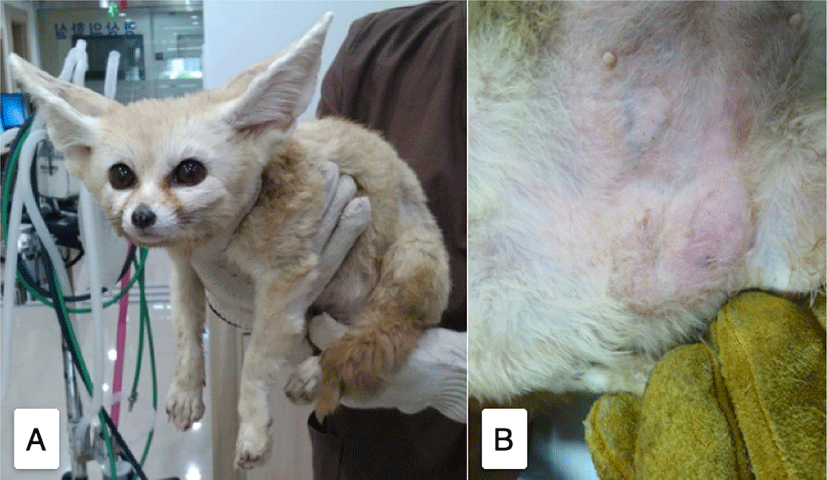
A 5-year-old female fennec fox weighing 1 kg was referred to the Veterinary Teaching Hospital, Chungbuk National University from Cheongju Zoo with swelling of the mammary gland, hematuria, and anorexia. On physical examination, all mammary glands, especially the fifth and sixth, were slightly distended (Fig. 1). A complete blood count (CBC), serum clinical biochemistry, hormone analysis, and abdominal ultrasonography were performed.
The results are shown in Tables 1 and 2. CBC, white blood count (WBC) differential count, and serum clinical biochemistry were done with ProCyte Dx (IDEXX, Westbrook, ME, USA), a manual method, HITACHI 7020 (Hitachi, Tokyo, Japan), and IMMULITE 1000 (Siemens, Germany), respectively. International Species Information System (ISIS, March 2002) reference intervals were used. WBC was increased with normocytic normochromic slight anemia (Table 1). Despite the increased amylase and lipase serum concentrations (Table 2), there was no evidence of pancreatitis.
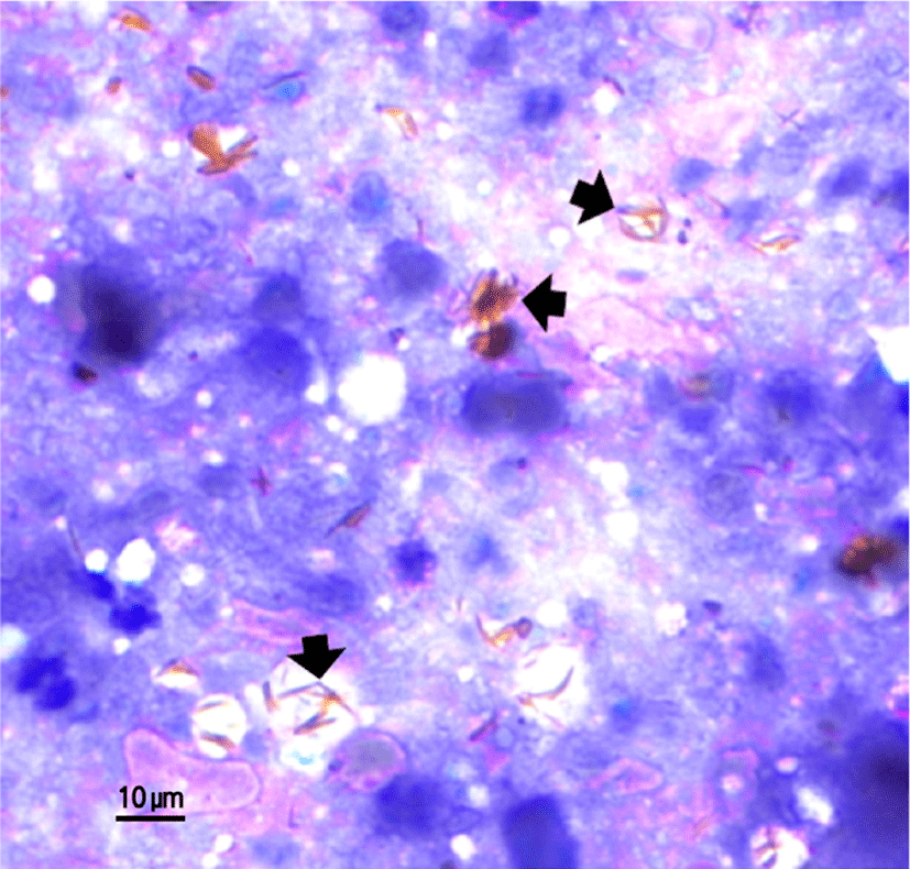
The serum progesterone concentration was 7.3 ng/mL, indicating mating during ovulation or just before parturition [1]. Cytology of the fluid from the uterus by fine needle aspiration showed many red blood cells (RBCs) and bilirubin crystals (Fig. 5). Bacterial culture of the uterine fluid was negative in both aerobic and anaerobic conditions of MacConkey broth culture. Urine analysis results were pH 7, protein 500 mg/dL, and WBC 500 /hpf. For the cytology, a uterine fluid sample that was collected after ovariohysterectomy was used. The fluid-smeared slide glass was stained with Wright-Giemsa. Cytologic examination showed low and high numbers of RBC and WBC, respectively. Many bilirubin crystals were observed upon microscopic examination, which implied a hemorrhagic condition in the uterus.
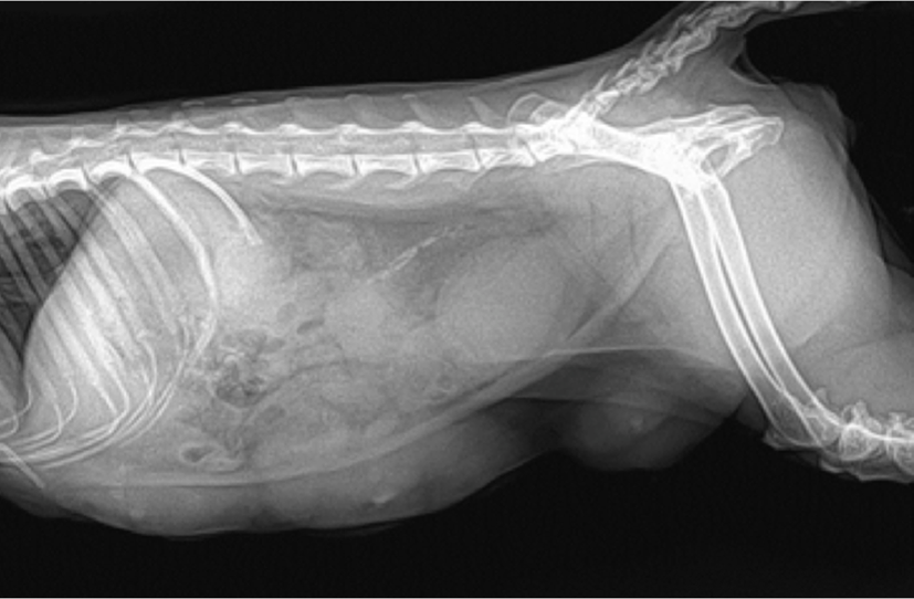
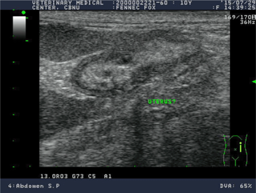
An abdominal ultrasound examination revealed that the uterus was enlarged and the mammary glands were swollen (Figs. 2 and 3). There was minimal free fluid in the abdominal cavity. The uterus was slightly distended, and both uterine horns were filled with hyperechoic floating material.
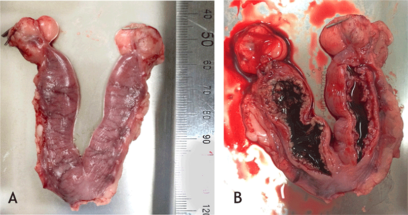
Ovariohysterectomy was performed in the fennec fox (Fig. 4) under inhalational anesthesia with isoflurane. Anesthesia was maintained using 2% isoflurane after induction using 4% isoflurane. Oxygen flow was set at 1.5 L/min. A histopathological examination was performed on the removed uterus. Postoperative care consisted of flunixin 2 mg/kg and enrofloxacin 10 mg/kg IM BID for 7 days.
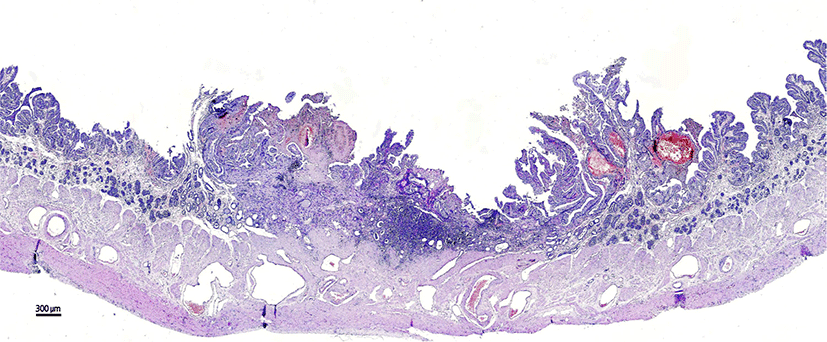
The zonary placenta papillae showed hyperplasia and enlargement change. A hyperplasia of the papillae hemorrhagic sign was observed upon histopathological examination. Moreover, mineralization was detected in the center of Fig. 6. Inflammatory cells had infiltrated the uterine tissue.
DISCUSSION
The reference interval of progesterone serum concentration for the fennec fox was limited [1]. However, the information of progesterone serum concentration change according to estrus cycle showed very similar patterns to those of the dog, wolf, and fennec fox [1, 5, 6]. The result of serum progesterone concentration in the fennec fox was 7.3 ng/mL, indicating that the fennec fox was at least in the post-ovulation phase.
Hematometra is an uncommon disease in Canidae. There are many reasons for hematometra, including uterine trauma, anticoagulant rodenticide toxicity and other acquired coagulation deficiencies, neoplasm, placental necrosis, and postpartum endometritis. Postpartum subinvolution of placental sites is also included among these reasons [7, 8].
Despite the fact that the clinical signs and physical examination results implied a pregnancy, a fetus was not discovered from the ultrasonography. A presumptive diagnosis of pyometra or uterine hyperplasia was made based on the physical examination and diagnostic findings. Ultimately, an ovariohysterectomy was performed. Canine pyometra may present clinically with inappetence, depression, polydipsia, lethargy, and abdominal distension, with or without vaginal discharge. Typically, the bitch is feverless and often has an elevated WBC. Prerenal azotemia commonly accompanies dehydration present with hyperproteinemia and hyperglobulinemia. The histopathological examination revealed endometrium hyperplasia from pyometra, hypertrophy, and small endometrial cysts scattered throughout the endometrium.
In this case, histopathological examination of the uterus indicated that the fennec fox had neither pyometra nor endometrium hyperplasia. Considering the results of several examinations, hematometra occurred from fetal death in the uterine cavity or pseudo-placental hyperplasia.
The canid has a zonary placenta, completely surrounding the fetus, and complex lamellar organization of the maternal and fetal tissues. It consists of the chorioallantoic membrane, the placental labyrinth, the necrosis zone, the maternal glandular chambers, the supraglandular layer, and the deep endometrial glands [9]. The amnion is avascular in the early stages but becomes vascularized by blood vessels of the internal allantoic membrane in the later stages of pregnancy [10].
In dogs, coagulation necrosis has been observed in two of the placentation sites early after fetal death. Edematous swelling of the maternal endothelium of blood vessels might contribute to hypoxia [9]. Hematometra is a very uncommon disease in pet animals like dogs and cats. Pyometra, endometrial hyperplasia, or fetal death in the uterus can possibly cause hematometra. However, in most cases of pets, the owners notice the clinical signs of pyometra and fetal death in the early stage and take appropriate action.
On the contrary, in wild animals, detecting clinical signs is very difficult because they tend to hide them. Thus, in this case, hematometra occurred because of fetal death. Hematometra from fetal death in canids and fennec foxes has not yet been studied. Thus, further investigations are needed for a more detailed understanding of the morphological processes occurring in hematometra after fetal death. In conclusion, we can assume that the fetus had died in the uterine cavity in the early stage and had been lysed by the uterus but that the hemorrhage had continued because the placenta remained after fetal absorption and that these events were the cause of the hematometra.
