Case Report
Oral leiomyosarcoma in a dog
Young Ju Kim
1,†
, Yeseul Jeon
1,†, Hyoena Bae
1, Sun Woo Shin
1, ARom Cho
1, Jae-Eun Hyun
1, Tae-Sung Hwang
1, Il-Hwa Hong
1, Hee-Chun Lee
1, Dong-In Jung
1, Kyu-Woan Cho
1, DoHyeon Yu
1,*
1College of Veterinary Medicine, Gyeongsang National University, Jinju 52828, Korea
*Corresponding author: DoHyeon Yu College of Veterinary Medicine, Gyeongsang National University, Jinju 52828, Korea, Tel: +82-55-772-2368, E-mail:
yudh@gnu.ac.kr
These authors contributed equally to this work.
© Research Institute of Veterinary Medicine, Chungbuk National University.
Received: Dec 17, 2021; Accepted: Dec 20, 2021
Abstract
Leiomyosarcoma is malignant mesenchymal tumor of smooth muscle and commonly encountered in the gastrointestinal tract of dogs. However, primary canine leiomyosarcoma in oral cavity is rare due to lack of smooth muscle in the oral tissue. A 13-year-old, neutered male Poodle presented a hard and immobile mass on the left maxilla. Imprinting cytology from the mass as well as fine-needle aspirated cytology from the left scapular lymph node revealed predominant spindle cells met malignancy criteria of the tumors, including coarse chromatin, high N/C ratio, nuclear molding, macro/multi-nucleoli with cigar-shaped nucleus. Radiography of the skull showed lysis of the nasopalatine bone, and mineral radiopacity in the mass. Computed tomography showed soft tissue attenuating mass from the left incisor teeth to the left retrobulbar space with loss of nasopalatine bone and medial wall of orbit. The histopathological examination showed irregularly arranged malignant spindle-shaped cells with oval or elongated nuclei. The nucleolus is distinct and moderate cellular polymorphism is observed. Mitotic figures are occasionally observed. The tumor cells are positive to vimentin, desmin, α-smooth muscle actin when immunohistochemistry was performed, and in Masson’s trichrome stain, tumor cells are stained as red. Overall, histopathologic exam and immunohistochemistry confirmed canine oral leiomyosarcoma. Because of the poor prognosis, the owner did not consent further treatment.
Keywords: leiomyosarcoma; mouth neoplasms; immunohistochemistry; vimentin; desmin
INTRODUCTION
Canine oral tumor is relatively common accounting for 6% of all tumors in dogs. The most common oral tumors in dogs include malignant melanoma, squamous cell carcinoma, fibrosarcomas [1-3]. Leiomyosarcoma is a malignant mesenchymal tumor originated from smooth muscle [4]. Leiomyosarcoma constitutes 20 to 30 per cent of all intestinal neoplasms in dogs [5], however, oral leiomyosarcoma is scare owing to paucity of smooth muscle structures in this location [6]. Therefore, no specific reference to their diagnosis, management and prognosis has been reported up to date [7]. Herein, we report maxillary leiomyosarcoma in a dog confirmed by immunohistochemistry.
CASE REPORT
A 13-year-old, neutered male Poodle was administered to veterinary medical teaching hospital in Gyeongsang National University for left maxillary mass. Initially, a small-sized mass (<1 cm) was found on the upper body of the maxilla, and it enlarged rapidly for the last 4 weeks. It was not responsive to antibiotics prescribed from the local veterinary clinic, and recently accompanied by dysphagia, hyporexia, and hemorrhage. Oral examination revealed a 4.8 cm × 2.5 cm-sized hard and immobile mass was observed on the left maxilla (Fig. 1A). Left prescapular lymph node was also enlarged, but no abnormalities in core temperature, purse rate, and respiratory rate were found. A complete blood count revealed normocytic normochromic regenerative anemia (hematocrit [HCT, 33.6%; reference range, 37.3%–61.7%; reticulocyte count, 117.6 × 10³/μL), leukocytosis (18.07 × 10³/ μL; reference range, 5.05–16.76 × 10³/μL), and thrombocytosis (651 × 103/μL; reference range, 148–484 × 103/μL). Serum biochemistry analysis revealed hyperglobulinemia (4.8 mg/dL; reference range, 2.5–4.5 mg/dL) (Table 1). Thromboelastgraphy (TEG) to determine the risk of hypercoagulability of the tumor revealed high MA, short K evaluated as hypercoagulable state. Cytological evaluation of the maxillary mass showed predominant spindle cells met malignancy criteria of tumor cells, including coarse chromatin, high N/C ratio, nuclear molding, macro/multi-nucleoli with cigar-shaped nucleus (Fig. 1B). Cytological evaluation of the enlarged left scapular lymph node by fine needle aspiration also revealed metastasis-associated malignant cells. Radiography of the skull showed lysis of the nasopalatine bone, and mineral radiopacity in the mass (Fig. 2A). Computed tomography showed soft tissue attenuating mass from the left incisor teeth to the left retrobulbar space with loss of nasal bone, palatine bone and orbital medial wall (Fig. 2B). The histopathological examination showed the irregularly arranged spindle-shaped cells, and oval or elongated nuclei of the tumor cells. The nucleolus is distinct and moderate cellular polymorphism is observed. Mitotic figures are occasionally observed (Fig. 3). The tumor cells are positive to vimentin, desmin, α-smooth muscle actin when immunohistochemistry was performed using anti-vimentin, anti-desmin, anti-alpha smooth muscle actin (α-SMA) (Santa Cruz Biotechnology, Dallas, TX, USA) antibodies. In Masson’s trichrome stain, tumor cells are stained as red (Fig. 4). Overall, histopathologic exam and immunohistochemistry confirmed canine oral leiomyosarcoma. However, owner did not consent surgical resection. Esophageal tubing for palliative therapy was applied and follow up was lost.
Table 1.
Clinicopathologic analysis in a dog with leiomyosarcoma
|
Complete blood count |
Results |
Reference intervals |
Serum biochemistry |
Results |
Reference intervals |
|
Red blood cells |
5.14 |
5.65–8.81 (× 109/L) |
Glucose |
103 |
74–143 (mg/dL) |
|
Hematocrit |
33.6 |
37.3–61.7 (%) |
Creatinine |
0.5 |
0.5–1.8 (mg/dL) |
|
Hemoglobin |
11 |
13.1–20.5 (g/dL) |
Urea nitrogen |
10 |
7–27 (mg/dL) |
|
Mean red cell volume |
65.4 |
61.6–73.5 (fL) |
Phosphorus |
4.9 |
2.5–6.8 (mg/dL) |
|
Mean cell hemoglobin concentration |
32.7 |
32–37.9 (g/dL) |
Calcium |
10 |
7.9–12 (mg/dL) |
|
White blood cells |
18.07 |
5.05–16.76 (× 109/L) |
Total protein |
7.5 |
5.2–8.2 (mg/dL) |
|
Neutrophils |
12.67 |
2.95–11.64 (× 109/L) |
Albumin |
2.8 |
2.3–4 (g/dL) |
|
Lymphocytes |
2.16 |
1.05–5.10 (× 109/L) |
Globulin |
4.8 |
2.5–4.5 (g/dL) |
|
Monocytes |
2.73 |
0.16–1.15 (× 109/L) |
Alanine aminotransferase |
26 |
10–125 (U/L) |
|
Eosinophils |
0.49 |
0.06–1.23 (× 109/L) |
Alkaline phosphatase |
57 |
23–212 (U/L) |
|
Basophils |
0.02 |
0.00–0.10 (× 109/L) |
Gamma-glutamyl transferase |
3 |
0–11 (U/L) |
|
Platelet count |
393 |
148–484 (× 109/L) |
Total bilirubin |
0.1 |
0–0.9 (mg/dL) |
|
|
|
|
Cholesterol |
139 |
110–320 (mg/dL) |
|
|
|
|
Triglyceride |
83 |
10–100 (mg/dL) |
|
|
|
|
Amylase |
812 |
500–1,500 (U/L) |
|
|
|
|
Lipase |
544 |
200–1,800 (U/L) |
Download Excel Table
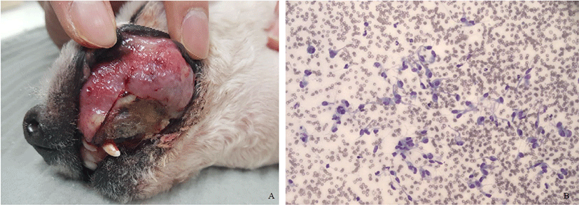
Fig. 1.
Macroscopic and microscopic examination of the oral leiomyosarcoma.
(A) A hard and immobile mass located on the left maxilla of the oral leiomyosarcoma. (B) Cytologic examination of the mass showing predominant malignant spindle-shaped cells satisfying malignancy criteria of tumor cells.
Download Original Figure
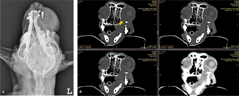
Fig. 2.
Diagnostic imaging analysis of the dog with oral leiomyosarcoma.
(A) Radiography of the skull showed bone lysis, and mineral opacity. (B) Computed tomography showed soft tissue attenuating mass from the left incisor teeth to the left retrobulbar space with loss of nasal bone, palatine bone and orbital medial wall (arrow).
Download Original Figure
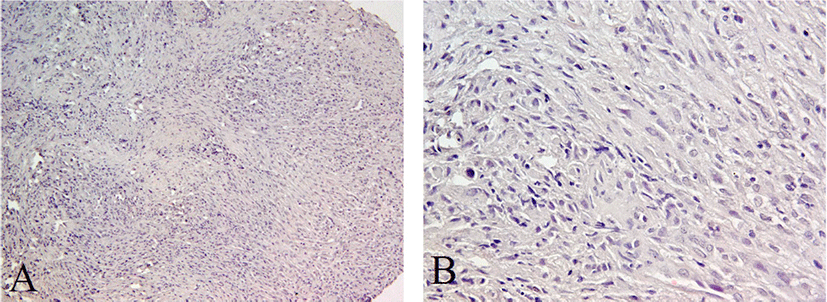
Fig. 3.
Histopathologic examination of the mass.
(A) the spindle-shaped cells are irregularly arranged. (B) The nuclei of the tumor cells are oval or elongated. The nucleolus of the tumor cells is distinct, and cellular polymorphism is moderated. Mitotic figures are occasionally seen. H&E, Original magnifications: 100 × (A), 400 × (B).
Download Original Figure
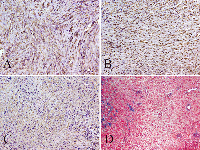
Fig. 4.
Immunohistochemistry and Masson’s trichrome stain of the mass.
(A) The tumor cells are positive to vimentin, (B) desmin, (C) α-smooth muscle actin. (D) In Masson’s trichrome stain, tumor cells are stained as red. A few Fibroblasts and some collagen in the vessels or connective tissue are stained as blue. Original magnifications: 100 × (D), 200 × (A, B, C).
Download Original Figure
DISCUSSION
Leiomyosarcoma is malignant mesenchymal tumor of smooth muscle. The most common site of the leiomyosarcoma is gastrointestinal tract, such as stomach, esophagus, small intestine, and colorectum in dogs [1, 8-11]. Oral leiomyosarcoma is rare in dogs due to lack of smooth muscle tissue in the oral cavity. Several reports suggested that the major sources of smooth muscle in the oral cavity are the walls of blood vessels or displaced embryonal tissue, the vascular system of bone or accessory salivary glands [6, 12]. Since it is not commonly encountered in veterinary clinic, leiomyosarcoma should be confirmed by histopathology and immunohistochemistry. The cells are typically composed of long, plump spindle cells, which have cytoplasm with oval blunt-ended nuclei, perinuclear vacuoles, and frequent nuclear chromatin crossbars, but collagenous stroma is little. Leiomyosarcomas are positive for vimentin, a-smooth muscle actin as well as desmin in the immunohistochemistry [13, 14]. The present case present oral mass on the left maxilla, and leiomyosarcoma was confirmed by immunohistochemistry. Sarcomas can be the top of the differential list for cigar-like nuclear morphology and spindle cells met malignant criteria of the tumor cells. In case of oral sarcomas, leiomyosarcoma can be considered if cytological analysis are consistent with smooth muscle-originated cells.
In human, the prognosis of leiomyosarcoma is poor, with a high percentage of recurrence or metastasis. The most common sites of metastasis include the lungs, bone, brain and the lymph node [15]. The most effective treatment of the leiomyosarcoma is radical surgical excision with adequate margins [10, 16]. For inoperable tumors or those in difficult sites such as the mandibular condyle or maxillary sinus, radiotherapy can be used in addition to surgery [17]. Chemotherapy appeared to be ineffective unless it was used as a palliative measure in patients with widespread metastatic disease [18]. Kapatkin et al. reported that the prognosis in dogs with leiomyosarcoma of the spleen, stomach, small intestine, and especially the cecum is good if surgery is performed [5]. Though it has not been sufficiently studied in dogs, the site of the leiomyosarcoma is more important for prognosis when compared to human study: those arising in a superficial cutaneous localization have the best prognosis [15] whereas bony involvement is a potential negative prognostic indicator in human [19]. In the present case, there was an extensive soft tissue mass from the left incisor teeth to the left retrobulbar space with bone invasion, thus complete excision with surgery was impossible. Unfortunately, proper treatment options were not applied, but the prognosis might not be good considering given the size and the location of the lesion.
Acknowledgements
This research was supported by Basic Science Research Program through the National Research Foundation of Korea (NRF) funded by the Ministry of Science, ICT & Future Planning (2020R1C1C1008675).
REFERENCES
Liptak J. Cancer of the gastrointestinal tract. In: Withrow and Macewen’s small animal clinical oncology. 6th ed. St. Louis: Elsevier; 2007. p. 462.
Vos JH, van der Gaag I. Canine and feline oral-pharyngeal tumours. Zentralbl Veterinarmed A 1987;34:420-427.


Todoroff RJ, Brodey RS. Oral and pharyngeal neoplasia in the dog: a retrospective survey of 361 cases. J Am Vet Med Assoc 1979;175:567-571.
Amarapala H, Tilakaratne WM. Leiomyosarcoma of the oral cavity: report of seven cases and review of literature. Oral Oncology Extra 2006;42:14-17.
Kapatkin AS, Mullen HS, Matthiesen DT, Patnaik AK. Leiomyosarcoma in dogs: 44 cases (1983–1988). J Am Vet Med Assoc 1992;201:1077-1079.
Takagi M, Ishikawa G. An autopsy case of leiomyosarcoma of the maxilla. J Oral Pathol Med 1972;1:125-132.
Boy SC, Van Heerden WFP, Steenkamp G. Diagnosis and treatment of primary intraoral leiomyosarcomas in four dogs. Vet Rec 2005;156:510-513.
Frost D, Lasota J, Miettinen M. Gastrointestinal stromal tumors and leiomyomas in the dog: a histopathologic, immunohistochemical, and molecular genetic study of 50 cases. Vet Pathol 2003;40:42-54.
LaRock RG, Ginn PE. Immunohistochemical staining characteristics of canine gastrointestinal stromal tumors. Vet Pathol 1997;34:303-311.
Penninck DG. Characterization of gastrointestinal tumors. Vet Clin North Am Small Anim Pract 1998;28:777-797.
Lo Muzio L, Favia G, Farronato G, Piattelli A, Maiorano E. Primary gingival leiomyosarcoma: a clinicopathological study of 1 case with prolonged survival. J Clin Periodontol 2002;29:182-187.
Izumi K, Maeda T, Cheng J, Saku T. Primary leiomyosarcoma of the maxilla with regional lymph node metastasis. Report of a case and review of the literature. Oral Surg Oral Med Oral Pathol Oral Radiol 1995;80:310-319.
Gross TL, Ihrke PJ, Walder EJ, Affolter VK. Smooth muscle and skeletal muscle tumors. In: Gross TL, Ihrke PJ, Walder EJ, Affolter VK (eds.). Skin diseases of the dog and cat: clinical and histopathologic diagnosis. 2nd ed. Ames: Blackwell; 2008. p. 778-785.
Andreasen CB, Mahaffey EA. Immunohistochemical demonstration of desmin in canine smooth muscle tumors. Vet Pathol 1987;24:211-215.
Pinheiro JJV, Alves SM, Okuda E, Jorge WA, Jaeger RG, de Araújo NS. Primary leiomyosarcoma of the mandible. A case report. Med Oral Patol Oral Cir Bucal 2007;12:56-59.
Goldschmidt PR, Goldschmidt JD, Lieblich SE, Eisenberg E. Leiomyosarcoma presenting as a mandibular gingival swelling: a case report. J Clin Periodontol 1999;70:84-89.
Schenberg ME, Slootweg PJ, Koole R. Leiomyosarcomas of the oral cavity. Report of four cases and review of the literature. J Craniomaxillofac Surg 1993;21:342-347.
Fields JP, Helwig EB. Leiomyosarcoma of the skin and subcutaneous tissue. Cancer 1981;47:156-169.
Recine F, Bongiovanni A, Casadei R, Pieri F, Riva N, De Vita A, Mercatali L, Liverani C, Spadazzi C, Miserocchi G, Fausti V, Amadori D, Ibrahim T. Primary leiomyosarcoma of the bone: a case report and a review of the literature. Medicine 2017;96:e8545.