Journal of Biomedical Translational Research
Research Institute of Veterinary Medicine, Chungbuk National University
Original Article

Osteological study of Sus scrofa relics excavated from the Gasan-Ri archaeological site in Jinju

Gunjan Thakur1https://orcid.org/0000-0002-9958-2148, Si-Joon Lee1https://orcid.org/0000-0002-5870-687X, Jong-Hyuk Choi2https://orcid.org/0000-0001-5426-494X, Chong-Sup Kim1https://orcid.org/0000-0001-6722-2634, Chung-Kil Won1,*https://orcid.org/0000-0002-3105-1869
1Institute of Animal Medicine & Department of Veterinary Medicine, College of Veterinary Medicine, Gyeongsang National University, Jinju 52828, Korea
2Gyeongnam Institute of Cultural Properties, Changwon 51402, Korea
*Corresponding author: Chungkil Won, Institute of Animal Medicine & Department of Veterinary Medicine, College of Veterinary Medicine, Gyeongsang National University, Jinju 52828, Korea Tel: +82-55-772-2351, E-mail: wonck@gnu.ac.kr

© Research Institute of Veterinary Medicine, Chungbuk National University.

Received: Oct 21, 2021; Revised: Nov 12, 2021; Accepted: Nov 15, 2021

Abstract

The natural relics excavated from the archeological site reveal the life, hunting, fishing, and gathering of mankind at the time, and the animal bones excavated from the remains become valuable data for the study of geography and bone diseases at the time. This study investigated the morphological structure, size, and osteometry of Sus scrofa bones excavated from the well area of the Gasan-Ri archaeological site in Jinju, presumed to be a relic of the Three Kingdoms period. The total weight of identified animal bones was 975.30 g and the number of bone fragments was 447. Among these 447 animal bones identified, the bone weight of Sus scrofa was 468.00 g (47.99%) and the number of bone fragments was 204 (45.64%). These 204 bone fragments of Sus scrofa consisted of 35 skulls (17.16%), 94 vertebrate axial skeletons (46.08%), 28 forelimb bones (13.73%), and 47 hindlimb bones (23.04%). The bones of Sus scrofa have been identified as those of two animals. The estimated age of these bones of Sus scrofa was 5-7 weeks. There was a fracture presumably caused by an artificial blow to the parietal bone located near the bregma in the skull.

Keywords: archaeological site; Gasan-Ri; Jinju; Sus scrofa

INTRODUCTION

유적지에서 출토된 자연유물은 당시 인류의 수렵, 어로, 채집 등의 생활양식과 동물분포상을 규명하는 데 중요한 단서가 되고, 출토된 동물뼈 유물은 당시의 사냥 동물의 종류와 인간의 문화적 수준을 이해하는 귀중한 자료로 활용할 수 있을 뿐만 아니라[17], 동물의 뼈 질병을 검색할 수도 있다[8, 9].

우리나라 유적지에서의 동물 뼈 유물에 관한 연구로는 김해 수가리패총에서 출토된 동물유물로는 연골어강(Chondrichthyes), 골어강(Osteichthyes), 양서강(Amphibia), 포유강(Mammalia) 동물뼈에 관한 보고[10], 진해용원패총 유적지에서 척추동물과 무척추동물의 뼈에 관한 보고[11], 백령도 말등유적의 뼈 유물에 관한 보고[12], 제주 곽지유적에서 출토된 동물뼈에 관한 보고[4, 13], 제주 종달리패총유적에 관한 보고[3] 및 제주 김녕리 궤내기동굴유적에서 출토된 동물뼈 유물에 관한 보고 등이 있다[5].

본 조사에서는 진주시 이반성면 가산리 우물지에서 출토된 동물뼈 중에서 동정된 멧돼지뼈의 특징과 크기를 측정하여 출토 당시 멧돼지뼈 유물의 해부학적 특징을 조사하였다.

MATERIALS AND METHODS

재료는 진주시 이반성면 가산리의 우물지에서 출토된 멧돼지뼈 유물을 이용하였다[14]. 출토된 멧돼지의 동물뼈는 부위별로 분류하였고, 현존 동물뼈와 비교 계측 조사를 위해 경상대학교 수의과대학 해부학 표본실에 보관 중인 동물 골격 표본을 이용하여 멧돼지 동물뼈 유물과 유사한 동물의 종, 나이, 크기 등을 고려한 동물의 골격 표본을 활용하여 비교 분석하였다.

동물뼈는 Schmid[15]의 방법에 따라 동물뼈 유물을 분류하였고, 골격을 몸 부위에 따라 머리뼈(skull), 척추(vertebrae), 갈비뼈(ribs), 앞다리뼈(bones of forelimb), 뒷다리뼈(bones of hindlimb)로 각각 구분하였다. 멧돼지뼈 유물의 계측은 Driesch [16]의 방법에 따라 Mitutoyo caliper (0.01 mm)로 측정하였다.

RESULTS

삼국시대 유적지로 추정되는 진주시 이반성면 가산리 우물지에서 출토된 동물뼈 중에서 동정된 동물뼈는 97.26%(975.30 g)로 뼈조각의 수는 447개(89.76%)였다. 그 중에서 멧돼지(Sus scrofa)의 뼈로 분류된 것은 47.99%(468.00 g)로 뼈조각의 수는 204개(45.64%)였다.

동정된 멧돼지의 뼈는 두 마리의 것으로 확인되었는데 멧돼지 유물뼈의 탈락치아 또는 젖니인 둘째작은어금니가 분출(eruption)되어 있어서 약 5–7주령의 새끼로 추정하였다. 멧돼지 유물뼈와 현존하는 50일령 랜드레이스 돼지 및 야생 멧돼지 뼈의 크기를 측정하여 비교 해부학적으로 분석하였다. 멧돼지 유물뼈의 두개지수는 각각 49.41, 50.13이었고, 현존하는 50일령 랜드레이스의 두개지수는 62.90이었으며 성체 멧돼지의 두개지수는 36.41이었는데 출토된 멧돼지 뼈는 현존하는 50일령 랜드레이스보다는 야생 멧돼지의 크기에 더 가까워 출토된 돼지 유물뼈는 멧돼지로 분류하였다(Table 1).

Table 1. Measurement of animal bones of Sus scrofa excavated from Gasan-Ri archeological site (unit: mm)
Classification of bone Measurement Ancient animal bones Present animal bones
(A) (B) 50 days landrace Wild boar (adult)
Skull Skull index 49.41 50.13 62.90 36.41
Profile length: Akrokranion-Prosthion 143.42 139.07 138.10 365.03
Opisthokranion-Prosthion 146.65 140.15 140.33 369.93
Greatest mastoid breadth 60.52 59.41 68.70 103.04
Greatest breadth of the occipital condyles 38.88 42.64 53.58
Greatest breadth of the foramen magnum 18.16 19.49 24.34
Height of the foramen magnum: Basion-Opisthion 20.44 19.97 27.25
Greatest breadth of the squamous part of the occipital bone. 37.36 43.52 64.75
Least breadth of the parietal-least breadth between the temporal lines. 26.17 31.66 19.75
Greatest frontal breadth - greatest breadth across the supraorbital process. 59.75 62.05 94.35
Zygomatic breadth=greatest breadth of skull: Zygion-Zygion. 70.87 69.71 86.87 132.92
Mandible Length from the angle: Gonion caudale-Infradentale. 108.94 101.85 261.17
Length from the condyle: Aboral border of the condyle process-Infradentale. 113.92 108.15 280.14
Aboral height of the vertical ramus: Gonion ventrale- highest point of the condyle process. 44.71 48.47 112.84
Middle height og the vertical ramus: Gonion ventrale- deepest point of the mandibular notch. 40.03 41.40 104.29
Oral height of the vertical ramus: Gonion ventrale- Coronion. 46.72 49.22 118.92
DP4 Length (L) and breadth (B) near the base of the crown L 22.47 20.84 18.95 M3 L 27.46
B 9.93 9.04 8.40 M3 B 19.83
Hip bone Greatest length of one half 101.71 97.32 94.53 232.48
Greatest breadth across the Tubera coxarum- Greatest breadth across the lateral angle 69.03 63.02 84.49 160.11
Smallest breadth of the shaft of ilium 7.98 7.07 8.58 13.86
Smallest height of the shaft of ilium 11.30 11.44 13.06 25.69
Greatest breadth across the acetabula 55.68 51.69 63.95 116.52
Smallest breadth across the bodies of the ischis 36.35 52.39 38.45 74.87
Greatest breadth across the tubera ischiadica 43.80 41.77 45.65 94.39
Length of the acetabulum including the lip 25.20 26.57 30.28 39.00
Humerus Greatest length 102.34 92.41 234.15
Greatest breadth of the proximal end 29.25 30.62 62.12
Greatest breadth of the distal end 28.79 28.07 53.32
Smallest breadth of the distal end 9.80 12.02 21.60
Ulna Greatest length 75.13 69.71 183.06
Greatest breadth across the coronoid process 13.94 16.48 25.75
Download Excel Table

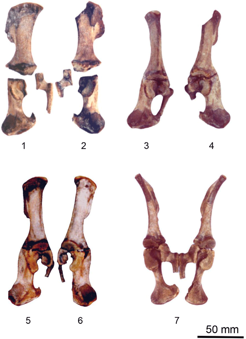
출토된 머리뼈 중에서 한 마리에서는 11개의 뼈조각을 확인하였고 다른 한 마리는 9개의 뼈조각을 접합시켜서 부분적으로 복원하였다(Fig. 1). 두 개체의 머리뼈는 모두 정수리점(bregma)에서 정중선을 따라 뒤통수뼈(occipital bone)와 왼쪽 마루뼈(parietal bone) 연결 부위가 파손되어 있었는데(Fig. 1. 1, 4의 화살표), 파손된 부위는 도축장에서 돼지를 기절시킬 때 사용되는 부위와 거의 일치하는 곳이었다. 멧돼지 유물의 머리뼈에서 옆면의 최대 길이는 각각 143.42 mm, 139.07 mm였고 뒤통수돌기의 중앙에서 최대 길이는 각각 146.65 mm, 140.15 mm였으며, 꼭지돌기 부위에서 최대폭은 각각 60.52 mm, 59.41 mm였고 광대뼈 부위에서 최대폭은 각각 70.87 mm, 69.71 mm였다(Table 1).

jbtr-22-4-149-g1
Fig. 1. Photographs of skulls of Sus scrofa excavated from the remains of Gasan-Ri. 1. skull (dorsal view); 2. skull (ventral view of Fig. 1); 3. skull (ventral view); 4. skull (dorsal view); 5. mandible; 6. mandible (right); 7. mandible (left).
Download Original Figure

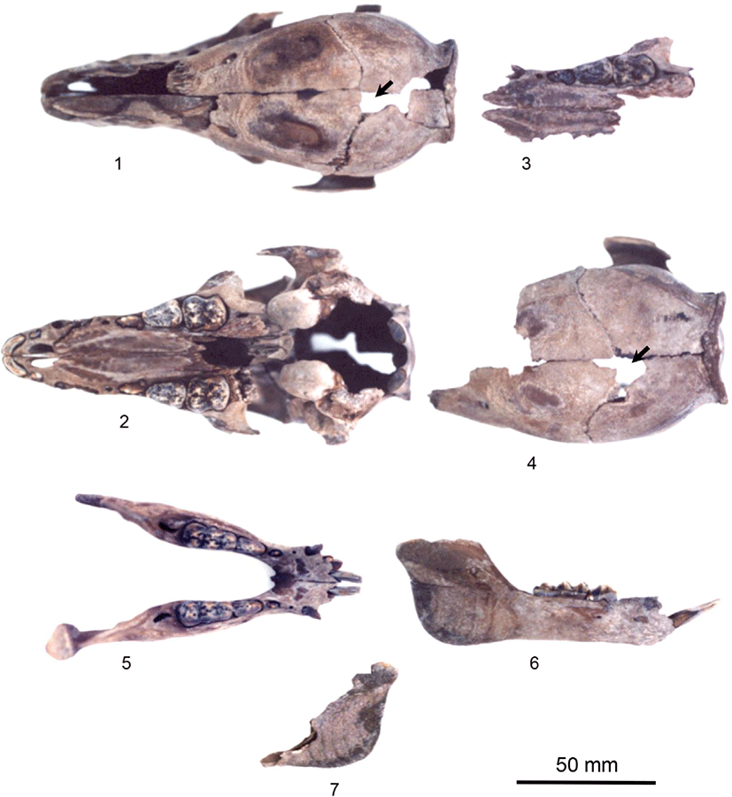
아래턱뼈(mandible)는 4개(왼쪽 2, 오른쪽 2; 이하 동일방법)로 두 마리의 것이었는데, 한 마리에서는 왼아래턱뼈의 근육돌기(coronoid process) 부분이 약간 파손된 것을 제외하고는 거의 완전한 형태로 출토되었다. 다른 개체는 오른아래턱뼈의 관절돌기(condylar process) 부분에서 수평으로 윗부분이 소실되었으며, 왼아래턱뼈는 젖니인 넷째작은어금니 뒤쪽에서 앞쪽으로 비스듬히 절단되어 있었다(Fig. 1. 5-7). 멧돼지 유물에서 아래턱뼈의 최대 길이는 113.92 mm였고 수직 부분의 최대 높이는 46.72 mm였다(Table 1).

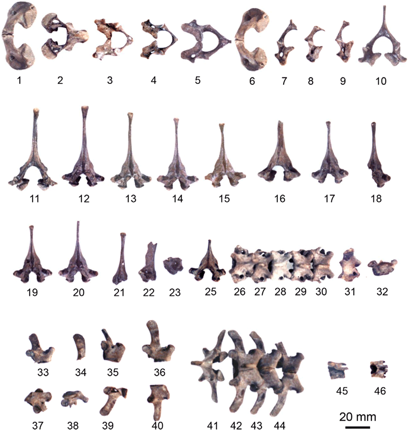
척추는 목뼈(cervical vertebrae)가 10개(Fig. 2. 1-10), 등뼈(thoracic vertebrae) 21개(Fig. 2. 11-32), 허리뼈(lumbar vertebrae) 12개(Fig. 2. 33-44), 엉치뼈(sacral vertebrae) 2개(Fig. 2. 45, 46)였으며 꼬리뼈는 발견되지 않았다.

jbtr-22-4-149-g2
Fig. 2. Photographs of vertebrae of Sus scrofa excavated from the remains of Gasan-Ri. 1. atlas; 2. axis; 3-5. cervical vertebrae; 6. atlas; 7-10. cervical vertebrae; 11-32. thoracic vertebrae; 33-44. lumbar vertebrae; 45, 46. sacral vertebrae.
Download Original Figure

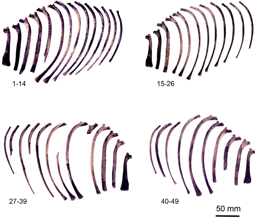
갈비뼈는 49개(26, 23)였고 갈비뼈머리의 최대 길이는 5.28(4.48-5.78) mm로 갈비뼈결절의 최대 길이 3.91(3.40-4.80) mm보다 길었다(Fig. 3).

jbtr-22-4-149-g3
Fig. 3. Photographs of ribs of Sus scrofa excavated from the remains of Gasan-Ri. 1-14. ribs (left); 15-39. ribs (right); 40-49.
Download Original Figure

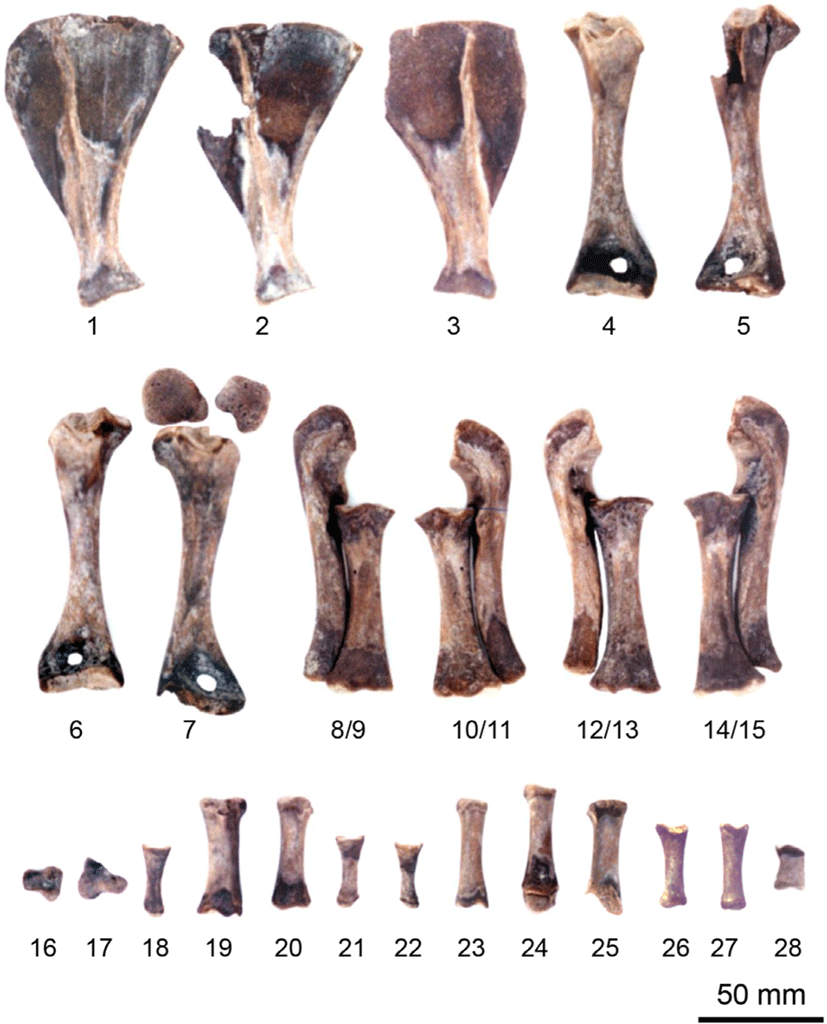
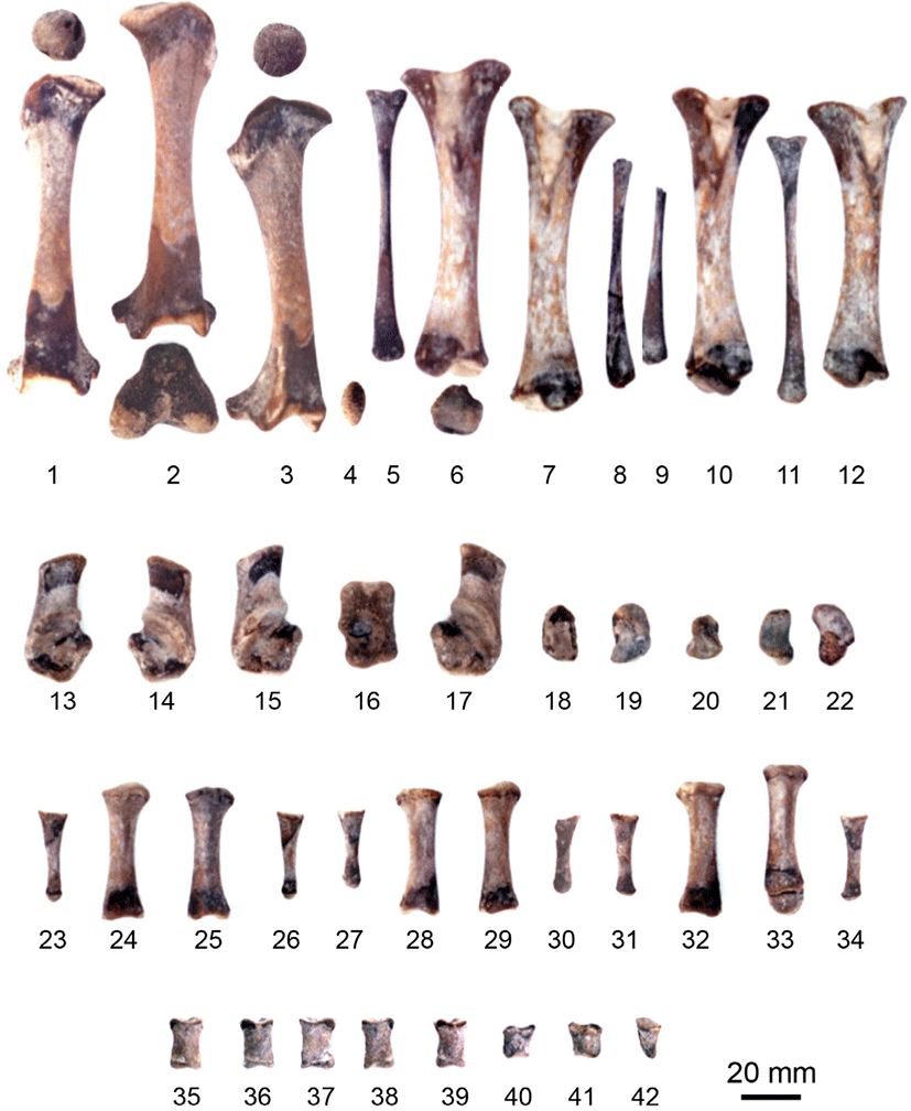
어깨뼈(scapula)는 3개(2, 1)였고(Fig. 4. 1-3), 상완뼈(humerus)는 4개(2, 2)였으며(Fig. 4. 4-7), 넙다리뼈 몸통의 최대 길이는 102.34 mm였고 몸쪽 부위의 최대폭은 29.25 mm, 30.62 mm였으며 최소폭은 9.80 mm였다(Table 1). 노뼈(radius)와 자뼈(ulna)는 각각 4개(4, 4)였다(Fig. 4. 8-15). 앞발목뼈(carpal bones)는 2개로 중간앞발목뼈와 첫째앞발목뼈가 각각 1개씩이었다(Fig. 4. 16, 17). 앞발허리뼈(metacarpal bones)는 10개였는데, 1개를 제외한 9개는 뼈끝이 소실되어 있었다(Fig. 4. 18-27). 앞발가락뼈(digital phalanges)는 중간마디뼈 1개뿐이었다(Fig. 4. 28).

jbtr-22-4-149-g4
Fig. 4. Photographs of forelimb bones of Sus scrofa excavated from the remains of Gasan-Ri. 1, 2. scapula (L); 3. scapula (R); 4. humerus (L); 5. humerus (R); 6. humerus (R); 7. humerus (L); 8. ulna (R); 9. radius (L); 10. radius (R); 11. ulna (L); 12. ulna (R); 13. radius (L); 14. radius (R); 15. ulna (L); 16. intermediate carpal bone; 17. first carpal bone; 18-27. metacarpal bones; 28. middle phalanx. R, right; L, left.
Download Original Figure

볼기뼈(hip bone)는 엉덩뼈(ilium), 궁둥뼈(ischium), 두덩뼈(pubis)가 각각 4개(2, 2)였고 두 마리의 것이었는데, 왼쪽 및 오른쪽 볼기뼈(Fig. 5. 1, 2)를 복원시켰고(Fig. 5. 3, 4), 왼쪽과 오른쪽 볼기뼈(Fig. 5. 5, 6)를 접합시킨 것이 Fig. 5. 7이다. 복원시킨 볼기뼈의 절반 최대 길이는 101.71 mm, 97.32 mm였고, 최대폭은 69.03 mm, 63.02 mm였다(Table 1).

jbtr-22-4-149-g5
Fig. 5. Photographs of hip bones of Sus scrofa excavated from the remains of Gasan-Ri. 1. hip bone (L); 2. hip bone (R); 3. hip bone (L); 4. hip bone (R); 5. hip bone (L); 6. hip bone (R); 7. hip bone (L and R). R, right; L, left.
Download Original Figure

넙다리뼈(femur)는 3개(1, 2)였으며(Fig. 6. 1-3) 무릎뼈(patella)는 1개만 보존되어 있었다(Fig. 6. 4). 정강뼈(tibia)와 종아리뼈(fibula)는 각각 4개씩이었다(Fig. 6. 5-12). 뒷발목뼈(tarsal bones)는 10개였는데(Fig. 6. 13-22), 뒷발꿈치뼈(calcaneus)가 4개(2, 2), 목말뼈(talus) 1개(1, 0)가 포함되었다(Fig. 6. 13-17). 뒷발허리뼈(metatarsal bones)는 12개였는데 먼쪽 뼈끝이 분리 소실된 것도 있었다(Fig. 6. 23-34). 뒷발가락뼈(pedal phalanges)는 8개였는데 첫마디뼈가 5개(Fig. 6. 35-39), 중간마디뼈가 2개(Fig. 6. 40, 41), 끝마디뼈가 1개였다(Fig. 6. 42).

jbtr-22-4-149-g6
Fig. 6. Photographs of hindlimb bones of Sus scrofa excavated from the remains of Gasan-Ri. 1. femur (R); 2. femur (L); 3. femur (R); 4. patella; 5. fibula (R); 6. tibia (R); 7. tibia (L); 8. fibula (L); 9. fibula (R); 10. tibia (R); 11. fibula (L); 12. tibia (L); 13. calcaneus (L); 14. calcaneus (R); 15. calcaneus (L); 16. talus (R); 17. calcaneus (R); 18. central tarsal bone; 19. fourth tarsal bone; 20. third tarsal bone; 21. central tarsal bone; 22. fourth tarsal bone; 23-34. metatarsal bones; 35-39. proximal phalanx; 40, 41. middle phalanx; 42. distal phalanx. R, right; L, left.
Download Original Figure

DISCUSSION

멧돼지(Sus scrofa)는 우제목(Artiodactyla) 멧돼지과(Suidae)의 멧돼지속(Sus)에 해당하는데, 멧돼지는 6개의 속(genus)으로 나누어져 있어 멧돼지속에 해당하는 것의 일부가 순화되어 집돼지가 되었다[17]. 류큐(琉球) 선사문화에서 돼지는 야생종과 가축종의 교배가 실제로 행해지고 있었으므로 형태상으로 야생종과 가축종을 구별하기는 어렵다[18]. 우리나라 고농서(高農書)에서는 고려 이전에 돼지를 가축의 종류로 보았고[19], 돼지의 사육연대를 적어도 2,000년 이상으로 보았는데 돼지의 사육은 삼국시대 이전인 고조선 시대로 보았다[20]. 동물뼈 유물에 대한 보고로 백령도 말등유적의 뼈 유물에서는 돼지의 왼쪽 뒤팔뼈(자뼈로 확인되었음)는 뼈의 크기와 앞팔뼈에 붙는 면의 상태로 보아 어린 돼지로 추정하였으나 멧돼지 또는 집돼지를 구분하기는 어렵다[12].

본 연구의 가산리 우물지에서 출토된 멧돼지의 뼈조각은 대체로 양호한 상태로 보존되어 있었고, 멧돼지의 뼈는 출토된 전체 골격의 45.64%를 차지하였는데, 제주 김녕리 궤내기동굴유적의 멧돼지 점유율(75%)보다는 낮았고[5], 제주 고내리유적의 점유율(48%)과 비슷하였으며[14], 제주 종달리패총유적의 멧돼지뼈 점유율(13.7%)[13]과 제주 곽지패총유적에서 멧돼지의 점유율(18.1%)[4] 및 진해 용원패총에서 출토된 멧돼지의 점유율(12.9%)보다는 높았다[11]. 따라서 본 연구의 가산리 우물지에서 출토된 멧돼지뼈는 삼국시대의 유적에서 출토된 것으로 멧돼지를 사냥하거나 가축으로 사육하였는지를 확인할 수 없지만 우리나라 남부지역에서 출토된 동물뼈에서 돼지가 차지하는 비율이 매우 높았던 보고와 비슷하였다.

본 연구의 진주 가산리 우물지에서 출토된 멧돼지를 해부학적으로 비교하기 위해 경상대학교 수의과대학 표본실에 보관 중인 버크셔(Berkshir)종과 야생 멧돼지의 성체 골격의 머리뼈 측정치를 비교하였다. 버크셔 성체 돼지와 야생 멧돼지에서의 옆 얼굴길이와 최대 머리폭의 비율은 각각 320 mm : 240 mm; 365 mm : 132 mm였는데 두개지수는 각각 버크셔가 75이고 야생 멧돼지는 36.41이었으며 현존하는 랜드레이스 50일령의 두개지수는 62.90이었는데, 본 연구에서 멧돼지뼈 유물 2마리의 두개지수는 각각 49.41과 50.13으로 확인되어 멧돼지뼈에 더 가깝다고 할 수 있다.

한편, 본 연구에서 멧돼지의 골격은 탈락치아 또는 젖니인 둘째작은어금니가 분출(eruption)되어 있어서 약 5-7주령의 새끼로 추정하였는데[21], 멧돼지뼈 유물에서 넙다리뼈의 최대 길이(102.34 mm)는 현존하는 50일령 랜드레이스의 길이(92.41 mm)보다 길었으나, 멧돼지 유물의 넙다리뼈 몸통의 최소폭(9.80 mm)은 랜드레이스 50일령의 최소폭(12.08 mm와)보다 훨씬 좁았다. 또한, 갈비뼈머리의 최대 길이(5.28 mm)는 갈비뼈결절의 최대 길이(3.91 mm)보다 길었는데, 대조군인 현존 야생 멧돼지 성체의 갈비뼈머리 최대 길이(9.87±0.74 mm)는 갈비뼈결절의 최대 길이(7.49±1.85 mm)보다 길었고 현존하는 야생 멧돼지 성체의 것보다 작았다. 이러한 결과에서 유물 멧돼지의 골격은 멧돼지에 더 근접하였다.

출토된 유적의 뼈에서 질병을 검색하였다는 보고가 있고[8, 9], 유적 멧돼지의 노뼈와 허리뼈에서 골류(骨瘤)가 관찰되어 고대에도 현대와 같은 뼈 관련 질병이 있었음을 보고하였는데[7], 본 연구에서 멧돼지 골격은 약 5-7주령의 새끼로 골류 등의 뼈 병변(病變)은 관찰되지 않았다.

돼지는 아주 오래 전부터 사람들과 아주 밀접한 관계를 가졌던 중요한 짐승으로 생각된다. 그런데 우리나라 유적에서 출토된 돼지가 거의 멧돼지로 분류되고 있는 점은 적지 않은 의문이 있지만[12], 돼지는 각지에 형질이 다른 야생종이 분포하였고, 야생종과 가축종과의 교배도 실제로 행하여 지고 있었기 때문에 형태만으로는 야생종과 가축종을 구별하기는 어렵다[18]. 따라서 가산리 유적에서 출토된 멧돼지의 뼈도 야생종인지 가축화된 돼지인지에 대한 형태학적인 분류는 한계가 있으며, 일부 동물종은 가축화되었을 가능성이 높은데, 옛 문헌 기록(三國志, 魏書 東夷傳)에 기원 전‧후 시기에 소와 돼지를 즐겨 길렀다는 내용으로 보아 이 시기에 소와 돼지가 가축화가 되었을 것으로 추측하였다[22]. 이처럼 유적에서 발굴된 자연유물로써 동물뼈 유물의 연구는 당 시대의 동물 분포상과 인간이 동물에게서 취한 여러 가지 경제적 가치와 생활양식을 따져 볼 수 있는데, 본 연구의 가산리 우물지에서 출토된 멧돼지의 머리뼈에는 이마뼈의 정수리점 부위가 파손되었고 어린 멧돼지의 뼈 말단 부위의 연골이 보존된 것으로 보아 이들 동물을 식용으로 이용한 것은 아닌 것으로 추정된다.

CONCLUSION

본 연구는 삼국시대 유물로 추정되는 진주 가산리 우물지 유적에서 출토된 멧돼지뼈 유물의 형태학적 구조와 크기를 현존하는 동물과 비교하여 조사하였다. 출토된 동물뼈 중에서 동정된 동물 뼈의 총 무게는 975.30 g으로 뼈조각의 수는 447개였다. 동정된 동물뼈 중에서 멧돼지뼈의 무게는 468.00 g(47.99%)이었는데 뼈조각의 수는 204개(45.64%)로 두 마리의 것으로 확인되었다. 204개의 멧돼지 뼈조각은 머리뼈 35개(17.16%), 몸통뼈 94개(46.08%), 앞다리뼈 28개(13.73%), 뒷다리뼈 47개(23.04%)로 구성되었다. 멧돼지뼈의 추정 연령은 생후 약 5-7주였다. 멧돼지의 머리뼈에서는 정수리점 부근에 위치한 마루뼈에 인위적인 타격으로 인한 골절이 관찰되었다.

Conflict of Interest

No potential conflict of interest relevant to this article was reported.

Acknowledgements

Not applicable.

Ethics Approval

Not applicable.

REFERENCES

1.

Kim HL, Kang CH, Shin TK. Comparative anatomical study on animal bones excavated from the Jongdal-Ri shell mound archaeological site Ⅳ on Jeju island. Korean J Vet Res 2002;42:309-320.

2.

Kim HC, Jung KS, Lee KH, Kang CH, Ahn MJ, Kang JC, Lee YD, Shin TK. Comparative anatomical study of animal bones excavated from the Konaeli archaeological site on Jeju Island. J Subtrop Agric Biotechnol (Jeju Natl Univ) 2002;18:129-141.

3.

Shin T. An osteological study of animal bones excavated from Jeju Jongdali shell-mount. Korean J Vet Res 2001;41:275-279.

4.

Shin TK, Kim SH, Yang KC, Koh JH, Lee BM, Ann SJ, Jin SH, Koh EM. Anatomical study of animal bones excavated from Kwakji historic site in Jeju island. Korean Antiq. 1992;40:31-32.

5.

Shin TK, Jin JK, Lee CS. Archaeological study of animal bones excavated from Cheju Kimnyungri cave site. Korean J Vet Res 1996;36:757-761.

6.

Nishinakagawa H, Hiji H, Matsumoto M, Otsuka J, Nakashima T. A study of animal bones from archaeological sites: VI. on the animal bones excavated at Muginoura shell-mound, Kagoshima Prefecture. Bull Fac Agric Kagoshima Univ 1987;37:105-113.

7.

Nishinakagawa H, Matsumoto M, Otsuka J, Kawaguchi S. A study of animal bones from archaeological sites: VI. on the animal bones excavated at Murokawa cave site, Kagoshima Prefecture. Bull Fac Agric Kagoshima Univ 1983;33:147-157.

8.

Baker J, Brothwell D. Animal diseases in archaeology. London: Academic Press; 1980.

9.

Chaplin RE. The study of animal bones from archaeological sites. London: Seminar Press; 1971.

10.

Katsumasa K, Ushizawa Y. Shell mound excavated from Suga-Ri in Gimhae, bone products and animal remains. In: Gimhae Suga-Ri Shell I. Busan: Pusan National University Museum; 1981. p. 258-291.

11.

Korea Land Corporation. Jinhae Yongwon ruins (1st and 2nd books). Busan: Dong-A University Museum; 1996. p. 213-271.

12.

Choi SY. A study on the bone relics of the Maldeung ruins in Baengnyeong-Do. Korean Neolithic Res 2001;2:51-62.

13.

Shin TG, Yang KC, Kim SH. An osteological finding of equine bones excavated from Kwakji archaeological site in Cheju-do. Cheju Univ J 1993;37:85-90.

14.

Gyeongnam Institute of Cultural Properties. Report on the results of excavation of relics in the Jinseong-Ibanseong road expansion and pavement construction area. Changwon: Gyeongnam Institute of Cultural Properties; 2005. p. 65-69, 103-105.

15.

Schmid E. Atlas of animal bones: for prehistorians, archaeologists and quaternary geologists. Amsterdam: Elsevier; 1972.

16.

Driesch A. A guide to the measurement of animal bones from archaeological sites. Cambridge, MA: Peabody Museum of Archaeology and Ethnology; 1976.

17.

Lee YM, Park YI. Small and medium domestic animal. Seoul: Advanced Progress in Culture; 1987. p. 21-49, 205-210.

18.

Akira M, Naoki I, Kazumi H, Masao M. Immigration and genealogy of pigs in Ryukyu prehistoric culture. In: The 4th Korea-Japan Neolithic Cultural Academic Seminar; 2001. p.1-6.

19.

Chang KY, Ha JK. Domestic and other animals shown in the ancient agricultural references (1300–1908). Korean J Anim Sci 1989;31:553-562.

20.

Chang KY. Pigs shown to the ancient agricultural references. Modern Pig Breed 1990;21:54-57, 22:110-113.

21.

Getty R. Sisson and Grossman’s: the anatomy of the domestic animals. 5th ed. London: Saunders; 1975.

22.

Lee CK. Jeju island remains. Jeju: Jeju National University Museum; 1986.