Case Report
A case of canine blepharoconjunctivitis associated with atopic dermatitis
Yoonji Kim
1,2
, Hyeon-Jin Kim
1,2, Ji-Hee Kim
1,2, Ji-Hye Lee
1, Soomin Kim
1,2, Se Eun Kim
1,2, Taejung Kim
1,*, Ha-Jung Kim
1,2,*
1College of Veterinary Medicine, Chonnam National University, Gwangju 61168, Korea
2BK21 Project Team, College of Veterinary Medicine, Chonnam National University, Gwangju 61168, Korea
*Corresponding author: Ha-Jung Kim, College of Veterinary Medicine, Chonnam National University, Gwangju 61168, Korea, Tel: +82-62-530-2836, E-mail:
kimhj614@jnu.ac.kr
*Corresponding author: Taejung Kim, College of Veterinary Medicine, Chonnam National University, Gwangju 61168, Korea, Tel: +82-62-530-2859, E-mail:
tjkim@chonnam.ac.kr
© Research Institute of Veterinary Medicine, Chungbuk National University. All rights reserved.
Received: Mar 01, 2021; Revised: Apr 17, 2021; Accepted: Apr 30, 2021
Published Online: Jun 30, 2021
Abstract
An 8-year-old, spayed female Maltese dog was presented with a one-month history of erythema, swelling and alopecia of periocular region with pruritus. The skin lesions were first detected at the age of three years, but this was the first time that symptoms had appeared in the eyes. Physical examination revealed markedly swollen and erythematous eyelids and conjunctiva including Meibomian glands. In addition, periocular alopecia and tears were identified. Slit lamp microscopy revealed erythema of conjunctiva and swelling of Meibomian glands. The Schirmer’s tear test was normal. Impression smear cytology of eyes revealed sterile neutrophils and corneal epithelial cells. There were no virus or bacterial infections in the eyes. On skin examination, generalized erythema was detected but there were no other skin lesions. A case of allergic blepharoconjunctivitis associated with canine atopic dermatitis was diagnosed based on history taking and skin examination. Treatment included cetirizine, cyclosporine, prednisolone and Forus eye drops®. The owner was instructed to wear an Elizabethan collar around his dog's neck at all times and restrict walking. And the diet was changed to hypoallergenic dog food. Medications and environmental restrictions significantly reduced erythema, edema and swelling of meibomian glands. Pruritus was also decreased. Six weeks later, the edema lesions of eyes disappeared.
Keywords: dog; eye; blepharitis; conjunctivitis; atopic dermatitis
Introduction
Blepharoconjunctivitis is a combination of blepharitis and conjunctivitis [1]. Major causes of blepharitis and conjunctivitis include infection, immune-mediated disease, allergic dermatitis and neoplasia [2, 3]. Clinical signs of blepharoconjunctivitis include erythema, periocular alopecia, crust, and pruritus [1, 3]. Blepharoconjunctivitis is commonly associated with atopic dermatitis in dogs [2, 4].
Canine atopic dermatitis is a common inflammatory skin disorder in genetically exposed dogs associated with IgE antibodies to allergens [5, 6]. Pruritus is most common clinical signs of atopic dermatitis and has the characteristics of relapse [7–9]. The face, neck, armpit, inguinal region, abdomen and tail are parts presenting lesions in general, but ocular involvement is not common in canine atopic dermatitis [10].
This case was diagnosed as allergic blepharoconjunctivitis caused by atopic dermatitis, excluding other diseases and was managed well.
Case Report
An 8-year-old, spayed female Maltese dog was a one-month history of presented with erythema, swelling and alopecia of periocular region with pruritus at the hospital. The history of skin lesions first appeared at the age of three years and the patient lived indoor. The skin lesions worsened in spring and fall. However, this was the first time that symptoms appeared in the eyes.
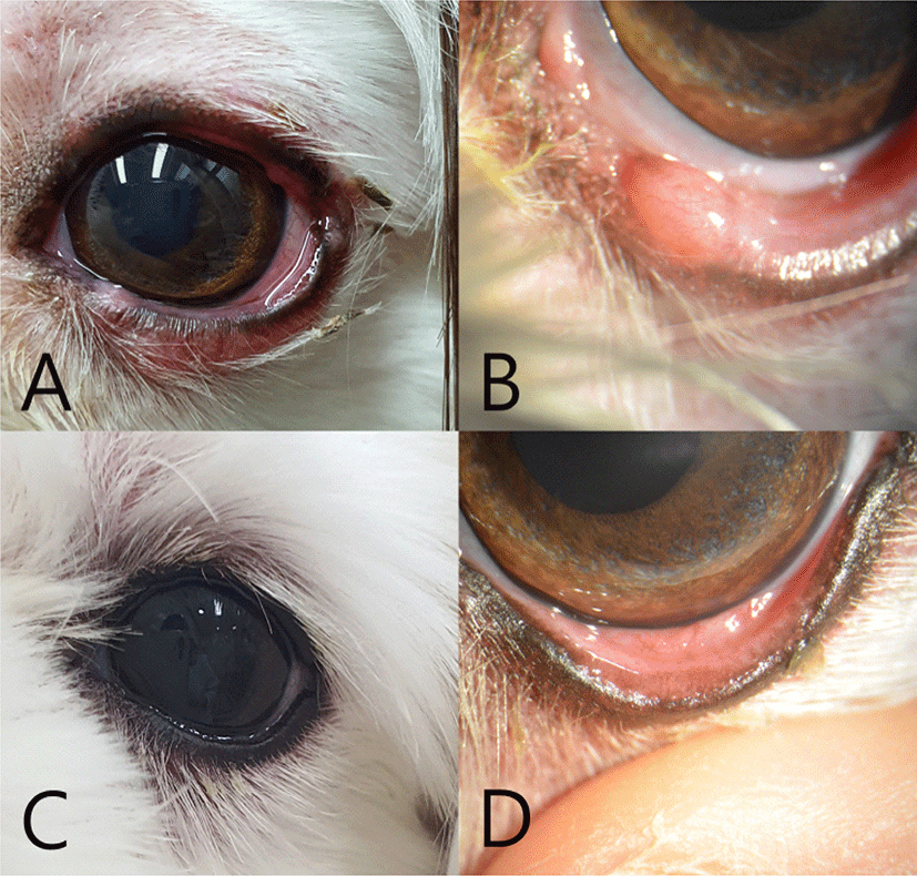
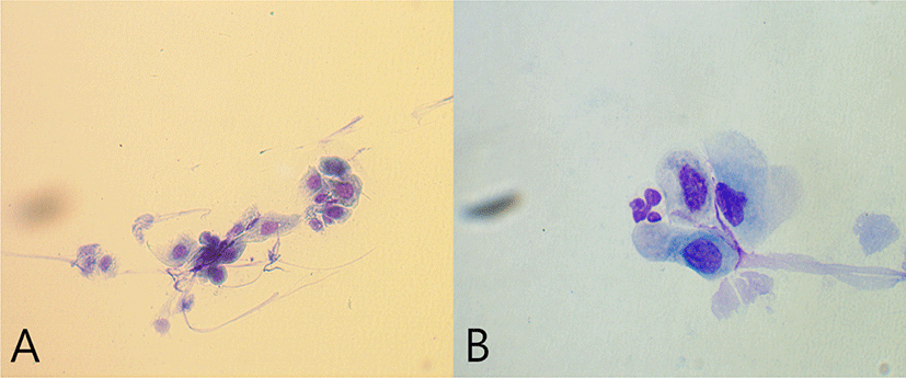
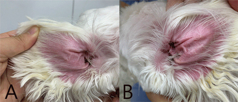
The eyelids and conjunctiva including Meibomian glands were markedly swollen and erythematous on physical examination (Fig. 1A). Erythema of conjunctiva and swelling of Meibomian glands were identified via slit lamp microscopic examination (Fig. 1B). The Schirmer’s tear test was normal. Impression smear cytology of eyes revealed sterile neutrophils with the corneal epithelial cells (Fig. 2). Generalized erythema was also detected on skin examination but there were no other skin lesions (Fig. 3).
Fig. 1.
Left eye at first visit (A, B). Ocular examination revealed erythema, swelling and alopecia of the periocular region. Slit lamp microscopic examination of the eyelids and conjunctiva revealed overall inflammatory findings and swollen Meibomian glands. Left eye 6 weeks later (C, D). Erythema, edema and swelling of Meibomian glands were significantly reduced.
Download Original Figure
Fig. 2.
Impression smear cytology of eyes (A, B). The corneal epithelial cells and few neutrophils are shown.
Download Original Figure
Fig. 3.
Identification of canine atopic dermatitis. Both ears revealed severe erythema but non-affected ear margin (A, B).
Download Original Figure
A real-time PCR tests using samples taken from pharynx and conjunctiva for Canine distemper virus and Canine herpesvirus 1 (IDEXX Reference Laboratories, Westbrook, ME, USA) were negative. The Canine Atopic Dermatitis Extent and Severity Index (CADESI)-04 revealed mild atopic dermatitis with a score of 18. The visual analog scale (VAS) score was 4 of 10, moderate pruritus. Based on history taking and skin examinations, an allergic blepharoconjunctivitis caused by atopic dermatitis was suspected.
Treatment included cetirizine (1 mg/kg PO bid; Zyrtec® tab, UCB Pharm, Brussels, Belgium), cyclosporine (5 mg/kg PO bid; Cipol-N® oral solution, CKD Pharm, Seoul, Korea), prednisolone (1 mg/kg PO bid; Solondo® tab, Yuhan Pharm, Seoul, Korea) and Forus eye drops® (dexamethasone 1 mg/mL, neomycin sulfate 3.5 mg/mL, polymyxin B sulfate 6,000 IU/mL q8h; Samil Pharm, Seoul, Korea). The dose of prednisolone was tapered over a month. The owner was instructed to wear an Elizabethan collar around his dog’s neck at all times and restrict walking. The diet was changed from adult dry dog food to hypoallergenic dog food. Medications and environmental restriction significantly reduced swelling of Meibomian glands, erythema and edema after 3 weeks. Also pruritus was decreased. Six weeks later, the edema lesions disappeared (Fig. 1C & D). For two month follow up, the symptoms did not recur.
Discussion
The dog showed erythema of eyelids and conjunctiva, swelling of Meibomian glands, alopecia of periocular region and pruritus in the present case. Other eye diseases were ruled out by ophthalmological examinations. The dog was evaluated canine atopic dermatitis extent, VAS, and severity index and skin condition [11, 12]. After treatment with medications including glucocorticoid, the pruritus of patient’s eyes was decreased. Finally, an allergic blepharoconjunctivitis caused by atopic dermatitis was tentatively diagnosed based on history taking, skin examinations and treatment response.
In human, eyes are commonly affected organ in allergic disease and blepharoconjunctivitis is commonly associated with atopic dermatitis [13, 14]. Atopic dermatitis can cause ocular complications such as blepharitis, keratoconjunctivitis, keratopathy, corneal ulcer and abnormal tear function and conjunctivitis causes abnormal structure of Meibomian glands in humans, but it is uncommon in dogs [10, 15, 16]. The present case showed swollen Meibomian glands, which were located partly in the eyelids. The glands can be affected by underlying diseases such as bacterial infections, tumors and allergic disorders [17]. In human with allergic blepharoconjunctivitis, conjunctival cytology includes inflammatory cells likes eosinophils, neutrophils and lymphocytes [18], whereas the corneal epithelial cells and only few neutrophils were shown in the impression smear cytology of eyes in this case. In histology, hyperkeratosis, acanthosis, and dermal infiltrates of eosinophils and mast cells along with deposition of major basic protein in human [19].
In humans, blepharoconjunctivitis is treated with antibiotics, steroids, antihistamines and mast cell inhibitors [14, 20]. This case shows a significant improvement using medications and restricting environmental factors. Antibiotics are recommended due to the increased susceptibility to infection resulting from compromised innate immunity in the eyes of atopic patients [21]. Steroids and antihistamine are used to reduce skin problems and pruritus in canine atopic dermatitis [22, 23]. In this case, the drugs relieved ocular erythema and swelling. In humans, cyclosporine is also used to treat blepharoconjunctivitis associated with atopic dermatitis [14, 24]. Cyclosporine is an immunosuppressive drug used to prevent the transcription of cytokine genes in activated T cells and mast cells [25, 26]. The drug is used to reduce the inflammation as a long term treatment sparing steroids. The owner was instructed to wear an Elizabethan collar around his dog’s neck at all time and restrict walking. The symptoms were improved with appropriate treatment.
This case report has a limitation. It is necessary to do biopsy to rule out other etiologies, in particular immune-mediated diseases. As the eyelids and conjunctiva were not easily taken for histopathology with owner’s compliance in the present case.
In conclusion, an allergic blepharoconjunctivitis was successfully managed with anti-allergic drugs and by avoidance of environmental exposure. The whole body must be evaluated to identify skin problems and/or diagnose canine atopic dermatitis when the patient has blepharoconjunctivitis not focus on only the eyes.
Acknowledgements
This research was supported by the Basic Science Research Program through the National Research Foundation of Korea (NRF), funded by the Ministry of Education (NRF-2020R1A2C2005364).
References
Fazal MI. Blepharoconjunctivitis. In: Patel BC (ed.). Florida: StatPearls; 2021.
Weingart C, Kohn B, Siekierski M, Merle R, Linek M. Blepharitis in dogs: a clinical evaluation in 102 dogs. Vet Dermatol 2019;30:222-e69.


Lourenço-Martins AM, Delgado E, Neto I, Peleteiro MC, Morais-Almeida M, Correia JHD. Allergic conjunctivitis and conjunctival provocation tests in atopic dogs. Vet Ophthalmol 2011;14:248-256.
Griffin CE, DeBoer DJ. The ACVD task force on canine atopic dermatitis (XIV): clinical manifestations of canine atopic dermatitis. Vet Immunol Immunopathol 2001;81:255-269.
Bizikova P, Santoro D, Marsella R, Nuttall T, Eisenschenk MNC, Pucheu-Haston CM. Review: clinical and histological manifestations of canine atopic dermatitis. Vet Dermatol 2015;26:79-e24.
Furue M, Yamamura K, Kido-Nakahara M, Nakahara T, Fukui Y. Emerging role of interleukin-31 and interleukin-31 receptor in pruritus in atopic dermatitis. Allergy 2018;73:29-36.
Rees CA. Canine and feline atopic dermatitis: a review of the diagnostic options. Clin Tech Small Anim Pract 2001;16:230-232.
Olivry T, DeBoer DJ, Favrot C, Jackson HA, Mueller RS, Nuttall T, Prélaud P. Treatment of canine atopic dermatitis: 2010 clinical practice guidelines from the International Task Force on Canine Atopic Dermatitis. Vet Dermatol 2010;21:233-248.
Olivry T, Saridomichelakis M, Nuttall T, Bensignor E, Griffin CE, Hill PB. Validation of the canine atopic dermatitis extent and severity index (CADESI)-4, a simplified severity scale for assessing skin lesions of atopic dermatitis in dogs. Vet Dermatol 2014;25:77-e25.
Reich A, Heisig M, Phan NQ, Taneda K, Takamori K, Takeuchi S, Furue M, Blome C, Augustin M, Ständer S, Szepietowski JC. Visual analogue scale: evaluation of the instrument for the assessment of pruritus. Acta Derm Venereol 2012;92:497-501.
Bielory L. Allergic and immunologic disorders of the eye. Part II: ocular allergy. J Allergy Clin Immunol 2000;106:1019-1032.
Pflugfelder SC, Karpecki PM, Perez VL. Treatment of blepharitis: recent clinical trials. Ocul Surf 2014;12:273-284.
Dogru M, Nakagawa N, Tetsumoto K, Katakami C, Yamamoto M. Ocular surface disease in atopic dermatitis. Jpn J Ophthalmol 1999;43:53-57.
Tilley LP, Smith FWK Jr.Blackwell’s five-minute veterinary consult: canine and feline. 5th ed. Hoboken: Blackwell; 2011.
Peuravuori H, Kari O, Peltonen S, Aho VV, Saari JM, Collan Y, Määttä M, Saari KM. Group IIA phospholipase A2 content of tears in patients with atopic blepharoconjunctivitis. Graefes Arch Clin Exp Ophthalmol 2004;242:986-989.
Saw VPJ, Leonard JN. Dermatoses of the eye, eyelids and eyebrows. In: Griffiths C, Barker J, Bleiker T, Chalmers R, Creamer D (eds.). Rook’s textbook of dermatology. 9th ed. Chichester, Wiley Blackwell; 2016.
Furiani N, Scarampella F, Martino PA, Panzini I, Fabbri E, Ordeix L. Evaluation of the bacterial microflora of the conjunctival sac of healthy dogs and dogs with atopic dermatitis. Vet Dermatol 2011;22:490-496.
Cook CP, Scott DW, Miller WH Jr., Kirker JE, Cobb SM. Treatment of canine atopic dermatitis with cetirizine, a second generation antihistamine: a single-blinded, placebo-controlled study. Can Vet J 2004;45:414-417.
Taszkun I. The evaluation of canine atopic dermatitis extent and severity index (CADESI) test in dogs with atopic dermatitis (AD) treated with cyclosporine or prednisone. Pol J Vet Sci 2010;13:681-688.
Cornish KS, Gregory ME, Ramaesh K. Systemic cyclosporin A in severe atopic keratoconjunctivitis. Eur J Ophthalmol 2010;20:844-851.
Olivry T, Rivierre C, Jackson HA, Murphy KM, Davidson G, Sousa CA. Cyclosporine decreases skin lesions and pruritus in dogs with atopic dermatitis: a blinded randomized prednisolone-controlled trial. Vet Dermatol 2002;13:77-87.