Journal of Biomedical Translational Research
Research Institute of Veterinary Medicine, Chungbuk National University
Original Article

Effects of cadmium on the behavior and regeneration of Dugesia japonica

Yong-Jin Kim1,https://orcid.org/0000-0002-9866-2648, Yeon-Hwa Kim2https://orcid.org/0000-0002-4374-1963, A. A. Dilki Indrachapa Adikari3https://orcid.org/0000-0002-1021-3814, Juneno So4https://orcid.org/0000-0002-1832-536X, Young-Joo Yi3,*https://orcid.org/0000-0002-7167-5123, Sang-Myeong Lee2,*https://orcid.org/0000-0002-3624-3392
1Division of Biotechnology, College of Environmental & Bioresource Sciences, Jeonbuk National University, Iksan 54596, Korea
2Laboratory of Veterinary Virology, College of Veterinary Medicine, Chungbuk National University, Cheongju 28644, Korea
3Department of Agricultural Education, College of Education, Sunchon National University, Suncheon 57922, Korea
4Department of Pharmaceutical Engineering, Woosuk University, Wanju 55338, Korea
*Corresponding author: Young-Joo Yi, Department of Agricultural Education, College of Education, Sunchon National University, Suncheon 57922, Korea, Tel: +82-61-750-3352, E-mail: yiyj@scnu.ac.kr
*Corresponding author: Sang-Myeong Lee, Laboratory of Veterinary Virology, College of Veterinary Medicine, Chungbuk National University, Cheongju 28644, Korea, Tel: +82-43-261-3356, E-mail: smlee@chungbuk.ac.kr

Current affiliation: Department of Veterinary Surgery, College of Veterinary Medicine and Biomaterial R&BD Center, Chonnam National University, Gwangju 61186, Korea

© Research Institute of Veterinary Medicine, Chungbuk National University. All rights reserved.

Received: Feb 15, 2021; Accepted: Mar 04, 2021

Abstract

This study was undertaken to evaluate the toxic effect of cadmium sulfate (Cds) on planarians (Dugesia japonica), and the suitability of planarians as an alternative animal model for toxicity studies. Planarians were exposed to varying concentrations of experimental solutions containing 0.1–25 mg/L Cds, and incubated for 1, 3, and 5 hrs. Motility, seizure-like behaviors (c-like, head-bop, snake-like, and screw-like) and regeneration ability of the amputated fragment were subsequently evaluated. Results showed decreased motility with increasing concentrations of Cds (p<0.05), with the lowest motility being observed at the highest concentration of 25 mg/L Cds. Results also indicate that seizure-like behavior patterns were significantly affected by increments in the Cds concentrations, especially the c-like and head-bop behaviors were notably increased. Compared to the control, the regeneration ability of the planarians was decreased in the experimental solutions containing Cds. Planarians exposed to Cds showed either delayed eye formation or no eye regeneration during incubation. Moreover, increased concentrations of Cds resulted in failure to regenerate and death of the planarians. In conclusion, this study confirm that the heavy metal Cds exerts a toxic effect on planarians. Furthermore, the performances of the planarians in the experimental period exhibit their suitability as an alternative animal model for toxicity studies.

Keywords: heavy metals; cadmium; behavior; regeneration; planarian

Introduction

Expeditious development in the fields of agriculture, medicine and manufacturing industries increases the environmental pollution due to anthropogenic activities [1]. Heavy metals are especially responsible for increasing the pollution due to improper waste handling, which ultimately results in undesirable consequences on the human health [2, 3]. Cadmium (Cd) is a naturally inhabited heavy metal, increasing in abundance due to anthropogenic activities of mankind, and subsequent increase in human exposure [4]. Humans can be exposed to Cd through the long-term consumption of contaminated food and water, and inhalation of the smoke [5]. Cd has the ability to replace calcium in minerals because of its similar ionic radius, identical charge, and similar chemical behavior [6]. Therefore, it can easily be ingested by the human body and accumulate in certain organs [7]. Cd is a known neurotoxin and targets organs such as the brain, lungs, liver and kidneys [5, 8, 9]. Moreover, it causes renal and hepatic dysfunction, testicular damage, osteomalacia, pulmonary edema, and damages the adrenals [10]. It is also a proven carcinogen, and is the causative agent of numerous lung, breast, pancreas, urinary bladder and nasopharyngeal malignancies [11].

Thus, there is an urgent necessity to evaluate the toxic effects of cadmium in the living body, and finding a suitable animal model is a critical factor to achieve this. Fresh water planarians are known to be free-living flatworms, having great anatomical suitability for toxicity studies [12]. They have the ability for both sexual and asexual reproduction as well as regeneration, and a simple lifecycle [12, 13]. They also encompass a well-developed central nervous system which is similar to humans [14]. Moreover, planarians are easy to manage, requiring less space, can easily be observed by the naked eye, and are comparatively inexpensive than other laboratory animal species [15, 16]. Most importantly, compared to other alternative toxic models like nematodes and zebrafish, planarians are highly sensitive to environmental chemicals [17, 18], especially their behaviors, regeneration ability and brain structure [19]. Considering all the above factors, the present study was performed to evaluate Cd toxicity in planarian (Dugesia japonica), and to determine the suitability of planarian as an animal model for toxicity studies.

Materials and Methods

Experimental animals and reagents

Approximately 1.0 cm long planarians (Dugesia japonica) were used as the animal model in this study. Planarians were acclimated in freshwater at 18°C–22°C, and fed finely homogenized beef liver twice a week. One week prior to the experiment, the planarians were starved to create a uniform metabolic state. Cadmium sulfate (3CdSO4 × 8H2O; Cds) was purchased from Sigma-Aldrich (Seoul, Korea). Chemicals were dissolved in distilled water (DW) before experimental application.

Examination of motility and seizure-like behaviors

Motility was measured by evaluating swimming ability of the planarian in its natural state. Briefly, petri dishes (p-dish) filled with treatment solutions were placed on a graph paper, which was divided into a grid of 0.5 cm intervals. Motility was evaluated by counting the number of grid lines that the planarians passed through. Simultaneously, planarian seizure-like behaviors were also observed, and the actions were classified into four categories: c-like, head-bop, snake-like, and screw-like [20, 21]. Planarians were incubated in 10 ml experimental solutions containing 0.1–25 mg/L cadmium sulfate (Cds) for 1–5 hrs, after which they were introduced into the respective petri dish for each treatment solution separately. Motility and seizure-like patterns were subsequently measured (number of grid lines crossed or re-crossed by each planarian) within 5 min.

Observation of regeneration pattern

Regeneration assay was conducted with a minimum number of planarians (n = 10) in each treatment solution. Planarians were paralyzed by chilling on ice, and the stretched bodies were cut by using an ethanol-sterilized blade. The head region on the auricular grooves was cut, and rest of the body was incubated in water until emergence of eye extrusion. Each amputated fragment was transferred to a 12-well plate filled with the treatment solutions, and maintained in a dark environment at 18°C–22°C. The imaging process of the regeneration fragments was conducted for 14 days, using a stereo-microscope (S8APO, Leica, Wetzlar, Germany).

Whole-mount immunohistochemistry

Immunofluorescence staining was conducted with slight modification [22]. Briefly, regenerating planarians were treated with 2% HCl diluted in DW for 5 min to remove excess mucus, after which they were fixed in Carnoy’s solution (ethanol : chloroform : acetic acid = 6 : 3 : 1) for 3 hrs at RT. Subsequently, samples were rinsed with 100% methanol and bleached overnight at RT in 6% H2O2 diluted in methanol under a light emitting diode (LED) lamp. Bleached samples were rehydrated through a graded methanol series (75%, 50%, and 25%) diluted with phosphate buffer saline (PBS), 15 min in each solution. Samples were then rinsed in PBS containing 0.3% Triton X-100 (PBST), and blocked in PBST containing 0.25% BSA (PBSTB) for 2 hrs, followed by incubation with rabbit polyclonal anti-arrestin antibody (LAgen Laboratories, 1:5,000 dilution) in PBSTB, overnight at 4°C. Probed samples were washed 3 times with PBSTB for 2 hrs, and subsequently incubated with goat rabbit anti- Alex488 IgG antibody in PBSTB (Invitrogen, 1:2,000 dilution) and 0.5 µg/mL 4’6’-diamidino-2-phenylindole (DAPI; DNA), overnight at 4°C in the dark. After incubation, samples were washed with PBST. Finally, the samples were mounted on a slide and observed using a fluorescence microscope (Nikon Eclipse Ci, Nikon, Seoul, Korea).

Statistical analysis

All experiments were performed in triplicate. Data were analyzed using a one-way ANOVA in GraphPad PRISM® (GraphPad software, San Diego, CA, USA) and expressed as Mean ± S.E.M. Values at *p<0.05, **p<0.01, and ***p<0.001 are considered statistically significant.

Results

Aberrant motility was observed in planarian exposed to Cds

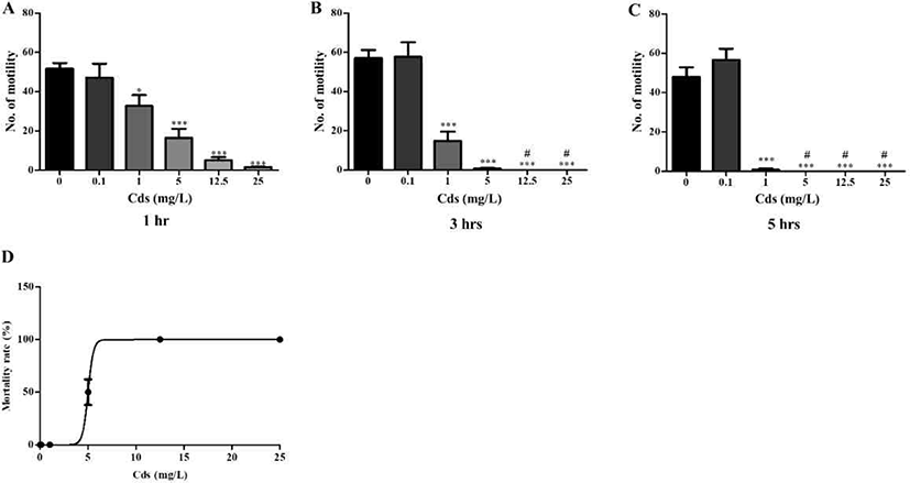
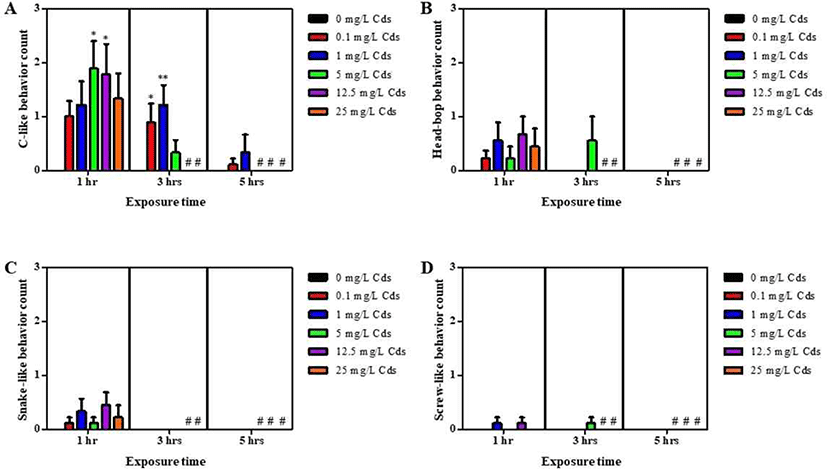
Planarians were incubated in water containing varying concentrations of Cds for 1–5 hrs (Fig. 1). Compared to the control (without Cds), motility was significantly and dose-dependently decreased in Cd concentrations at varying incubation times (p<0.05 & p<0.001; Fig. 1A–C). After incubation, the lethal concentration (3–5 hrs LC50) of Cds was estimated at 5 mg/L (Fig. 1D). Also, seizure- like behaviors were observed in planarians exposed to Cds (Fig. 2). Higher numbers of c-like behavior were observed in planarian exposed to varying concentrations of Cds for 1–3 hr incubation, compared to the control without Cds (p<0.05 & p<0.01; Fig. 2A). Behavior patterns, including head bop, snake-like and screw- like movements, were observed in planarian exposed to Cds for 1–3 hrs incubation, whereas either no seizure- like behaviors were observed or most of them died after 5 hrs incubation in the presence of Cds (Fig. 2B–D). These results indicate that normal behavioral patterns are altered in planarians incubated in the presence of Cds.
jbtr-22-1-42-g1
Fig. 1. Motility patterns in planarian exposed to varying concentrations of Cds (0.1–25 mg/L) at different incubation times (1–5 hrs; A–C). (D) Lethal concentration (LC50) of Cds on planarians after incubation. Values are expressed as Mean ± S.E.M. The different superscripts in each group of columns denote a significant difference at *p<0.05 and ***p<0.001. All dead planarian is indicated as # in the columns. Cds, cadmium sulfate.
Download Original Figure
jbtr-22-1-42-g2
Fig. 2. Seizure-like behaviors (A: c-like, B: head-bop, C: snake-like, D: screw-like) in planarians exposed to varying concentrations of Cds and incubation times. Values are expressed as Mean ± S.E.M. The different superscripts in each group of columns denote a significant difference at *p<0.05 and **p<0.01. All dead planarian is indicated as # in the columns. Cds, cadmium sulfate.
Download Original Figure
Eye formation of planarians in the presence of Cds

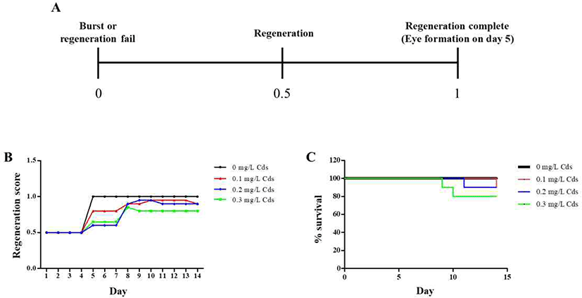
In order to observe eye regeneration from amputated fragments, the head region on the auricular grooves was cut, and rest of the body was incubated in water until emergence of eye extrusion. The degree of regeneration was calculated by indicating a score of 0, 0.5, and 1, depending on burst, regeneration and completion of regeneration, respectively, as shown in Fig. 3A.
jbtr-22-1-42-g3
Fig. 3. Observation of eye formation in the absence/presence of Cds. Amputated fragments were incubated in water containing Cds (0.1–0.3 mg/L). (A) The degree of regeneration was scored as “0–1” according to the shape of regeneration. Regeneration (B) and survival (C) were observed for 14 days. Cds, cadmium sulfate.
Download Original Figure

On day 5, the eye spot was observed in planarians incubated in water without Cds; however, planarians exposed to Cds showed either no formation or delayed formation of eye spots (Fig. 3B). This pattern lasted in planarians incubated in the presence of Cds until the 8th day, after which the eye spots appeared. In particular, planarians cultured at concentrations above 0.3 mg/L showed low eye formation score for 14 days (Fig. 3B). Moreover, planarians incubated in the absence of Cds showed 100% survival for 14 days, but the lifespans of planarians exposed to Cds were decreased, and survival was less than 80% in planarians incubated in the presence of 0.3 mg/L Cds (Fig. 3C).

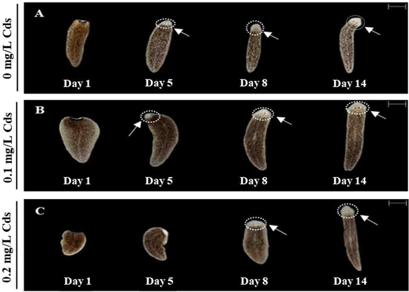
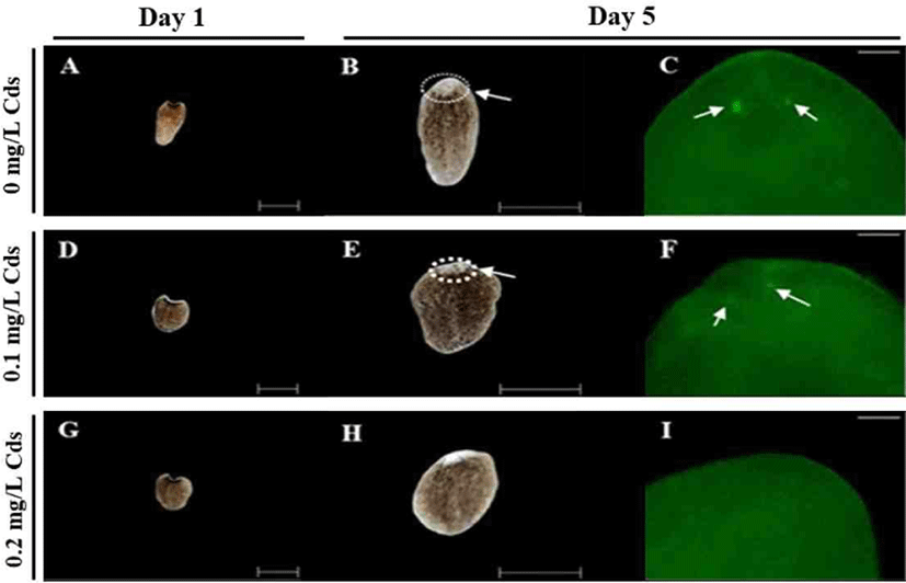
As presented in Fig. 4, eye formation was clearly observed in water without Cds or with 0.1 mg/L Cds on day 5 (Fig. 4A and B), whereas 0.2 mg/L Cds exposed planarian was unable to form eye spots on day 5, but finally formed eye spots on day 8 (Fig. 4C). Planarians in all treatment groups completed eye formation on day 14; thus, higher concentrations of Cds induced delayed eye regeneration.
jbtr-22-1-42-g4
Fig. 4. Amputated fragments were incubated in the absence (A) /presence of 0.1 mg/mL (B) or 0.2 mg/mL Cds (C). Eye regeneration of D. japonica was observed by stereo-microscope for 14 days (magnification at × 10). The white arrow and round dotted line indicate eye formation. The scale bar in the upper right represents a length of 1 mm. Cds, cadmium sulfate.
Download Original Figure

Optic nerves were observed by immunohistochemistry using anti-arrestin antibody (Fig. 5). Amputated fragments in each treatment were cultured for 14 days (Fig. 5A, D & G), and eye spots appeared in planarian cultured without or with 0.1 mg/L Cds on day 5 (Fig. 5B & E). Delayed eye formation was observed in planarian cultured in 0.2 mg/L Cds on day 5 (Fig. 5H). Eye spots (blue; DNA) and nerves (green; arrestin) were observed in planarian incubated without or with 0.1 mg/L (Fig. 5C & F), but there was no nerve formation in planarian exposed to 0.2 mg/L Cds (Fig. 5I).
jbtr-22-1-42-g5
Fig. 5. Nerve formation on the eye spot was observed by immunohistochemistry of anti-arrestin antibody. Amputated fragments in each treatment were initiated after incubation with/without Cds (A, D & G). The white arrows and round dotted lines indicate eye spots on day 5 (B & E). Eye formation was delayed in planarian exposed to 0.2 mg/L Cds on day 5 (H). Eyes (blue; DNA) and nerves (green; arrestin) were observed under fluorescence microscope (C & F); no nerve formation was observed in planarian exposed to 0.2 mg/L Cds (I). Cds, cadmium sulfate.
Download Original Figure

Discussion

Rapid development in the economic sector has resulted in numerous natural and man-made toxins being discharged into the natural environment [1]. Heavy metal pollution is increasing due to the industrialization process and anthropogenic activities of humans [1, 23, 24]. Among the heavy metals, Cd is one of the most ejected metals in a wide range of industries [25]. Human exposure to Cd occurs through the occupational environment or the long-term in-contact with common goods available in their immediate environment, such as Cd-containing common products like batteries and artist paints [26].

Cd is toxic at low concentrations and has the ability to act as a carcinogen. It is mainly absorbed through the respiratory tract, while relatively smaller amounts are absorbed through the gastro-intestinal tract and the skin [27]. In the present study, planarians exhibit a dose-dependent decline in motility and increase in seizure-like behaviors (Fig. 1 & 2). Moreover, their ability to regenerate was reduced when exposed to Cd (Fig. 35). Exposing fresh water planarians to high concentrations of Cd also resulted in altered neurobehaviors and changes in the morphology (shape and body elongation). These results are similar to a previous study, which reported that exposing planarians to Cd causes disturbances to their neurotransmission system [28].

Cd accumulation in the body induces cardiovascular diseases, kidney disease, metabolic syndrome, and cancer [29]. Calcium is an essential element for biochemical reactions in the body. Due to similar chemical characteristics (ionic radius, charge and behavioral pattern) [5], Cd competes with calcium ions in the body, and causes oxidative stress by inducing reactive oxygen species (ROSs) [30]. Moreover, when Cd enters the body, it induces increased levels of ROSs, which ultimately induces DNA damage and interferes with the DNA repair mechanism [30]. Cd exerts its toxic effect by blocking the electron transfer chain of mitochondria, where Cd ions impair the electron flow [5]. Numerous experiments related to heavy metal toxicity have been studied in invertebrate animals. The long-term exposure or high dose intake of trace metals alters the antioxidant activity of the body by inhibiting the antioxidant enzymes, resulting in decreased antioxidant capacity in the body [31, 32], which subsequently leads to accumulation of ROS, causing tissue damage and cell apoptosis [33], inhibition in the activity of total superoxide dismutase (SOD), and an increased malonaldehyde (MDA) content [34]. Several research articles on crustaceans have proved that Cd exposure leads to an increase in the MDA content [3335]. Although examination of the antioxidant activity was excluded in this study, it is presumed that Cd is involved in antioxidant functions and delayed cell regeneration. These experiments will be done in subsequent studies.

Taken together, results obtained in this study indicate that behaviors of planarians are altered after exposure to Cds, along with delayed eye formation. This study therefore presents the risks of direct toxicity and heavy metal exposure that are likely to occur when exposed to Cd. This study further validates the suitability of planarians as an alternative animal model in toxicity studies.

Conflict of Interest

No potential conflict of interest relevant to this article was reported.

Ethics Approval

This article does not require IRB/IACUC approval because there are no human and animal participants.

References

1.

Briffa J, Sinagra E, Blundell R. Heavy metal pollution in the environment and their toxicological effects on humans. Heliyon 2020;6:e04691.

2.

Wei X, Gao B, Wang P, Zhou H, Lu J. Pollution characteristics and health risk assessment of heavy metals in street dusts from different functional areas in Beijing, China. Ecotoxicol Environ Saf 2015;112:186-192.

3.

Meena RAA, Sathishkumar P, Ameen F, Yusoff ARM, Gu FL. Heavy metal pollution in immobile and mobile components of lentic ecosystems-a review. Environ Sci Pollut Res 2018;25:4134-4148.

4.

Yin J, Wang AP, Li WF, Shi R, Jin HT, Wei JF. Sensitive biomarkers identification for differentiating Cd and Pb induced toxicity on zebrafish embryos. Environ Toxicol Pharmacol 2017;56:340-349.

5.

Genchi G, Sinicropi MS, Lauria G, Carocci A, Catalano A. The effects of cadmium toxicity. Int J Environ Res Public Health 2020;17:3782.

6.

Franklin NM, Glover CN, Nicol JA, Wood CM. Calcium/cadmium interactions at uptake surfaces in rainbow trout: waterborne versus dietary routes of exposure. Environ Toxicol Chem 2005;24:2954-2964.

7.

Pan J, Plant JA, Voulvoulis N, Oates CJ, Ihlenfeld C. Cadmium levels in Europe: implications for human health. Environ Geochem Health 2010;32:1-12.

8.

Antonio MT, Corpas I, Leret ML. Neurochemical changes in newborn rat’s brain after gestational cadmium and lead exposure. Toxicol Lett 1999;104:1-9.

9.

Järup L, Åkesson A. Current status of cadmium as an environmental health problem. Toxicol Appl Pharmacol 2009; 238:201-208.

10.

Tinkov AA, Filippini T, Ajsuvakova OP, Skalnaya MG, Aaseth J, Bjørklund G, Gatiatulina ER, Popova EV, Nemereshina ON, Huang PT, Vinceti M, Skalny AV. Cadmium and atherosclerosis: a review of toxicological mechanisms and a meta-analysis of epidemiologic studies. Environ Res 2018;162:240-260.

11.

Mezynska M, Brzóska MM. Environmental exposure to cadmium-a risk for health of the general population in industrialized countries and preventive strategies. Environ Sci Pollut Res 2018;25:3211-3232.

12.

Best JB, Morita M. Planarians as a model system for in vitro teratogenesis studies. Teratog Carcinog Mutagen 1982;2:277- 291.

13.

Izawakatsu M. On the ecology and distribution of freshwater planarians in the Japanese islands, with special reference to their vertical distribution. Hydrobiologia 1965;26:349-408.

14.

Ross KG, Currie KW, Pearson BJ, Zayas RM. Nervous system development and regeneration in freshwater planarians. Wiley Interdiscip Rev Dev Biol 2017;6:e266.

15.

Raffa RB. Planaria: a model for drug action and abuse. Boca Raton: CRC Press; 2008.

16.

Noreña C, Damborenea C, Brusa F. Phylum platyhelminthes. In: Thorp JH, Rogers DC (eds.). Thorp and covich’s freshwater invertebrates. 4th ed. Amsterdam: Academic Press; 2015. p. 181-203.

17.

Poirier L, Ghigo É, Daudé D, Chabrière É. Planarian, an emerging animal model for toxicology studies. Med Sci 2019;35:544-548.

18.

Pagán OR, Rowlands AL, Urban KR. Toxicity and behavioral effects of dimethylsulfoxide in planaria. Neurosci Lett 2006;407:274-278.

19.

Hagstrom D, Cochet-Escartin O, Zhang S, Khuu C, Collins EMS. Freshwater planarians as an alternative animal model for neurotoxicology. Toxicol Sci 2015;147:270-285.

20.

Raffa RB, Holland LJ, Schulingkamp RJ. Quantitative assessment of dopamine D2 antagonist activity using invertebrate (Planaria) locomotion as a functional endpoint. J Pharmacol Toxicol Methods 2001;45:223-226.

21.

Raffa RB, Desai P. Description and quantification of cocaine withdrawal signs in Planaria. Brain Res 2005;1032:200- 202.

22.

Kobayashi C, Saito Y, Ogawa K, Agata K. Wnt signaling is required for antero-posterior patterning of the planarian brain. Dev Biol 2007;306:714-724.

23.

He ZL, Yang XE, Stoffella PJ. Trace elements in agroecosystems and impacts on the environment. J Trace Elem Med Biol 2005;19:125-140.

24.

Dhuldhaj U, Pandya U. Combinatorial study of heavy metal and microbe interactions and resistance mechanism consort to microbial system. Geomicrobiol J 2021;38:181-189.

25.

Sall ML, Diaw AKD, Gningue-Sall D, Aaron SE, Aaron JJ. Toxic heavy metals: impact on the environment and human health, and treatment with conducting organic polymers, a review. Environ Sci Pollut Res 2020;27:29927-29942.

26.

Duruibe JO, Ogwuegbu MOC, Egwurugwu JN. Heavy metal pollution and human biotoxic effects. Int J Phys Sci 2007; 2:112-118.

27.

Satarug S. Dietary cadmium intake and its effects on kidneys. Toxics 2018;6:15.

28.

Wu JP, Lee HL, Li MH. Cadmium neurotoxicity to a freshwater planarian. Arch Environ Contam Toxicol 2014;67: 639-650.

29.

Fatima G, Raza AM, Hadi N, Nigam N, Mahdi AA. Cadmium in human diseases: it’s more than just a mere metal. Indian J Clin Biochem 2019:34:371-378.

30.

Xu B, Chen S, Luo Y, Chen Z, Liu L, Zhou H, Chen W, Shen T, Han X, Chen L, Huang S. Calcium signaling is involved in cadmium-induced neuronal apoptosis via induction of reactive oxygen species and activation of MAPK/ mTOR network. PloS One 2011;6:e19052.

31.

Ding Z, Kong Y, Shao X, Zhang Y, Ren C, Zhao X, Yu W, Jiang T, Ye J. Growth, antioxidant capacity, intestinal morphology, and metabolomic responses of juvenile Oriental river prawn (Macrobrachium nipponense) to chronic lead exposure. Chemosphere 2019;217:289-297.

32.

Das S, Tseng LC, Chou C, Wang L, Souissi S, Hwang JS. Effects of cadmium exposure on antioxidant enzymes and histological changes in the mud shrimp Austinogebia edulis (Crustacea: Decapoda). Environ Sci Pollut Res 2019;26: 7752-7762.

33.

Lin Y, Huang JJ, Dahms HU, Zhen JJ, Ying XP. Cell damage and apoptosis in the hepatopancreas of Eriocheir sinensis induced by cadmium. Aquat Toxicol 2017;190: 190-198.

34.

Zhang C, Jin Y, Yu Y, Xiang J, Li F. Cadmium-induced oxidative stress, metabolic dysfunction and metal bioaccumulation in adult palaemonid shrimp Palaemon macrodactylus (Rathbun, 1902). Ecotoxicol Environ Saf 2021;208: 111591.

35.

Ren X, Wang X, Liu P, Li J. Bioaccumulation and physiological responses in juvenile Marsupenaeus japonicus exposed to cadmium. Aquat Toxicol 2019;214:105255.