Journal of Biomedical Translational Research
Research Institute of Veterinary Medicine, Chungbuk National University
Original Article

Effect of dyslipidemia on vascular smooth muscle cell proliferation in a porcine coronary restenosis model

Kang Jin Jeong1,, Philyong Kang1,, Hwal-Yong Lee1,, Hee Chang Son1, Sung Hyun Park1, Won Seok Choi1, Bon Sang Koo1, Chang-Yeop Jeon1, Seug Ho Baek1, You Jung An1, Hyun Kuk Kim2http://orcid.org/0000-0002-4554-041X, Sung Soo Kim2http://orcid.org/0000-0002-5190-227X, Jun Kyu Park3http://orcid.org/0000-0002-1587-5673, In Ho Bae4,5http://orcid.org/0000-0002-7298-6329, Dae Sung Park4,5,6http://orcid.org/0000-0002-9948-7546, Jae Won Shim4,5http://orcid.org/0000-0003-3453-0416, Jae Un Kim4,5http://orcid.org/0000-0001-5962-0325, Nan Yeol Kim7, Sun-Uk Kim1http://orcid.org/0000-0002-5168-6976, Kyung Seob Lim1,*http://orcid.org/0000-0002-9117-887X, Myung Ho Jeong4,5,*http://orcid.org/0000-0003-4173-1494
1Futuristic Animal Resource and Research Center & National Primate Research Center, Ochang 28116, Korea
2Department of Internal Medicine and Cardiovascular Center, Chosun University Hospital, Gwangju 61453, Korea
3CGBio Co. Ltd, Jangseong 57248, Korea
4Cardiovascular Research Center, Chonnam National University Hospital, Gwangju 61469, Korea
5Korea Cardiovascular Stent Research Institute, Jangseong 57248, Korea
6Research Institute of Medical Sciences, Chonnam National University, Gwangju 61186, Korea
7Departments of Thoracic and Cardiovascular Surgery and Cardiology, Wonkwang University School of Medicin, Iksan 54538, Korea
*Corresponding author: Kyung Seob Lim Futuristic Animal Resource and Research Center & National Primate Research Center, Ochang 28116, Korea Tel: +82-43-240-6308, Fax: +82-43-240-6309, E-mail: dvmlim96@kribb.re.kr
*Corresponding author: Myung Ho Jeong Cardiovascular Research Center, Chonnam National University Hospital, Gwangju 61469, Korea Tel: +82-62-220-6243, Fax: +82-62-228-7174, E-mail: mhjeong@chonnam.ac.kr

These authors equally contribute to this work.

© Research Institute of Veterinary Medicine, Chungbuk National University. All rights reserved.

Received: Jul 19, 2019; Revised: Sep 20, 2019; Accepted: Sep 20, 2019

Abstract

This study aimed to examine the effect of a mild elevation in serum cholesterol level in a porcine coronary overstretch restenosis model using a balloon angioplasty catheter or drug-eluting coronary stent. Pigs were divided into two groups and were fed a commercial normal diet (CND, n = 4) or a high-fat diet (HFD, n = 4) for 5 weeks. Coronary overstretch injury by balloon angioplasty or stent implantation was induced in the left anterior descending and left circumflex artery after 1 week of feeding. Histopathological analysis was performed at 4 weeks after coronary injury. During the experiment, the total cholesterol level in the HFD group increased by approximately 44.9% (from 65.9 ± 3.21 mg/dL at baseline to 95.5 ± 9.94 mg/dL at 5 weeks). The lumen area in the CND group was reduced in comparison with that in the HFD group after balloon angioplasty. After stent implantation, the injury score showed no significant difference. There were significant differences in the neointimal area (2.7 ± 0.33 mm2 in the CND group vs. 3.3 ± 0.34 mm2 in the HFD group, p<0.05), lumen area (2.6 ± 0.54 mm2 in the CND group vs. 2.0 ± 0.33 mm2 in the HFD group, p<0.05), and percent area stenosis (52.0 ± 7.96% in the CND group vs. 62.4 ± 5.15% in the HFD group, p<0.05). Body weight change was not different between the two groups. Increased serum cholesterol level activated vascular smooth muscle cell proliferation in the porcine coronary overstretch model.

Keywords: cholesterol; balloon angioplasty; coronary stents; percutaneous coronary intervention; restenosis

Introduction

Worldwide, coronary artery disease (CAD) is the principal cause of death among patients [1]. Vascular smooth muscle cell (vSMC) proliferation, commonly known as neointima hyperplasia, is a major side effect after balloon angioplasty and/or coronary stent implantation for treating patients with obstructive CAD [2]. Dyslipidemia is a well-known risk factor for cardiovascular disease. vSMC proliferation in severe hypercholesterolemia has already been studied [3, 4]. However, there has been no study on the effects of mild elevation in serum cholesterol level on vSMC proliferation after vessel injury in a large animal model such as a porcine model.

Therefore, the present study aimed to evaluate the effect of mild elevation in serum cholesterol level on vSMC proliferation in a porcine coronary overstretch restenosis model using a balloon angioplasty catheter or drug-eluting coronary stent.

Materials and Methods

Ethics statement for animal experiments

The animal study was approved by the Ethics Committee of Chonnam National University Medical School and Chonnam National University Hospital (CNU IACUC-H-2016-04) and conformed to the Guide for the Care and Use of Laboratory Animals published by the US National Institutes of Health (NIH Publication No. 85-23, revised 1996).

Diet

Pigs were divided into two groups according to the diet they were fed: commercial normal diet (CND; 3.6% fat content; n = 4) and a high-fat diet (HFD; 10.2% fat content; n = 4). The diets were fed at day 0 before coronary artery injury was induced using balloon angioplasty or stent implantation (day 7) and continued for the entire duration of this experiment for 35 days (Fig. 1).

jbtr-20-4-91-g1
Fig. 1. Study protocol. HFD, high-fat diet; DES, drug-eluting stent; FU, follow-up; CAG, coronary angiography; CND, commercial normal diet.
Download Original Figure
Serum cholesterol analysis

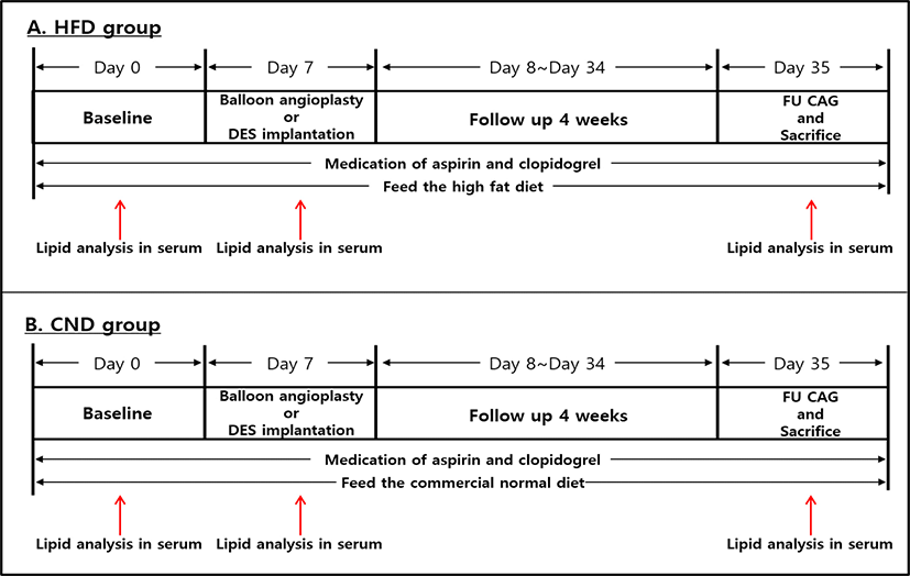
Whole blood samples were collected from pigs at baseline (before diet administration, day 0), immediately after balloon angioplasty or stent implantation (day 7), and at follow-up and sacrifice (day 35). Serum cholesterol levels were measured on days samples were collected (Alere Cholestech LDX® System, Abbott, Santa Clara, CA, USA; Fig. 1).

Animal preparation and stent implantation

Study animals were castrated male pigs weighing 20–25 kg. To prevent acute thrombosis after balloon angioplasty or stent implantation, premedication with aspirin 100 mg and clopidogrel 75 mg per day was administered for 7 days before the procedure. On the day of the procedure, pigs were anesthetized with zolazepam and tiletamine (2.5 mg/kg, Zoletil50®, Virbac, Caros, France), xylazine (3 mg/kg, Rompun®, Bayer AG, Leverkusen, Germany), and azaperone (6 mg/kg, Stresnil®, Janssen-Cilag, Neuss, Germany). Supplemental oxygen was continuously provided through an oxygen mask. Subcutaneous 2% lidocaine at the cut-down site was administered; the left carotid artery was surgically exposed, and a 7-Fr sheath was inserted.

Continuous hemodynamic and surface electrocardiographic monitoring was performed throughout the procedure. Then 5,000 U of heparin was intravenously administered as a bolus prior to the procedure; the target coronary artery was engaged using standard 7-F guide catheters, and control angiograms of both coronary arteries were performed using a nonionic contrast agent in two orthogonal views.

The balloon catheter or coronary stent was deployed by inflator, and the resulting balloon or stent-to-artery ratio was 1.3:1. Coronary angiograms were obtained immediately after balloon angioplasty or stent implantation. All equipment was then removed, and the carotid artery was ligated.

Follow-up, sacrifice, and sampling

Four weeks after balloon angioplasty or stent implantation, follow-up angiography was performed in the same orthogonal views previously used; the animals were then sacrificed by an intracoronary injection of 20 mL of potassium chloride.

The hearts were removed, and the coronary arteries were pressure-perfusion fixed at 110 mmHg in 10% neutral buffered formalin overnight. Step sectioning of each artery was performed, followed by routine processing under light microscopy and staining for histological analysis.

Histopathological analysis

Histopathological evaluation of each artery was performed by an experienced cardiovascular pathologist. The specimens were embedded; sections of 3–4 μm thickness at approximately 1 mm apart were obtained and stained with hematoxylin-eosin for histological analysis. Measurements of the histopathological sections were performed using a calibrated microscope, digital video imaging system, and microcomputer program (Visus 2000 Visual Image Analysis System, IMT Tech, CA, USA). Borders were manually traced for lumen area, area circumscribed by the internal elastic lamina, and innermost border of the external elastic lamina (external elastic lamina area). Morphometric analysis of the neointimal area for a given vessel was calculated as the measured internal elastic lamina area subtracted by the lumen area. The measurements were performed for five cross-sections from the proximal and distal ends and the three midpoints of each stented segment. Percent area stenosis was calculated as follows: 100 × [1 – (lesion lumen area/lesion internal elastic lamina area)] [5]. All results were interpreted by two independent pathologists in a blinded manner.

Evaluation of injury score

Arterial injury at each strut site was determined by anatomical structures penetrated by each strut. A numerical value was assigned as follows [5]: 0 = no injury; 1 = break in the internal elastic membrane; 2 = perforation of the media; and 3 = perforation of the external elastic membrane to the adventitia. The average injury score of each segment was calculated by dividing the sum of the injury scores by the total number of struts at the examined section.

Evaluation of inflammation scores and fibrin score

Regarding the inflammation score for each individual strut, the grading was as follows: 0 = no inflammatory cells surrounding the strut; 1 = light, noncircumferential lymphohistiocytic infiltrate surrounding the strut; 2 = localized, moderate-to-dense cellular aggregate surrounding the strut noncircumferentially; and 3 = circumferential dense lymphohistiocytic cell infiltration of the strut. The inflammation score of each cross-section was calculated by dividing the sum of the individual inflammation scores by the total number of struts at the examined section [6]. Ordinal data for fibrin were collected for each stented section using a scale of 0 to 3 [7].

Statistical analysis

Statistical analysis was performed using a commercially available software (SPSS Version 15, Chicago, IL, USA). Data are presented as mean ± SD. Unpaired student’s t test was used for the comparison of the groups. p<0.05 was considered statistically significant.

Results

Procedure outcomes after balloon angioplasty and stent implantation

Coronary artery injury using balloon catheter inflation was induced in the left anterior descending and left circumflex artery in 2 pigs (1 from each group). Two coronary stents were implanted in two coronary arteries in each pig. A total 12 stents, including 6 in the HFD group and 6 in the CND group, were placed in the middle to proximal left anterior descending and circumflex artery in 6 pigs (3 in each group). Mortality rate was zero until sacrificed in this study.

Alteration in serum lipid profile

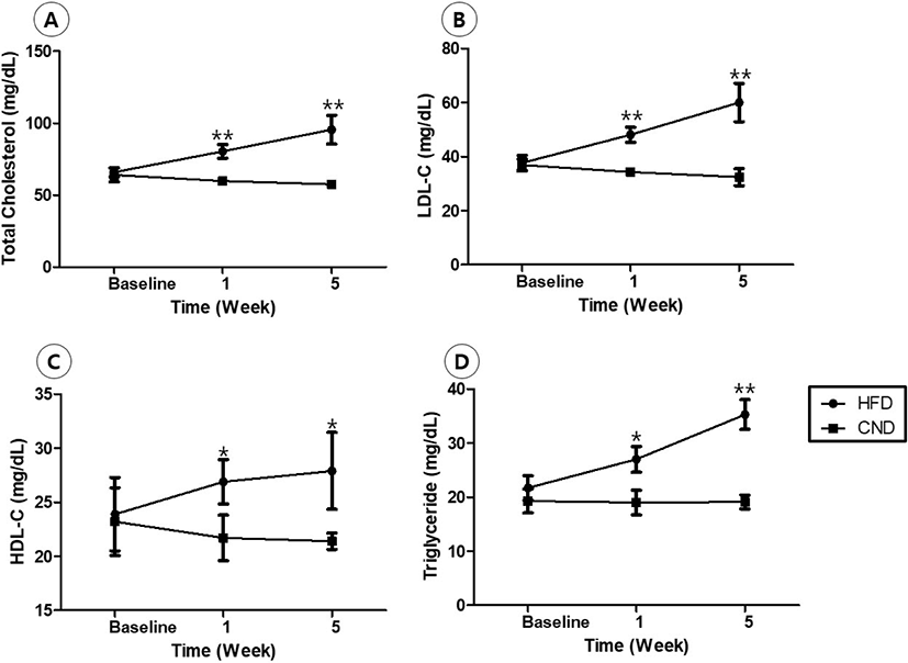
At 1 and 5 weeks after feeding, total cholesterol, low-density lipoprotein cholesterol, high-density lipoprotein cholesterol, and triglyceride levels were significantly higher in the HFD group than in CND group (Fig. 2).

jbtr-20-4-91-g2
Fig. 2. Alteration in lipid profile. Total cholesterol (A), LDL-C (B), HDL-C (C), and triglyceride (D) levels. *p<0.05 versus the CND group, **p<0.001 versus the CND group. HFD, high-fat diet; CND, commercial normal diet; LDL-C, low-density lipoprotein cholesterol; HDL-C, high-density lipoprotein cholesterol.
Download Original Figure
Difference in vessels after balloon angioplasty

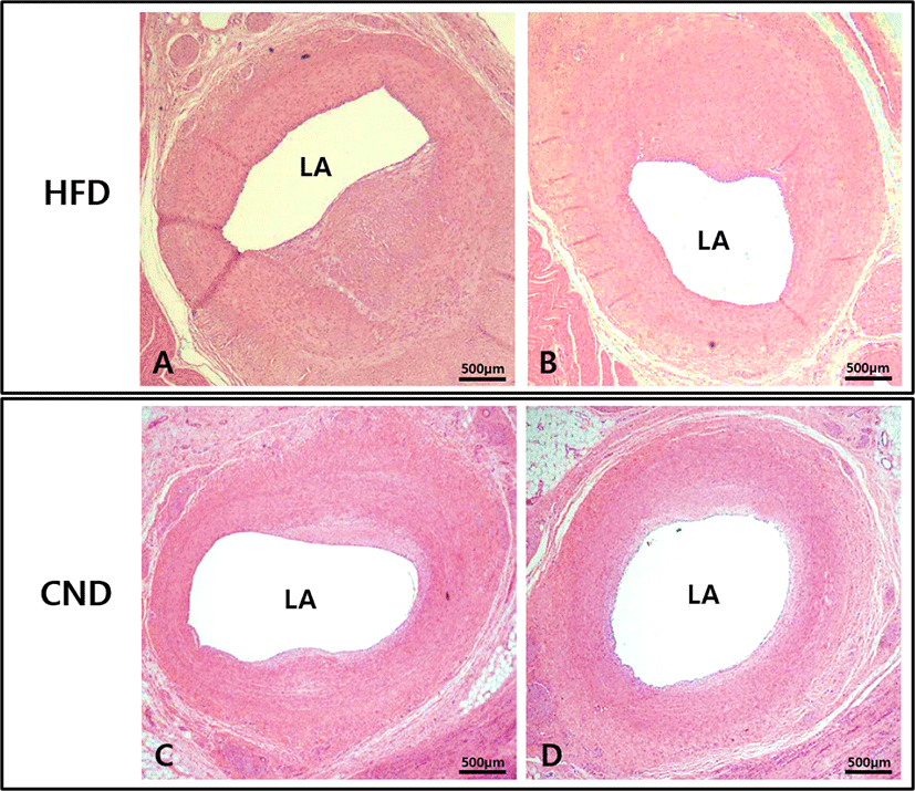
The lumen area was significantly lesser in the HFD group (1.8 ± 0.51 mm2 in HFD group vs. 2.8 ± 0.32 mm2 in CND group, p<0.01; Fig. 3). Internal elastic lamina area, neointimal area, and percent area stenosis could not be measured because the intima line was not clear.

jbtr-20-4-91-g3
Fig. 3. Representative images of hematoxylin and eosin staining at 4 weeks after balloon angioplasty: HFD group (A and B, 20×) and CND group (C and D, 20×). HFD, high-fat diet; CND, commercial normal diet; LA, lumen area.
Download Original Figure
Histopathologic analysis of stented arteries

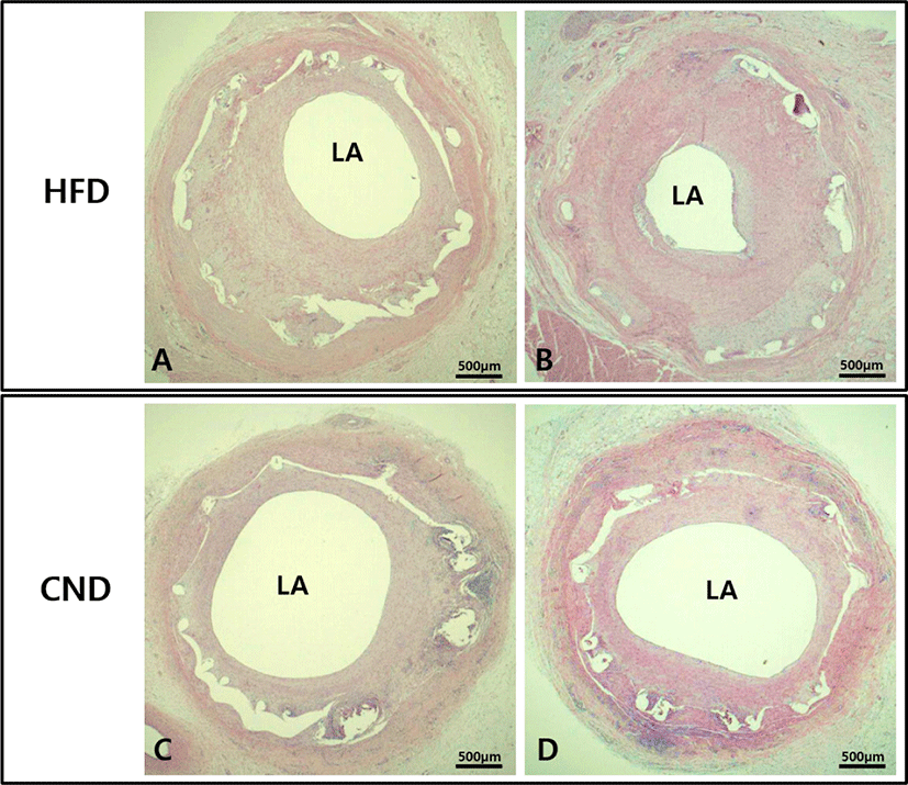
There were no significant differences in the injury score (1.3 ± 0.49 in the HFD group vs. 1.3 ± 0.52 in the CND group, p = NS) and internal elastic lamina area (5.3 ± 0.41 mm2 in the HFD group vs. 5.3 ± 0.42 mm2 in the CND group, p = NS) between the two groups. There were significant differences in the neointimal area (3.3 ± 0.34 mm2 in the HFD group vs. 2.7 ± 0.33 mm2 in the CND group, p<0.05), lumen area (2.0 ± 0.33 mm2 in the HFD group vs. 2.6 ± 0.54 mm2 in the CND group, p<0.05), fibrin score (1.1 ± 0.38 in the HFD group vs. 1.2 ± 0.41 in the CND group, p = NS), inflammation score (1.6 ± 0.53 in the HFD group vs. 1.5 ± 0.55 in the CND group, p = NS), and percent area stenosis (62.4 ± 5.15% in the HFD group vs. 52.0 ± 7.96% in the CND group, p<0.05) between the two groups (Fig. 4, Fig. 5A, B, C).

jbtr-20-4-91-g4
Fig. 4. Histology of the stented arteries. Representative images of hematoxylin and eosin staining at four weeks after coronary stenting. Specimens of the HFD group (A and B, 20×) and CND group (C and D, 20×). HFD, high-fat diet; CND, commercial normal diet; LA, lumen area.
Download Original Figure
jbtr-20-4-91-g5
Fig. 5. Comparison of histopathological analysis and body weight changes. Neointimal area (A), lumen area (B), percent area stenosis (C), and body weight (D) in the HFD and CND group. HFD, high-fat diet; CND, commercial normal diet.
Download Original Figure
Alteration in body weight

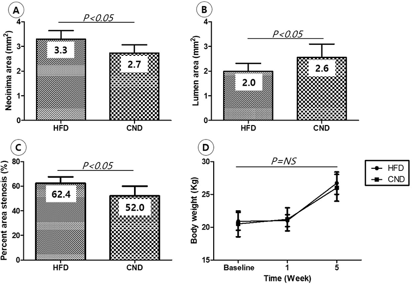
There was no difference in body weight between the two groups until the end of the experiment (Fig. 5D).

Discussion

Our study was conducted to investigate the effect of a mild elevation in serum cholesterol level on vSMC proliferation in a porcine coronary overstretch restenosis model. Our study demonstrated that the mild elevation in serum cholesterol level promoted neointimal hyperplasia after vascular injury in the HFD group.

Previous studies have shown that the consumption of a high-cholesterol and/or HFD is associated with an increase in the prevalence of vascular diseases [810]. Hypercholesterolemia is one of the most important risk factors for cardiovascular diseases.

A large animal model such as a porcine model provides enables researchers to physiologically, pathologically, and preclinically understand diseases. Based on this, knowledge of the prevention or inhibition of the progression of diseases is gained. These models are also important tools for evaluating novel therapeutic drugs and medical devices. In previous studies, the degree of vSMC proliferation after vessel injury in pigs was consistent with the degree in human trials [11, 12]. In addition, pigs have similarities to humans in terms of coronary artery anatomy, vessel physiology, disease progression, lipoprotein profile, and lipid metabolism [1316]. Therefore, our findings can be considered to be applicable in humans.

The main difference of our study from previous studies is the timing of vessel injury. Previous studies using HFD-induced hypercholesterolemia models have been conducted where serum cholesterol level rise to their peak and then balloon angioplasty or stent implantation was performed to induce injury [1720]. One more important difference is that cholesterol levels did not exceed the standard limit [21].

Coronary arterial injury induced by either balloon angioplasty or stent implantation leads to neointimal hyperplasia of intimal smooth muscle cells in a porcine model [22]. There was a significant correlation between arterial injury and vSMC proliferation [23]. It is necessary to confirm the intima layer of the injured arteries to determine the injury score. It was more difficult to accurately measure the injury score after balloon angioplasty than after stent implantation when injury was induced. After stent implantation, the exact injury score can be measured based on the position of the stent strut in the vessel wall. If there is no difference in the injury score between the two groups, vSMC proliferation may be a useful histopathological comparison criterion. Therefore, we compared the lumen area in each after balloon angioplasty.

This study has some limitations. First, hematoxylin and eosin staining was used for estimating vSMC proliferation after injury was induced. Immunohistochemistry techniques using a specific antibody such as α-smooth muscle actin is the gold standard for estimation [24]. Second, we did not determine the complete blood count during the systemic inflammatory reaction in hypercholesterolemia [25, 26]. Third, this was not a long-term study. Finally, the sample size was small.

However, our results showed that mild elevation in serum cholesterol level was related to a higher degree of vSMC proliferation.

This study demonstrated that patients with cardiovascular disease with a mild elevation in serum cholesterol level requires a more aggressive treatment for cholesterol reduction. In particular, the serum cholesterol levels of patients undergoing percutaneous transluminal coronary angioplasty using balloon angioplasty and coronary stenting should be carefully managed.

Conclusion

Patients undergoing percutaneous transluminal coronary angioplasty require more aggressive cholesterol management because mild elevation if serum cholesterol levels induced vSMC proliferation (neointimal hyperplasia) in a porcine model.

Acknowledgments

This study was supported by grants from the Basic Science Research Program through the National Research Foundation of Korea (NRF) funded by the Ministry of Education, Science and Technology (NRF-2017R1C1B1001956 and NRF-2018R1D1A1B07040783) and the KRIBB (Korea Research Institute of Bioscience and Biotechnology) Research Initiative Program (KGM4251824), Korea.

References

1.

Low Wang CC, Hess CN, Hiatt WR, Goldfine AB. Clinical Update: cardiovascular disease in diabetes mellitus: atherosclerotic cardiovascular disease and heart failure in type 2 diabetes mellitus-mechanisms, management, and clinical considerations. Circulation 2016;133:2459-2502.

2.

Tahir H, Niculescu I, Bona-Casas C, Merks RMH, Hoekstra AG. An in silico study on the role of smooth muscle cell migration in neointimal formation after coronary stenting. J R Soc Interface 2015;12:20150358.

3.

Theilmeier G, Quarck R, Verhamme P, Bochaton-Piallat ML, Lox M, Bernar H, Janssens S, Kockx M, Gabbiani G, Collen D, Holvoet P. Hypercholesterolemia impairs vascular remodelling after porcine coronary angioplasty. Cardiovasc Res 2002;55:385-395.

4.

Weidinger FF, McLenachan JM, Cybulsky MI, Fallon JT, Hollenberg NK, Cooke JP, Ganz P. Hypercholesterolemia enhances macrophage recruitment and dysfunction of regenerated endothelium after balloon injury of the rabbit iliac artery. Circulation 1991;84:755-767.

5.

Schwartz RS, Huber KC, Murphy JG, Edwards WD, Camrud AR, Vlietstra RE, Holmes DR. Restenosis and the proportional neointimal response to coronary artery injury: results in a porcine model. J Am Coll Cardiol 1992;19: 267-274.

6.

Schwartz RS, Edelman E, Virmani R, Carter A, Granada JF, Kaluza GL, Chronos NAF, Robinson KA, Waksman R, Weinberger J, Wilson GJ, Wilensky RL. Drug-eluting stents in preclinical studies: updated consensus recommendations for preclinical evaluation. Circ Cardiovasc Interv 2008;1: 143-153.

7.

Suzuki T, Kopia G, Hayashi S, Bailey LR, Llanos G, Wilensky R, Klugherz BD, Papandreou G, Narayan P, Leon MB, Yeung AC, Tio F, Tsao PS, Falotico R, Carter AJ. Stent-based delivery of sirolimus reduces neointimal formation in a porcine coronary model. Circulation 2001; 104:1188-1193.

8.

Kumar A, Datusalia AK. Metabolic stress and inflammation: implication in treatment for neurological disorders. CNS Neurol Disord Drug Targets 2018;17:642-643.

9.

Crunkhorn S. Metabolic disorders: breaking the links between inflammation and diabetes. Nat Rev Drug Discov 2013;12:261.

10.

Hotamisligil GS. Inflammation and metabolic disorders. Nature 2006;444:860-867.

11.

Huckle WR, Drag MD, Acker WR, Powers M, McFall RC, Holder DJ, Fujita T, Stabilito II, Kim D, Ondeyka DL, Mantlo NB, Chang RS, Reilly CF, Schwartz RS, Greenlee WJ, Johnson RG. Effects of subtype-selective and balanced angiotensin II receptor antagonists in a porcine coronary artery model of vascular restenosis. Circulation 1996;93: 1009-1019.

12.

Matsumoto K, Morishita R, Moriguchi A, Tomita N, Aoki M, Sakonjo H, Matsumoto K, Nakamura T, Higaki J, Ogihara T. Inhibition of neointima by angiotensin-converting enzyme inhibitor in porcine coronary artery balloon-injury model. Hypertension 2001;37:270-274.

13.

Marzetta CA, Rudel LL. A species comparison of low density lipoprotein heterogeneity in nonhuman primates fed atherogenic diets. J Lipid Res 1986;27:753-762.

14.

He Q, Ren P, Kong X, Wu Y, Wu G, Li P, Hao F, Tang H, Blachier F, Yin Y. Comparison of serum metabolite compositions between obese and lean growing pigs using an NMR-based metabonomic approach. J Nutr Biochem 2012;23:133-139.

15.

Skold BH, Getty R, Ramsey FK. Spontaneous atherosclerosis in the arterial system of aging swine. Am J Vet Res 1966;27:257-273.

16.

Lee JM, Kim HK, Lim KS, Park JK, Choi KH, Park J, Hwang D, Rhee TM, Yang JH, Shin ES, Nam CW, Doh JH, Hahn JY, Koo BK, Jeong MH. Influence of local myocardial damage on index of microcirculatory resistance and fractional flow reserve in target and nontarget vascular territories in a porcine microvascular injury model. JACC Cardiovasc Interv 2018;11:717-724.

17.

Xia J, Qu Y, Yin C, Xu D. Optical coherence tomography assessment of glucose fluctuation impact on the neointimal proliferation after stent implantation in a diabetic/hypercholesterolemic swine model. Int Heart J 2017;58:608-614.

18.

Llano R, Winsor-Hines D, Patel DB, Seifert PS, Hamamdzic D, Wilson GJ, Wang H, Keane MG, Huibregtse BA, Wilensky RL. Vascular responses to drug-eluting and bare metal stents in diabetic/hypercholesterolemic and nonatherosclerotic porcine coronary arteries. Circ Cardiovasc Interv 2011;4:438-446.

19.

Jenkins JS, Webel R, Laughlin MH, Rowland SM, Yoklavich MF, Amann JF, Branson K, Myers PR. Restenosis following placement of an intracoronary heparin treated tantulum stent in the hyperlipidemic miniature swine model. J Invasive Cardiol 1995;7:173-182.

20.

Grinstead WC, Rodgers GP, Mazur W, French BA, Cromeens D, Van Pelt C, West SM, Raizner AE. Comparison of three porcine restenosis models: the relative importance of hypercholesterolemia, endothelial abrasion, and stenting. Coron Artery Dis 1994;5:425-434.

21.

Grundy SM, Stone NJ. 2018 American heart association/ american college of cardiology/multisociety guideline on the management of blood cholesterol-secondary prevention. JAMA Cardiol 2019;4:589-591.

22.

Karas SP, Gravanis MB, Santoian EC, Robinson KA, Anderberg KA, King SB. Coronary intimal proliferation after balloon injury and stenting in swine: an animal model of restenosis. J Am Coll Cardiol 1992;20:467-474.

23.

Kornowski R, Hong MK, Tio FO, Bramwell O, Wu H, Leon MB. In-stent restenosis: contributions of inflammatory responses and arterial injury to neointimal hyperplasia. J Am Coll Cardiol 1998;31:224-230.

24.

Malik N, Gunn J, Holt CM, Shepherd L, Francis SE, Newman CM, Crossman DC, Cumberland DC. Intravascular stents: a new technique for tissue processing for histology, immunohistochemistry, and transmission electron microscopy. Heart 1998;80:509-516.

25.

Busnelli M, Manzini S, Froio A, Vargiolu A, Cerrito MG, Smolenski RT, Giunti M, Cinti A, Zannoni A, Leone BE, Forni M, Bacci ML, Biasi GM, Giovannoni R, Lavitrano M. Diet induced mild hypercholesterolemia in pigs: local and systemic inflammation, effects on vascular injury-rescue by high-dose statin treatment. PLOS ONE 2013;8:e80588.

26.

Busnelli M, Froio A, Bacci ML, Giunti M, Cerrito MG, Giovannoni R, Forni M, Gentilini F, Scagliarini A, Deleo G, Benatti C, Leone BE, Biasi GM, Lavitrano M. Pathogenetic role of hypercholesterolemia in a novel preclinical model of vascular injury in pigs. Atherosclerosis 2009;207:384-390.