Introduction
The Bispectral Index (BIS), a derived variable of the electroencephalography (EEG), has also been suggested to reflect the level of sedation and hypnosis with intravenous and inhalational anesthesia [1,2]. This electrophysiological variable is considered to provide useful information regarding brain stem function because it is a marker reflecting the activity degree of cerebral cortex. It provides values of a numerical expression quantified over level of consciousness, enabling objective assess- ment of patients’ brain condition. There have been some studies investigating that the BIS can reflect the level of consciousness in psychiatric patients during ECT. These studies are manly about the changes and the accuracy of the BIS in predicting the hypnotic degrees in those patients who undergone ECT that may make the BIS bizzare (EEG abnormality) [3,4]. It was also found that ECT may decrease the BIS during ECT under propofol anesthesia. ECT has been broadly used in the treatment of schizophrenia and major depressive disorders which are resistant to pharmacologic therapy and the effect of ECT are alleged to correlate with seizure duration. This prospective and observational study was performed with the aim of determining if there is any correlation between these Pre-ECT BIS and seizure duration in the patients who undergo ECT.
Patients and Methods
Eleven patients (aged 21-67 years) with schizophrenia and major depressive disorders which are resistant to pharmacologic treatments were prospectively evaluated for the BIS and seizure duration. All patients fasted for at least 6 hours before ECT. All other aspects of pre-procedural care were routine. Before the induction of anesthesia routine monitorings were initiated and an intravenous line secured. The EEG electrodes were applied to the forehead, temple and mastoid of the patient, then connected to the BIS monitor. Anesthesia was induced with a propofol (100-120 mg i.v.) and mechanical ventilation using 100% oxygen was adjusted for end-tidal concentration of carbon dioxide to be maintained between 30 and 35 mmHg, and an pneumatic tourniquet was applied on an arm for isolated forearm technique (IFT) [5-11] for the detection of arm movement during ECT. Before ECT started the tourniquet was inflated to a pressure of 200 mmHg and succinylcholine (1 mg/kg i.v.) was administered for neuromuscular blockade. At the end of procedures patients were verbally prompted to open and squeeze the fingers of patient’s hand in 30 seconds intervals. ECT was done 8-10 times per patient every other day depending on the clinical responses. But the data were obtained from the first five electroconvulsive therapies in a row. The BIS was measured at four specific time points (T1=baseline before the start of induction, T2=before ECT (Pre-ECT), T3=at eye opening to verbal command and T4=at 5 minutes after eye opening) with a BIS monitor, Model A-3000 vistaTM (Aspect Medical Systems, Norwood, USA). The correlation between the BIS and the seizure duration was determined using Pearson’s correlation coefficient. Each variable was recorded simultaneously. Mean values of measurements of the BIS made over the study period were compared for each specific time points. Statistical analysis was performed on an IBM-compatible computer with standard statistical software packages (SPSS For Windows, version 22.0). Data were analyzed using Kruskal-Wallis test and Mann-Whitney test. A value of P<0.05 was considered statistically significant. All patients were interviewed in the recovery area for any memory about procedures. Information concerned with the monitoring modalities was provided to patients’ family or next of kin and informed consent signed after obtaining approval of Institutional Review Board before the commencement of study.
Results
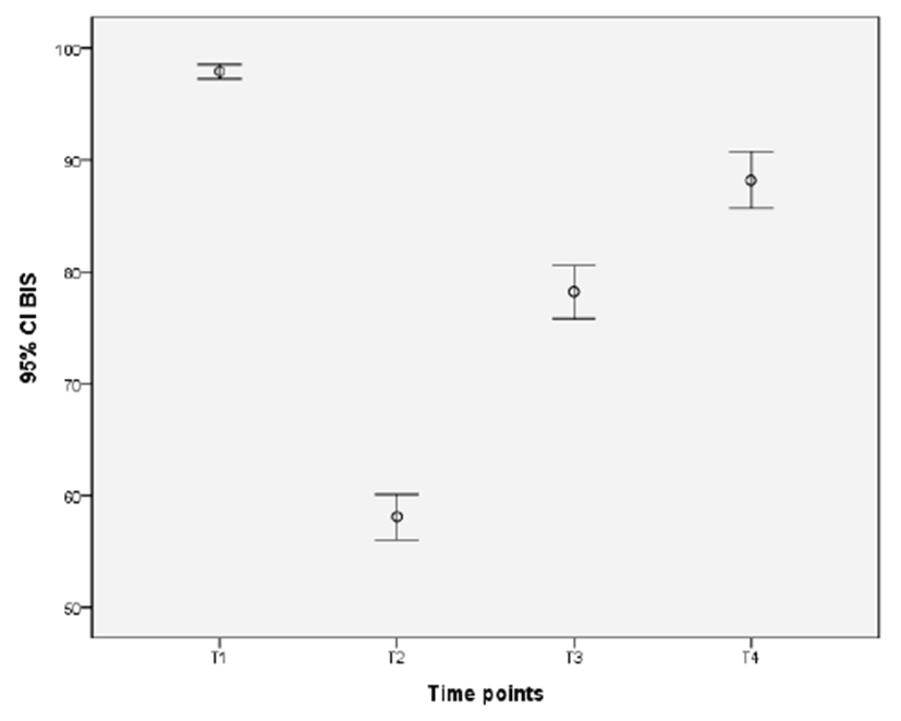
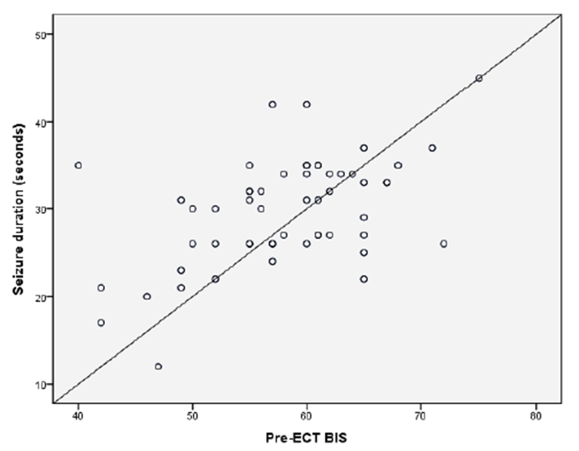
Awake baseline value (T1) of the BIS showed significant time dependent changes from 97.92 ± 2.3 to 58.07 ± 7.5 before ECT (T2, Pre-ECT), 78.20 ± 8.7 at eye opening to verbal command (T3) and 88.19 ± 9.2 at 5 minutes after eye opening (T4). (p<0.001, Fig. 1). The Pre- ECT BIS was positively correlated with seizure duration (p<0.001, r=0.507, Fig. 2).
Discussion
Patients requiring ECT need effective sedation and neuromuscular blockade to prevent discomfort and the possibility of fractures of bones during ECT. Seizure duration is considered as a major determinant of treatment efficacy in ECT. This study was based on a simple clinical protocol to monitor motor seizure of the patients with psychiatric disorders such as schizophrenia and major depressive disorders.
BIS is calculated from a multivariate regression model depending on three variables (burst suppression ratio, relative α/β ratio and bicoherence of the EEG). The BIS, as an electroencephalographic variable, provides continuous data between 0 and 100 indirectly reflecting cerebrocortical and subcortical activities as it is based on EEG. EEG has effectively been used to evaluate the depth of consciousness of the patients with mental change and to predict prognosis according to EEG patterns [12,13]. BIS was developed to be applicable to normal surgical patients. Monitoring of the BIS during ECT may not reliably reflect the clinical degrees of consciousness. Furthermore ECT may increase electromyographic (EMG) activity, especially in frontalis muscle activity. For this reason, during ECT the BIS may be fluctuating, however, it may not be artifacts but rather clinical state changes [14,15].
In our study, after patients lost consciousness, succinylcholine was given to prevent bone or tooth fractures by muscle jerk with ECT. There have been some reports investigating the effects of neuromuscular blocking agents on the depth of anesthesia monitored by BIS. Flaishon and his colleagues suggested that BIS might be able to provide a reliable method in predicting the probability of recovery of consciousness from anesthesia using thiopental or propofol in the patients paralyzed with neuromuscular blockers [16]. In their report, although they did not mention the direct effect of neuromuscular blocking agent on the BIS, it was recognizable that the capability of BIS to predict the probability of a patient being aware was not affected by neuromuscular blockade. On the contrary, Messner and his colleagues were very doubtful of the capability of the BIS indicating the level of consciousness in paralyzed state because in their study using volunteers neuromuscular relaxation decreased the BIS to very low values in fully conscious state, which were generally accepted as unconscious state [17,18]. It implies that BIS might not be a sensitive measure of the depth of sedation and detector of awareness in subjects who were totally paralyzed with a neuromuscular blocking agent. The current study was not designed to clarify these conflicting results about the capability of the BIS in detecting awareness in paralyzed condition, however, it may be carefully recognizable that the BIS could be applied to detect awareness of paralyzed patients undergoing ECT. Electromyographic activity may influence surface EEG and the BIS. In our study spontaneous facial muscle EMG activity was abolished by succinylcholine given before ECT, therefore the BIS was not affected by EMG activity.
There has been some research investigating the relationship the BIS, and motor and EEG seizure durations [19,20]. In their studies positive correlations were found between Pre-ECT BIS, and motor and EEG seizure duration in spite of wide correlation degrees between 0.4- 0.73. In the present study we also found that the BIS was positively correlated with the Pre-ECT BIS. In conclusion, the BIS monitoring might be useful in predicting seizure duration in psychiatric patients requiring ECT.