Introduction
Rheumatoid arthritis (RA) represents cartilage and bone destruction, persistent synovitis and systemic inflammation [1]. Among several mouse RA models, the collagen antibody-induced arthritis (CAIA) model shows good similarity with human RA and is usually used to investigate pathogenic mechanisms of RA [2, 3]. A cocktail of collagen type II-specific monoclonal antibodies has been used to induce an acute form of arthritis in experimental animals [4]. Modification of the cocktail to enhance its arthritogenicity has induced severe and consistent arthritis, even in low responder strains [5].
For in vivo monitoring of arthritis progression, it is necessary to visualize the real presentation of pathological lesions in experimental animals. Modern several imaging technologies enable the detection and visualization of biological processes [6], and provide the data related to several disease progression [7, 8] and 3D structures of tissues [9], and reduce the number of animals required for experiments [10]. Also, they even supply quantitative results [11]. High-resolution micro-computed tomography (micro-CT) has advantage over other modalities for the imaging of bony structures and is primarily used to detect bony changes in experimental animals [12]. However, it is limited in its ability to detect slight or minimal alterations in joints, especially in cartilage [13]. To overcome this, optical imaging using fluorescence probes has been used for the detection of altered biomolecules [14]. Nearinfrared fluorescence imaging agents have been utilized to detect several pathological changes such as apoptosis [15] and oxidative stress [16].
Even though near-infrared spectroscopic probing could be used to detect changes in the composition of the subchondral bone and might distinguish healthy cases from osteoarthritis in rats [17], there have been a few reports about in vivo monitoring of mouse RA using near-infra red probes. The purpose of this study was to examine CAIA lesions using fluorescence imaging and to compare the results with those of micro-CT and histopathological examination.
Materials and Methods
Male BALB/c mice were obtained from Nara Biotech Co. Ltd. (Seoul, Korea) at 6 weeks old of age. The mice were kept in a room with 22 ± 3°C, 55 ± 5% and a 12-h light/dark cycle. Mice were fed filtered water ad libitum and unrefined chlorophyll-containing ingredients in an alfalfa free diet (Harlan Laboratories, Inc., Madison, WI). After one week for acclimation, twelve mice were randomly divided into three groups: group 1 (G1) as control animals, group 2 (G2) as fluorescence probe control animals and group 3 (G3) as animals with collagen antibody-induced arthritis. The mice of G3 intravenously received anti-type II collagen 5-clone antibody cocktail (Chondrex, Redmond, WA) (2 mg/mouse) on day 0 and intraperitoneally received lipopolysaccharide (LPS, Chondrex) (50 μg/mouse) on day 3. On the while, the mice of G1 and G2 received 0.9% saline in equal volumes at equivalent times.
Veet cream was used for removal of hair before the process of fluorescence bioimaging. Mice in G1 received equal volumes of 0.9% saline. Mice in G2 and G3 were injected intravenously with OsteoSenseⓇ 680 EX (PerkinElmer, Waltham, MA) at the dose of 2 nmol/100 μl/ mouse on day 7 post collagen cocktail treatment. Fluorescence bioimaging was taken by IVIS Lumina Series III (PerkinElmer) at 48 h after near-infrared probe treatment under anesthesia with avertin (Sigma, Saint Louis, MO).
All mice were sacrificed at day 9, and the knee joints were fixed in 10% neutral phosphate-buffered formalin. This study was approved by the animal experiment committee of Namseoul University based on the Animal Protection Act.
A micro-CT system (Skyscan1173, Bruker, Kontich, Belgium) was used for analysis of the knee joints. The specimens were scanned with an X-ray source of 75 kV/106 μV and pixel size of 9.94 μm using an aluminum 1.0-mm filter. After scanning, cross-sectional slices were reconstructed by NRecon software Ver.1.6.9.16 (Bruker). Three-dimensional analysis was performed using CT Analyser Ver. 1.14.4.1 (Bruker).
Results
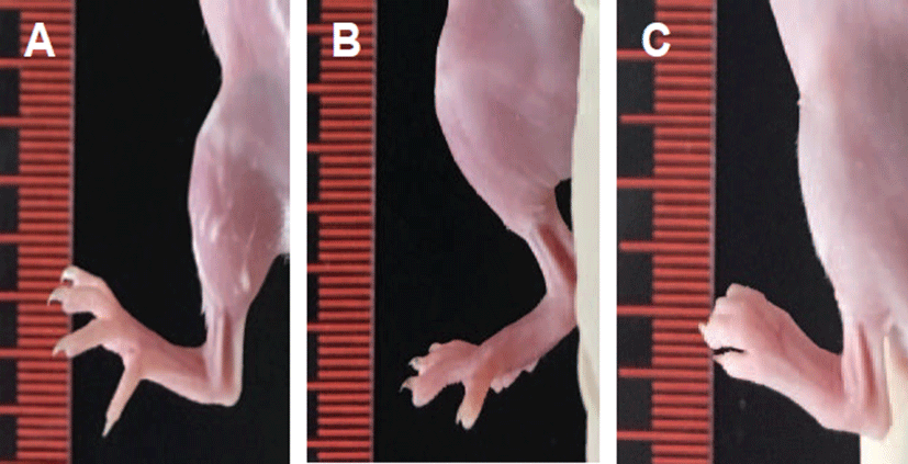
Measurable macroscopic changes were determined by paw thickness measurement. Normal paw appearance was shown in G1 and G2. However, the paw thickness of G3 was increased on day 6 after the treatment with anti-type II collagen 5-clone antibody cocktail (Fig. 1).
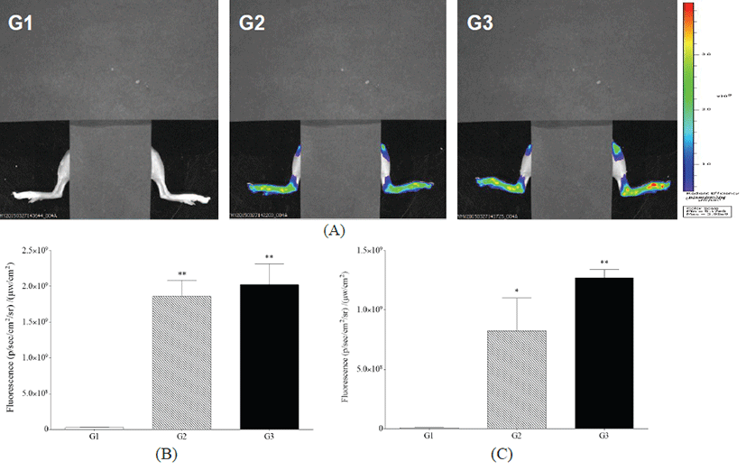
Analysis of fluorescence bioimaging showed that no fluorescence was observed in G1. Fluorescence intensity was observed in G2 and G3, with G3 showing greater intensity than G2 at 24 h after OsteoSenseⓇ 680 EX treatment (Fig. 2).
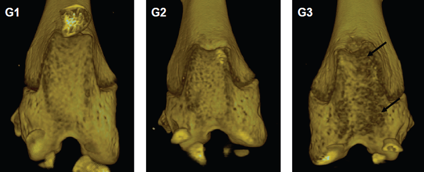
Micro-CT analyses showed that joint surfaces in G1 and G2 represented normal articular appearance. However, in G3, micro-CT images showed irregular articular appearance (Fig. 3). It showed a diffuse pattern mainly located on the articular surface.
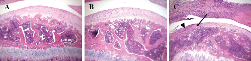
No cartilage destruction or inflammation was found in G1 and G2. However, G3 showed destruction of the cartilage with degeneration of chondrocytes. Furthermore, the destruction of articular surfaces of joints in G3 showed destruction of bony structures, as well as synovial hyperplasia and infiltration of inflammatory cells, mainly consisted of neutrophils (Fig. 4).
Discussion
In this study, fluorescence bioimaging and micro-CT provided information for the assessment of arthritis lesions, and these results generally agreed with histopathology assessment.
When examining arthritis lesions in vivo, bioimaging enables detection of several molecules and visualization of biological processes [6]. In this study, OsteoSenseⓇ 680EX as fluorescence bioimaging probe was tested in vivo. A near-infrared bioimaging agent, OsteoSenseⓇ 680EX, is basically designed to emit the light by binding to hydroxyapatite for the detection of microcalcification areas and bone remodeling lesions. Compared to G1, fluorescence intensity in G2 and G3, with G3 showing higher intensity than G2. Furthermore, this fluorescencebased imaging has some limitations in its ability to detect the anatomical location of lesions. Although bioimaging using near-infrared probe(s) may provide tissue detection and potential quantification [18], in this study, a high background level of fluorescence signals was observed in G2, even though G3 showed higher intensity compared to G2. Further studies will be warranted to develop the specific fluorescence probe(s) capable of detecting the disease progression of arthritis, with low background level.
This study used micro-CT to analyze alterations in bone structures. Micro-CT analyses showed that joint surfaces in G3 showed degeneration of articular surface and alteration of bony structures. This was consistent with disruption of synovial membrane and structural changes in bony tissues revealed by HE examination. Although micro- CT analysis identified damaged areas in the articular surface, it may have the possibility not to detect the pathological lesions, especially when the lesions are restricted to small areas. And as there may be some limitations to current methods for quantification of pathological lesions, the development of new techniques is necessary [19]. Three-dimensional visualization of lesions may be one of these new approaches [20]. Further studies into the examination of arthritis lesions via different bioimaging techniques, especially 3D approaches, are warranted.
Among several RA animal models, the CAIA is an acute arthritis model that induces rapid onset of disease, usually with an experimental period of two weeks [2]. In this study, arthritis lesions occurred from day 5 to 9 after collagen antibody injection. However, the CIA model induces gradual onset of disease, usually with an experimental period of four weeks, showing hypertrophy of the synovial lining in early inflammatory arthritis and formation of fibrovascular pannus in the later stage of the disease [21]. For the rapid screening, the acute RA model is preferred. However, animal variation should be considered and the proper RA model should be selected according to research purpose and target molecules.
Taken together, analyses of fluorescence bioimaging using OsteoSense® 680EX probe made it possible to evaluate CAIA lesions, comparable with micro-CT and histopathological examination in mice. However, it had some limitations when used with fluorescence owing to a high background level of fluorescence signals.