Introduction
Neuron-specific enolase (NSE) is a brain specific isoenzyme of the glycolytic enzyme enolase and is considered a specific marker for neuronal elements in various regions of brain [1-3]. Immunoreactive NSE in mammalian taste buds is related to gustatory function [4]. The gustatory cells in the taste buds are one typical sensory paraneurons and have been demonstrated as immunoreactive for some neuron-specific proteins, including NSE [5]. The levels of NSE are very low in embryonic rat brain and are increased with the morphological and functional maturation of neurons [6]. Immunohistochemical studies have shown that antibodies directed against specific proteins in nervous tissues are much more definitive in clarifying the origins and detailed morphology of the structural components of nervous system in embryos [7, 8].
In our previous reports, we described the morphological changes of lingual papillae during development of goat, examined by scanning electron microscopy [9, 10] and light microscopy [11]. However, no studies have been reported in the literature that has described the immunohistochemical localization of NSE in the developing tongue of Korean native goat. Therefore, to understand the expression of NSE-immunoreactivity (IR), we investigated the localization of NSE-IR in the developing tongue in Korean native goat.
Materials and Methods
The tongues from three fetuses and neonate of Korean native goat (Capra hircus) were used in this study. The day of mating was designated as embryonic day 0 (E0), and the goat embryos were obtained at different embryonic ages. To investigate the changes in NSE-IR during prenatal development of the tongues, the tongues were removed from 60, 90, 120, and 150-day-old fetuses (neonates) originated from 2 to 4 years old Korean native goats with body weight ranging from 23 to 33 kg, by caesarean section performed under general anesthesia using xylazine hydrochloride (10 mg/kg, i.v.; Bayer Korea Ltd. Seoul, Korea). The expression of NSE in the developing tongue of goat fetuses (60, 90, 120, 150 days) was studied using an immunohistochemistry. All animal experiments were performed according to a protocol set out in the guidelines of the Animal Experiments Ethics Committee at Gyeongsang National University (Approval No. GNU-LA-10).
Serial sectioned tissues (5-7 μM) were mounted on charged slides and pre-treated in microwave (800 Watt) in citric acid buffer (pH 6.0) for 20 min. The sections were incubated in 0.3% Triton X-100 (Sigma-Aldrich, USA) 0.1 M phosphate buffered saline (PBS, pH 7.4) overnight at 4℃ and in a blocking solution containing 10% normal goats serum to reduce non-specific reactions overnight at 4℃. After washing with PBS three times, the sections were exposed to primary antibody, comprising of NSE (Dako) diluted in PBS (pH 7.2) (1:10) containing 0.5% bovine serum albumin (BSA) for 2 h at room temperature. The slides were again washed with PBS and incubated using appropriate anti-mouse biotinylated secondary antibodies for 1 h at room temperature. The reactions were visualized with avidin-biotin-peroxidase complex kit (ABC, Vector Co, Burlingama, CA, USA) for 2 h followed by incubation with AEC (3-amino-9-ethylcarbazole) as a substrate for 10 min. Reacted sections were briefly counter-stained with Meyer's hematoxylin, and then immunoreactive cells were observed under light microscope. Substituting blocking solution with primary antiserum was employed as controls.
Results
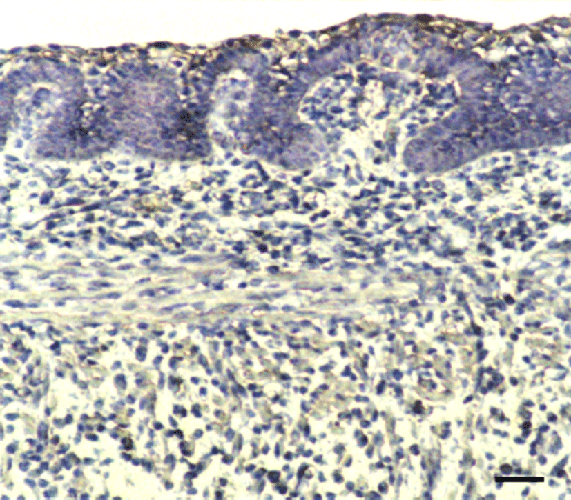
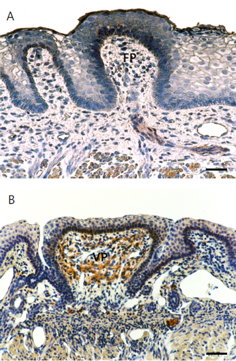
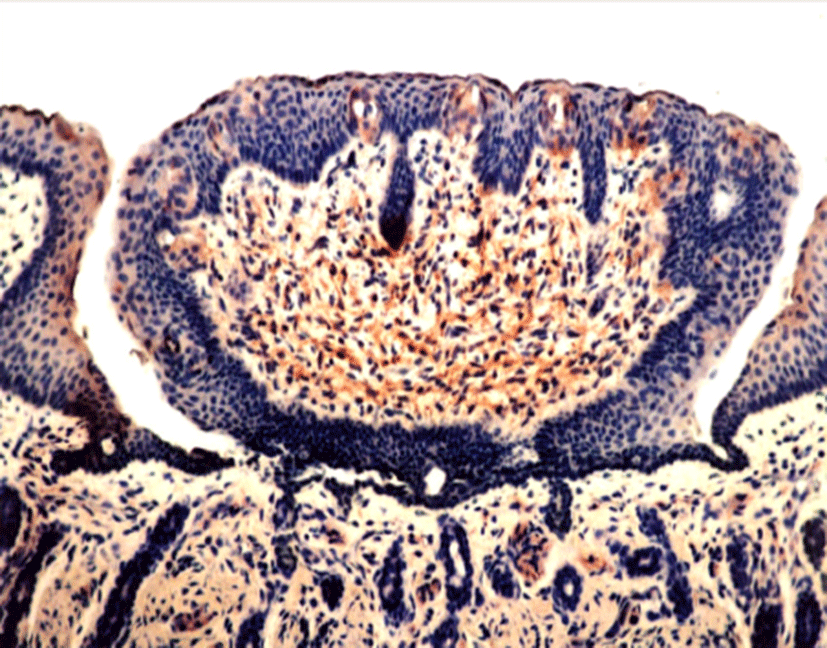
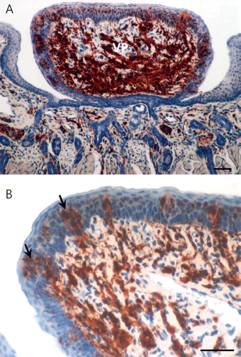
The tongue tissues were examined using immunohistochemical techniques. In this study, NSE was used as a neural marker to understand the development of gustatory nerve innervations and taste buds in the developing tongue. In 60-day-old fetuses, NSE-IR weakly appeared in lamina propria of the basal portion and basilar aspects of the apical epithelium of the tongue (Fig. 1). In 90-dayold fetuses, NSE positive cells were observed in laminar propria and extended in the core part of connective tissue in vallate papillae. Especially, taste buds in the primordia of the papilla were moderately positive (Fig. 2). In 120-day-old fetuses, taste buds in the vallate papillae were strongly positive for NSE. Interestingly, NSE-IR nerve fibers more precisely appeared in connective tissues of the papilla (Fig. 3). In neonates, the taste buds of vallate papillae were strongly positive for NSE and nerve fibers synapsed with taste buds of vallate papilla were demonstrated as strongly positive for NSE (Fig. 4a, b). Thus, based on this study, we demonstrated that tastesensing ability of neonate was well developed at the time of its birth.


Discussion
The present study demonstrates the sequential morphologic pattern of NSE-nerve fibers in developing tongue of Korean native goat. NSE is located within neurons in the mammalian central and peripheral nervous systems [1]. There have been a few reports describing the presence of NSE-cells of taste organs. NSE-like immunoreactivity has been found in discrete cells within the gustatory epithelium of guinea pig [4], frogs [12, 13], and rat [7]. Especially, NSE has been used as a neuronal marker for the development of lingual papillae and synapse of gustatory nerves in the taste bud and its distribution for a variety of animals including rat [7, 14, 15], human [16, 17], mouse [18], and guinea pig [5, 19].
NSE-IR nerve fibers were observed in the median lingual sulcus at the base in the tongue of 15 to 16-day-old rat fetuses and appeared on round-shaped undifferentiated NSE-taste bud cells in 18 to 21-day-old rat fetuses [7]. NSE-IR neurons were first spotted in taste buds of trench wall epithelia of vallate papillae in 5 to 10-day-old mouse and were expressed on the taste cells of vallate papillae in a 14-day-old mouse. Moreover, the shape was similar to that of an adult NSE-IR neuron [18].
Witt and Reutter [17] reported that NSE-IR nerve fibers approached towards the basal membrane of lingual epithelia in 7-week-old human fetuses and first appeared on taste buds in the primordia of fungiform and vallate papillae in 10 to 11-week-old human fetuses. NSE-IR nerve cells were well developed in the primordia of taste buds of fungiform papillae in 12 to 13-week-old human fetuses. Hirata and Kanaseki [7] reported that NSE-IR nerve fibers were also observed in the core of connective tissue of vallate papillae in 16- to 17-day-old rat fetuses. Furthermore, NSE-IR nerve cells first appeared in the taste buds of vallate papillae in 3 to 4-day-old rat, thus stating that NSE-IR penetrated into the taste buds, which were connected with sensory nerves.
NSE-IR was positive in the taste buds and taste cells of lingual gustatory papillae in mice [18, 19], rats [14, 20], cat [21], guinea pigs [20, 22], and hamsters [23], and NSE-IR nerve fibers formed dense nerve plexus in the connective tissue of gustatory papillae. Montavon and Lindstrand [19] reported in their study on the expression of NSE in the gustatory papillae of the pig that NSE reacted in the form of large neuronal network under the epithelium of all gustatory papillae, and that a strongly positive reaction to NSE was seen both in the inner part of taste buds and the lower part of taste buds, which seemed to be related to synaptogenesis.
Measurement of isoenzyme by specific radioimmunoassay during the course of brain development in rat showed very low levels of NSE in embryonic brain and the increase at a time coincident with the morphological and functional maturation of neurons [6]. The appearance of NSE in neurons has often been correlated with neuronal differentiation [24], and the expression pattern of NSE may correlate with developmental stages of taste receptor cells [15].
In this study, examination of tongue tissues revealed weak appearance of NSE-IR in lamina propria and basilar aspects of the apical epithelia of gustatory papillae in 60-day-old fetuses. However, NSE-immunoreactive nerve fibers were positive in the core of connective tissue in gustatory papillae in 90-day-old fetuses. NSE-IR nerve cells also appeared in taste buds. Judging from appearance of the part where nerve fibers synapse with cells in taste buds, 120-day-old fetuses were suspected to have prior gustatory sensitivity. In neonates, a large number of nerve fibers were synapsed with taste buds; therefore, it was supposed that the sensing ability of neonate in Korean native goats for gustation was well developed at the time of its birth. Taken together, these findings suggest that NSE may be a useful neural marker to understand the development of gustatory nerve in the taste buds.