Introduction
Growth disturbances resulting in alignment deformities of the canine radius are common and well documented [1]. Asymmetric distal radial physeal closure can result in radial shortening, elbow subluxation, and carpal varus or valgus deformities [1]. Premature closure of the distal ulnar physis is the most common cause of radial growth deformation [1]. Angular deformities are also caused by local thickening of the growth plate associated with osteochondrosis in the radius and ulna [2].
Choice of surgical treatment for radial angular limb deformities is dependent upon residual growth potential within the radial and ulnar physis. Reports have described antebrachial angular limb deformity correction by using osteotomies and many different fixation modalities, including internal fixation with bone plates [3, 4], external skeletal fixation with linear external skeletal fixators [5-7], circular external skeletal fixators [8, 9, 10] and hybrid linear-circular external skeletal fixator constructs [11]. Because these surgical treatments include soft-tissue dissection and bone cutting, there are complications concerning wound closure, infection, delayed union or malunion.
Guided growth through temporary hemiepiphysiodesis is a minimally invasive treatment, which has a lower morbidity than traditional osteotomies [12, 13]. In human, early guided growth in pathologic knee deformities produces qualitative improvement in the physis of not only the knees, but also the hips and ankles [14]. Traditionally, hemiepiphysiodesis has been performed with staples. The technique using staples might have some complications such as the premature closure of the growth plate and implant migration or breakage [15]. The tension-band plating technique has been preferred to stapling because of preventing the growth plate from compression and reducing mechanical failures [16]. With this technique, a small plate is applied extraperiosteally and fixed to the metaphysis and epiphysis with two screws. The screws are not tightly fixed in the plate for angulating progressively while the deformity is corrected. An approximately 30% faster correction rate was noted in a recent paper which is retrospective research investigating 34 children with 65 deformities [16].
Hemiepiphysiodesis has only been reported in four dogs with tibial deformities, however, measurement using the Center of Rotation of Angulation (CORA) method and follow-up were not reported [17]. The purpose of this study was to describe the outcome of hemiepiphysiodesis with a tension band plate for treatment of angular deformity resulting from osteochondrosis by using CORA method to evaluate the effect of hemiepiphysiodesis.
Case Report
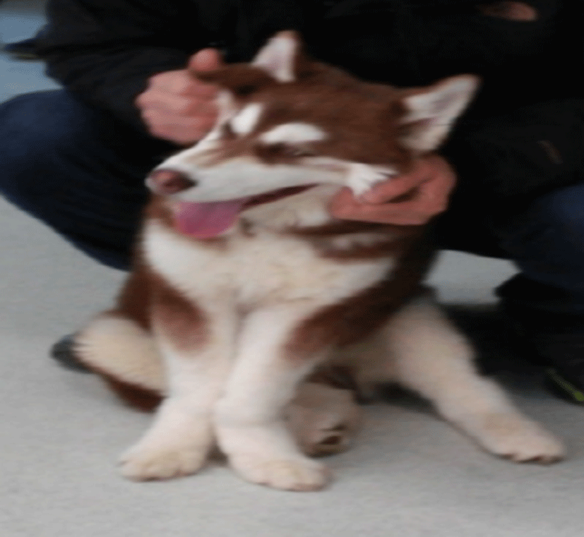
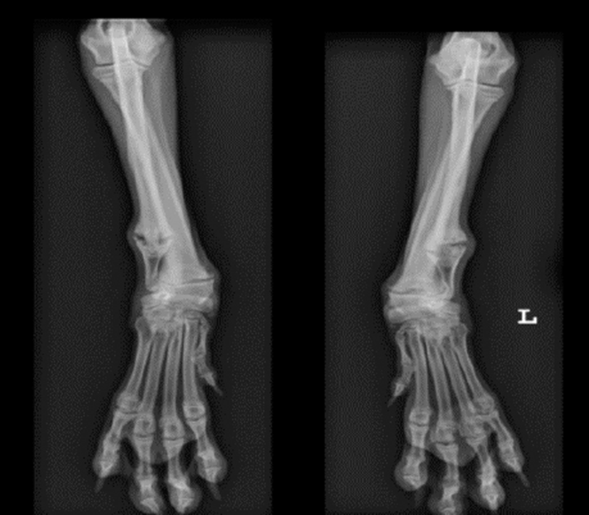
A 4-month-old, intact male Siberian husky dog, weighing 14.6 kg, was presented to the Gyeong-Sang National University Animal Medical Center (GAMC) with a history of bilateral fore-limb lameness and angular deformity. The patient had a history of trauma, which had occurred after jumping over a fence one month prior to referral. Physical examination revealed moderate forelimb lameness (Grade 2/5) and valgus deformities (Fig 1). Pain was elicited upon palpation of the carpal joint. Complete blood count revealed no significant issues whereas serum biochemical parameters indicated hyperphosphatemia (8.6 mg/dL; reference range 2.9 to 6.6 mg/dL). Radiography showed the distal growth plate to be conical shaped, radiolucent, thickened and irregular at both distal ulnar physis. A thickened distal growth plate also was revealed at both the distal radial physis with osteophytes presented. Bilateral valgus and antecurvatum deformity of the radius was observed (Fig 2). Based on these findings, a diagnosis of angular deformity caused by a shortened ulna associated with osteochondrosis was made.
Analgesics and carprofen (2.2 mg/kg PO BID), misoprostol (5 μg/kg) and famotidine (0.5 mg/kg PO BID) were prescribed for two weeks. Following this treatment, although pain was dramatically relieved and lameness had improved, angular deformity had deteriorated, indicating a requirement for surgical correction. Surgical implantation of a temporary hemiepiphysiodesis using a DCP plate between the epiphysis and the metaphysis was considered as an appropriate intervention.
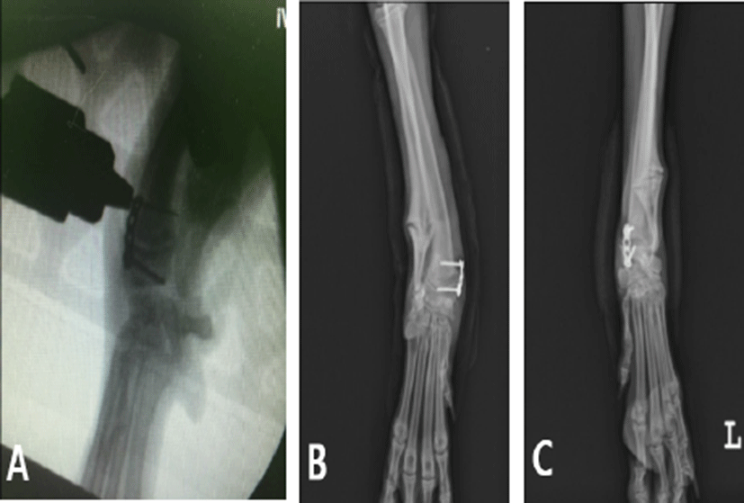
Atropine (0.04 mg/kg, subcutaneous [SC] administration), butorphanol (0.2 mg/kg, SC), and diazepam (0.2 mg/kg, intravenous [IV] administration) were used as pre-anesthetic drugs for surgical treatment, and cefazolin (25 mg/kg, IV) was used as a prophylactic antibiotic. After induction with etomidate (1.5 mg/kg, IV), anesthesia was maintained with inhaled isoflurane. A two cm incision was made over the physis, located by using a C-arm. A 24G needle was passed into the physis under C-arm guidance. Care was taken to avoid damage to the physis. The DCP plate was placed extraperiosteally, and after drilling with a pin, the plate was fixed to the metaphysis and epiphysis with two cortical screws (Fig 3, A). The plate was placed craniomedially at the left physis to correct the valgus and antecurvatum and medially at the right physis with the primary purpose of correcting the valgus (Fig 3, B, C). The final placement of the plate and screws was confirmed under the C-arm in AP (anteroposterior) and lateral views. The skin was sutured with non-absorbable sutures. The plates were maintained in situ for six weeks.
Butorphanol (0.2 mg/kg, SC, SID [once daily]) for immediate pain management and cefazolin (25 mg/kg, SC) and vitamin K (1 mg/kg, SC, SID) were administered. Cefadroxil (25 mg/kg, PO, BID [twice daily]), carprofen (2.2 mg/kg, PO, BID), misoprostol (5μg/kg, PO, BID), and famotidine (0.5 mg/kg, PO, BID) were administered for 2 weeks postoperatively.
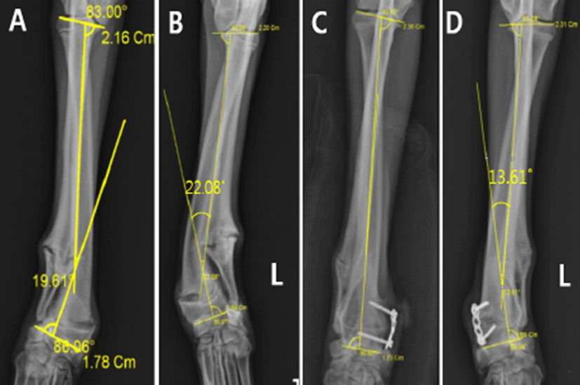
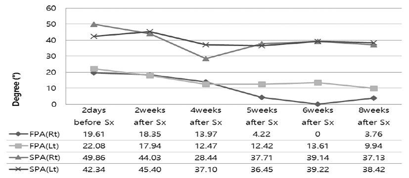
Radiographs were obtained pre-operatively and at 2, 4, 5, 6 and 8 weeks post-operatively to enable monitoring of the healing process. Forelimb alignment corrections were determined by comparing the angle of frontal plane alignment (FPA) and sagittal plane alignment (SPA) measurements with previously established reference ranges (normal FPA range, 0 – 8˚; normal SPA range, 8 – 35˚) using the Center of Rotation of Angulation (CORA) method [4]. The preoperative angle of FPAs were 19.61˚ and 22.08˚ at the right and left limbs, respectively (Fig 4, A, B). At six weeks post-surgery, correction of the right forelimb into the normal range for FPA was apparent, (Fig 4, C) and the left forelimb was close to reference range (Fig 4, D). The preoperative angles of SPAs were 49.86˚ and 42.34˚ for the right and left limbs, respectively (Fig 5). Six weeks following surgery, both forelimb SPAs were close to the reference range (Fig 5). The preoperative radiography showed the radiolucent, thickened and irregular distal growth plate at both distal ulnar and radial physis (Fig 4, A, B). At 6 weeks, these lesions associated with osteochondrosis at both distal ulnar and radial physis had almost completely been healed (Fig 4, C, D). A lameness score was 1.5/5 at the 6-week postoperative examination by the surgeon. Removal of DCP plates was perfomed at 6 weeks. At the 8-week radiograph assessment, the right forelimb FPA was increased, and the right, left forelimb SPA and the left forelimb FPA were decreased compared with 6 weeks (Fig 5). A lameness score improved to 1/5 and there is no pain upon palpation of the carpal joint at the 8-week postoperative examination by the surgeon. On telephone interviews with the owners at 10 weeks, the owner reported that the patient had begun to return to pre-injury activity level. At 14 weeks, the patient maintained normal gait patterns and had no pain without non-steroidal anti-inflammatory drugs administration based on telephone interviews. Since then, patient follow-up has been lost.
Discussion
Various surgical methods have been developed to treat angular deformity in humans. Osteotomies have traditionally been the preferred method for treating bone deformities. However, these involve extensive soft-tissue dissection and have associated complications relating to wound closure, infection, delayed union or malunion and prolonged immobilization, which increase patient morbidity [18-20]. In dogs, although corrective osteotomy is the gold standard for angular deformity, this treatment method has many complications and should not be performed on skeletally immature dogs. Being a minimally invasive technique and of lower morbidity than osteotomy in humans [12, 13], guided growth such as hemiepiphysiodesis using a tension band plate is an appealing option and the preferred primary treatment for pediatric lower limb deformities. In this case, we applied DCP plate to a growing dog at four months of age, with minimal soft-tissue dissection and a short hospitalization period. There were no complications of wound closure, infection, postoperative immobilization or malunion. Furthermore, pain elicited upon palpation of the carpal joint was reduced continuously after surgery, without any requirement for non-steroidal anti-inflammatory drugs administration. We propose that hemiepiphysiodesis with a tension band plate to allow “guided growth” is an attractive alternative for correcting angular deformity in the growing dog.
Stevens reported a retrospective review of 14 patients with pathological physis undergoing temporary hemiepiphysiodesis [21]. This review confirmed improvement not only in limb alignment, but also in the pathological state of the physis. Recovery was hastened by elimination of an eccentric loading of an already compromised physis. Studies by Novais and Stevens and Boero et al. showed safe implementation of temporary hemiepiphysiodesis in pathologic physis with the preferred implant being a tension band plate and screw construct [22, 23]. In the case presented here, we observed that, following the restoration of the mechanical axis using guided growth, the quality and appearance of both physis improved dramatically. We expect that hemiepiphysiodesis using a tension band plate resulted in enlargement of the growth cartilage caused by osteochondrosis in radius and ulna, which gradually turned into bone.
Our approach in this case was to apply plates craniomedially and also medially at the left and right physis. The reason for placing the plate craniomedially was to correct both the valgus and the antercurvatum. However, our study showed that the postoperative angle of FPA, which is used to evaluate the valgus in medial applications, was 3.76˚ at 8 week, but for the craniomedial application, it was 9.94˚, and the postoperative angles of SPAs, which is used to evaluate antecurvatum, were 37.14 and 38.42 in medial applications and craniomedial application respectively (Fig 5). Furthermore, medial plate attachment improved the quality and appearance of the physis. As a result, prognosis following medial application was superior to that of craniomedial application.
Determining the timing of surgical intervention for angular deformities can be challenging, and each case must be evaluated independently with consideration of the patient’s age, growth potential, and the nature of the angulation. If a young patient presents with a pathological distal ulnar and radial physis which results in radial angulation, hemiepiphysiodesis can be attempted to release the bowstring effect of the affected ulna and radius. However, the timing of implant removal and application have not been prospectively studied in a large number of juvenile dogs. In our case, because FPA and SPA were corrected to, or close to, the normal ranges, and almost complete healing of the lesions associated with osteochondrosis was achieved based on radiologic assessment, plates were removed six weeks post-operatively. Following plates removal, both forelimb SPAs and the left forelimb FPA were decreased, but right forelimb FPA did increased at eight weeks post-surgery (Fig 5). It was demanding to determine whether elimination of the plate at 6 weeks is proper choice because there is little data indicating best time for removal and application of plates in dogs applying hemiepiphysiodesis using tension band plate. In people, growth after implant removal is unpredictable and highly variable in instance and intensity [24]. Therefore, successive radiographs are advised to decide the best time for removal of plates. Because additional radiograph was not taken after 8 weeks with owner’s choice, the optimal timing for adequate correction could not be determined in our study. Further investigation of hemiepiphysiodesis in relation to the timing of implant removal and application is needed for the successful correction of angular deformity.
In conclusion, hemiepiphysiodesis by treatment with a tension band plate is a technique that was safely performed in dogs. The technique allowed for a significant reduction in SPA, and FPA evaluated through CORA method and should be option as an early treatment for immature dogs that are presented with angular deformities caused by osteochondrosis. Further studies are required with respect to best time for removal and application of plates.