Introduction
The inflammation of external ear canal (otitis externa) of dogs is frequently seen in veterinary practice and thought to affect 5 to 20% of the total dog population, and the condition is difficult to cure [1] . There are mainly three types of causes of otitis in dogs. One of them is the predisposing factor which includes ear anatomy, systemic disease and inappropriate treatment. The second cause is the primary factor that includes ectoparasites, foreign bodies, abnormal growth like tumor, allergies, autoimmune disease, and third cause is the secondary factor which plays its role in provoking the inflammation of ear along with other factors such as microbes like bacteria, fungus, yeast and histopathological changes [2]. Yeasts are considered as one of the main causes of ear canal infection wherein mainly four genera of Yeast are associated with otitis in dogs i.e., Malassezia, Candida, Saccharomyces and Rhodotorula. Malassezia species are the part of normal biota of human and animal skin and mucous membranes [3, 4]. Almost 50% of healthy dogs are carriers of Malassezia which can be found in the external ear canal [5]. However, it may become an opportunistic pathogen due to alterations of the ear canal microclimate [6]. Presently, 13 species of genus Malassezia have been discovered, but M. pachydermatis has been believed to be one of the several etiologies in developing otitis externa in dogs [7] and responsible for 30~80% cases of otitis externa in dogs [8]. Mostly, the chronic M. pachydermatis otitis in dogs is accompanied by redness, thickening of the medial surface of the ear due to excessive itching and rubbing as well as abundant foul smelling brownish discharge of thicker cerumen from the ear canal [9]. It is difficult to diagnose and treat otitis externa caused by M. pachydermatis based on clinical signs alone because they are quite variable and easily confused with other causes of otitis externa as predispos ing and primary factors. Treatment of ear canal infection in canines is usually topical rather than systemic therapy [1]. Many topical antifungal drugs like ketoconazole, miconazole, itraconazole, nystatin, terbinafine, clotrimazole, and pimaricin etc. are commonly used worldwide [10]. But still it is challenging to choose most efficacious drugs among these. Keeping in view the importance of otitis externa due to M. pachydermatis in dogs and degree of damage it causes, the present study was designed to find out the presence of M. pachydermatis in dogs, its clinical symptoms and comparative efficacy of two most commonly available medicines. This study could help to inform field veterinary practitioners regarding the selection of most appropriate treatment of otitis externa.
Materials and Methods
The dogs selected for the study were handled according to animal welfare regulations of Pet center, University of Veterinary and Animal Sciences, Lahore, Pakistan. The present study was conducted during October 2012 to June 2014 at the University of Veterinary and Animal sciences, Lahore, Pakistan. A total of 200 dogs suffering from otitis externa, of different age, sex and breeds were randomly selected from different private and government pet clinics. Temperature and humidity during this study period were 28°C to 40°C and 60%, respectively, in the region under study. The cerumen samples were taken from the dogs showing the following peculiar signs of otitis externa, dark pungent discharge from the ear canal, swollen and painful ear canal mucosa, intense pruritus, scratching and rubbing of the ears. After the clinical examination, a sterile cotton-tipped applicator was employed to acquire specimens of auricular exudates from the external ear canal. The applicator was placed in the plastic storage tube. The samples were labeled for identification and transferred immediately to Medicine Laboratory, UVAS, Lahore for cytological examination and yeast culture and isolation. The applicator was rolled on a clean glass slide to make the direct smear and then stained with Gram staining for microscopic examination. The morphological identification criteria used to identify Malassezia spp. were the cell shape, size, and the budding pattern [11, 12]. The population of yeast for the severity of disease was calculated from consecutive microscopic fields at 40× magnification by using a semiquantitative scale [1].
To isolate the yeast, samples were inoculated onto the plates of Mycobiotic agar containing Sabouraud dextrose agar without lipid enrichment, chloramphenicol (0.05 g/L) and cycloheximide (0.5 g/L) and incubated at 32°C for 7 days [13]. Identification of Malassezia yeast was done at the species level by catalase test and tryptophan assimilation test [14].
The animals positive for the M. pachydermatis were divided into two groups; group A and B and consequently treated with Clotrimazole and Nystatin, respectively. In group A, Clotrimazole (Dermosporin drops 1%, Nabiqasim Industries Pvt. Ltd Pakistan) was applied topically for 3 weeks, and group B was treated with Nystatin (Monilstat drops 100,000 I.U/ml, Ellahi Pharma, Pvt. Ltd. Pakistan) topically 4 drops < 15 kg and 8 drops ≥ 15 kg body weight, thrice a day for 3 weeks. Samples of both groups were collected at 0, 7, 14 and 21 days [15]. Efficacy of both antifungals was determined on the basis of reversal of clinical signs such as scoring and cytological examinations at days 7, 14 and 21 post- treatment.
Data on gender, age, breed and therapeutic trials were analyzed by completely randomized design through analysis of variance technique. The means were compared for significance according to the least significant differences and therapeutic trials data were also analyzed by one-way ANOVA using Statistical Package for Social Sciences (SPSS); P < 0.05 was considered significant.
Results
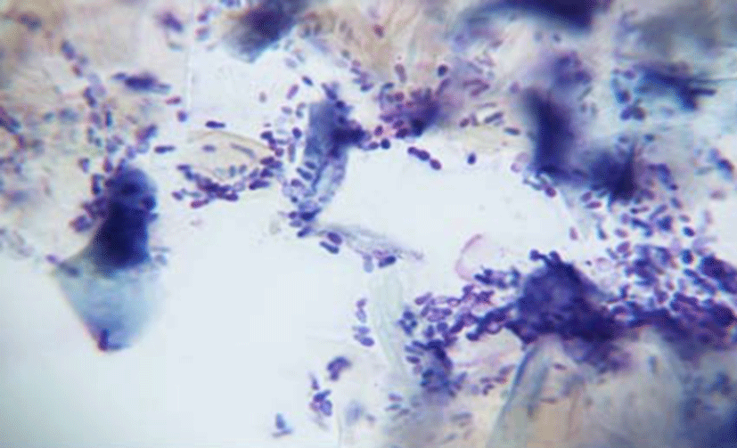
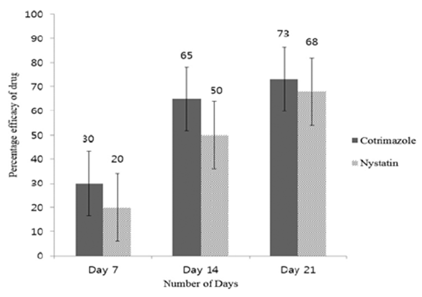
Out of the total 200 animals, 23% (46) were found positive for M. pachydermatis infection, whereas 154 dogs were negative. The highest prevalence (86.95%) of M. pachydermatis was observed in more than one year of age, and 13.04% was observed in less than one year of age. The prevalence of M. pachydermatis was higher in male than female dogs but statistically, the association between the two sexes was non-significantly different (P>0.05). The morphological identification criteria for the Malassezia spp. were the cell shape, size, and the budding pattern at 40× magnification by using a semiquantitative scale (Fig. 1). The prevalence of Malassezia otitis was significantly higher (P<0.05) in dogs possessing pendulous ears such as St. Bernard and Cocker spaniel than the ones having erected ears like German shepherd and Russian breeds groups (Table 1). The dogs of group A were treated with Clotrimazole, and group B were treated with Nystatin. Efficacy of drugs was measured on the basis of reversal of clinical signs and cytology examination. The efficacy of Clotrimazole and Nystatin was recorded as 73% and 68%, respectively at the completion of therapeutic trial. There was no significance difference (P>0.05) between two treatment groups on the completion of the trial (Fig. 2).


Discussion
In the present study, 46 (23%) cases of otitis externa were found positive for M. pachydermatis infection. Similar findings were reported by the previous studies [16-18] showing 24.6% prevalence in dogs. Another study described comparable prevalence of M. pachydermatis in cats i.e. 23.1% in Spain. This resembling level of infection in dogs and cats might be due to the matching climatic conditions in the areas of study.
In the present study, a non-significant difference (P>0.05) was observed between the pendulous ear and erected ear dogs. The highest percentage of Malassezia otitis externa was recorded in drooping ear dogs (56.52%) than in dogs with erected ears and medium hair on the ears (43.47%). Our study was strengthened by the results of [19] which also pointed out identical findings in pendulous ear (55.2%) and in erect ear breed (53.6%) groups. They also concluded that pendulous ears entrap more humidity within the ear canal along with ceruminous discharges which become infected secondarily and predispose the ears to Malassezia organisms’ replication. Likewise, Conkova and coworkers [20] found higher prevalence of M. pachydermatis in samples from longhaired (51.5%) and short-haired (45.9%) dogs compared to smooth haired (21.4%) dogs. The higher prevalence i.e. 86.9% in the current study was observed in age group of more than one years, which is very similar to the results of [11-13] who observed the peak incidence of otitis externa in dogs of the same age group, while a lower incidence was seen in animals less than one year old (13.04%).
In the present investigation, higher efficacy (73%) was recorded for Clotrimazole which showed a significant (P<0.05) reduction in yeast population and reversal of clinical signs of otitis, whereas Nystatin showed an efficacy of 68% as compared to Clotrimazole. This is in agreement with the study [2] which observed the efficacy of Clotrimazole to be 58.3% on day 14. In terms of availability, both drugs are readily available in the market with a number of products by different companies at different economical costs. Clotrimazole is a costly product as compared to Nystatin, but it showed better curative performance than Nystatin from mycological infection in Dogs. Moreover, it is concluded that Malassezia otitis is predominantly present in young age and pendulous ear dog breeds against which Clotrimazole is the most effective drug of the two.