Introduction
Tenosynovial giant cell tumor (TGCT) represents a group of neoplastic process that usually arises from the synovium of joints, bursae, and tendon sheaths. They can be classified as intra-articular or extra-articular by the anatomic location and as localized or diffuse by the growth pattern [1]. Localized TGCT most commonly involves the hand or wrist, followed by the ankle and foot region. Diffuse TGCT usually affects the knee and hip region. Histologically, TGCT consists of small or large mononuclear cells, multinucleated giant cells, foamy macrophages, and siderophages [2]. Cartilaginous metaplasia in TGCT is rare, with 10 reported cases of diffuse TGCT involving variable regions including the temporomandibular joint (TMJ), elbow, and hip joint [1-3]. There has been no report of chondroid metaplasia in localized TGCT involving the toe. Herein, we describe a case of chondroid TGCT arising in the flexor digitorum tendon sheath of the right second toe.
Case report
A 57-year-old male patient was hospitalized in January 2013 due to a palpable mass on the right second toe, which caused pain while walking. The pain began 1 year earlier, without history of trauma. Magnetic resonance imaging (MRI) revealed a well-defined, ovoid mass (2.2 cm in diameter) in the flexor digitorum tendon sheath of the second toe (Fig. 1). The resected mass, which measured 2.2 × 2.0 × 1.0 cm, was a well-demarcated nodule, with a lobulated outer surface and no invasion of the adjacent bone and tendon. The cut section was solid, grayish-white with focal red-brown areas. Microscopically, the tumor was multinodular and embedded in a partially circumscribed collagenous pseudocapsule on low power magnification, indicating the localized growth pattern. The tumor consisted of extensive cartilaginous metaplasia with focal, but typical areas of TGCT. The conventional TGCT areas consisted of mononuclear cell proliferation, multinucleated giant cells, and hemosiderin deposition (Fig. 2A). Specifically, the mononuclear cell population was composed of small histiocytic cells with round to oval nuclei, and large mononuclear cells with abundant eosinophilic or amphophilic cytoplasm, eccentric nuclei, distinct nucleoli, and intracytoplasmic hemosiderin. Foamy macrophages were not observed. Areas of chondroid metaplasia were noted throughout the tumor and comprised at least 90% of the tumor volume. The chondroid matrix was myxoid, basophilic stained, and had foci of calcification. The cellular components of the chondroid areas were morphologically similar to the large mononuclear cells in the conventional TGCT areas (Fig. 2B). There was no evidence of cellular pleomorphism, atypical mitoses, or necrosis.
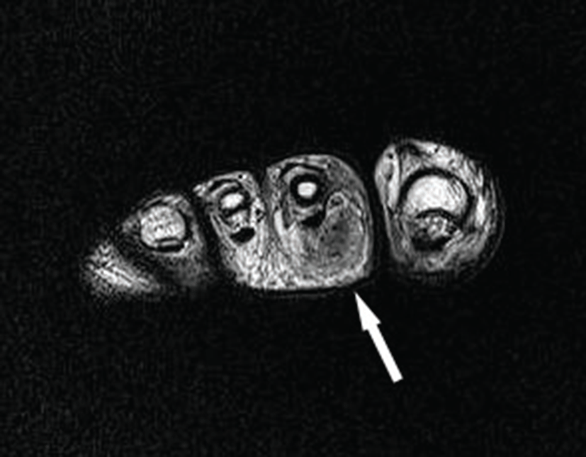
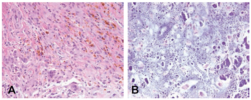
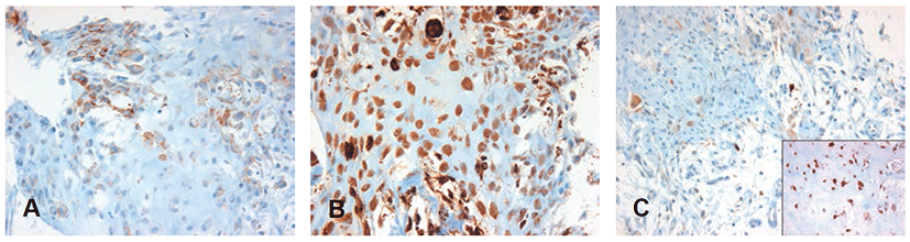
Table 1 summarizes the antibodies, their sources, and dilutions of antibodies against podoplanin, CD68, S100 used in commercially available immunohistochemical kits. Immunohistochemical staining showed focal cytoplasmic staining for podoplanin in the mononuclear cells in both the chondroid and conventional TGCT components (Fig. 3A). CD68 staining was diffuse and strong in mononuclear cells of both components, as well as in multinucleated giant cells (Fig. 3B). The mononuclear cells in the chondroid components were uniformly positive for S100 protein, whereas few cells in the conventional TGCT areas showed S100 protein positivity (Fig, 3C). Desmin-positive cells were not observed.
| Antibody | Clone | Dilution | Source |
|---|---|---|---|
| Podoplanin | D2-40 | 1:50 | Dako |
| CD68 | KP-1 | 1:200 | DiNonA |
| S100 | S100 | 1:800 | Dako |
Discussion
Chondroid metaplasia in TGCT is a rare phenomenon in both localized (giant cell tumor of tendon sheath) and diffuse (pigmented villonodular synovitis) type. Since the first report by Pignatti et al. [3] in 1990, 10 cases of chondroid metaplasia have been reported [1-4]. Clinical features of the previously described cases are summarized in Table 2. Of the 10 patients, 6 were male and 4 were female; their ages ranged from 36 to 70 years. Clinically, all cases presented with symptoms that were associated with the tumor. As described by Weiss and Goldblum [5], cartilaginous and osseous metaplasia is rare and focal finding in localized TGCT. However, there have been no reports of extensive chondroid metaplasia in localized TGCT specifically arising in the tendon sheath of the toe. Of the previously reported cases of chondroid TGCT, the degree of chondroid metaplasia was extensive in 4 cases and focal in 5 cases. All of the 10 cases of chondroid TGCT presented a diffuse growth pattern. Eight of the cases involved the TMJ, with the other 2 cases involving the hip and elbow joint, respectively. The toe is not an uncommon site of the conventional form of localized TGCT; however, the case presented herein is the first case of a chondroid variant of localized TGCT involving the tendon sheath of the toe.
| Reference | Age (range) | Sex | Involving site | Symptom | Size (range) | Growth pattern | Extent of chondroid metaplasia |
|---|---|---|---|---|---|---|---|
| Pignatti et al.3 (n=1) | 54 | M | Elbow joint | Pain and limited motion | 3 cm | Diffuse | Diffuse |
| Oda et al.4 (n=3) | 52 | M | TM joint | Ear tightness | 2 cm | Diffuse | 20% |
| 67 | M | TM joint | Pain | NA | Diffuse | 30% | |
| 52 | F | Hip joint | Coxalgia | 3 cm | Diffuse | 30% | |
| Hoch et al.5 (n=5) |
49 (36~70) |
M(n=3) F (n=2) |
TM joint | Mass effect | 3.9 cm (3.1~4.5) | Diffuse |
90% (n=3) 30%(n=2) |
| Fisher et al.6 (n=1) | 50 | F | Temporal bone | Aural fullness and otalgia | 0.5 cm | Diffuse | NA |
Histopathologically, chondroid TGCT was defined as a variant of TGCT by the recognition of conventional TGCT component within the tumor [1]. In the present case, the growth pattern was nodular. There was proliferation of small and large mononuclear cells, scattered multinucleated giant cells, and deposition of hemosiderin as a component of conventional TGCT. Notably, foamy macrophages, which are often present in conventional TGCT, were not identified in our case. This is consistent with the previous case reports, suggesting that foamy macrophages are not common in the chondroid variant of TGCT [1-4].
The presence of extensive chondroid metaplasia and calcification in the tumor need to be distinguished from other chondroid lesions, such as chondroblastoma and chondrosarcoma. Brien et al. [6], investigated chondroblastoma in a nonepiphyseal site and reported 15 cases of giant cell tumor of the tendon sheath or pigmented villonodular synovitis with extensive chondroid differentiation that were indistinguishable from chondroblastoma. In the present case, the pure basophilic stained chondroid matrix and poorly formed coarse calcification differed from the characteristics of chondroblastoma. In addiaddition, the cells within the chondroid matrix were distinguishable from chondroblasts, as the cells had larger and more abundant cytoplasm without longitudinal nuclear grooves.
Immunohistochemical studies were helpful in distinguishing TGCT from chondroblastoma. Mononuclear cells in the conventional TGCT components showed immunoreactivity for CD68 and were focal positive for S100 protein. In contrast, the staining pattern of chondroblastoma is negative for CD68 and positive for S100 protein. In the present case, mononuclear cells in both conventional and chondroid components showed focal cytoplasmic staining for podoplanin, a marker of synovial differentiation that is expressed in conventional TGCT [7]. The mononuclear cells in the chondroid areas were uniformly positive for S100 protein, whereas the mononuclear cells in the conventional TGCT areas showed focal positivity for S100 protein. There were no desmin-positive cells. Overall, the present case showed an immunophenotype that is consistent with the previously reported 10 cases of chondroid TGCT [1-4].
The tumor described herein could be distinguished from chondrosarcoma, as the growth pattern was not infiltrative. In addition, atypical nuclei, enlarged and hyperchromatic nuclei, and mitoses were not observed. Although, relatively large nuclei and prominent nucleoli were observed in mononuclear cells, the nuclear features were uniform, in contrast to what is observed in chondrosarcoma.
In conclusion, we described a rare case of localized TGCT with extensive chondroid metaplasia, that involved the tendon sheath of the toe. The presence of widespread chondroid matrix often leads to the misdiagnosis of other chondroid lesions; however, the recognition of the conventional TGCT areas with appropriate immunohistochemical analyses supports the diagnosis of the chondroid variant of TGCT.